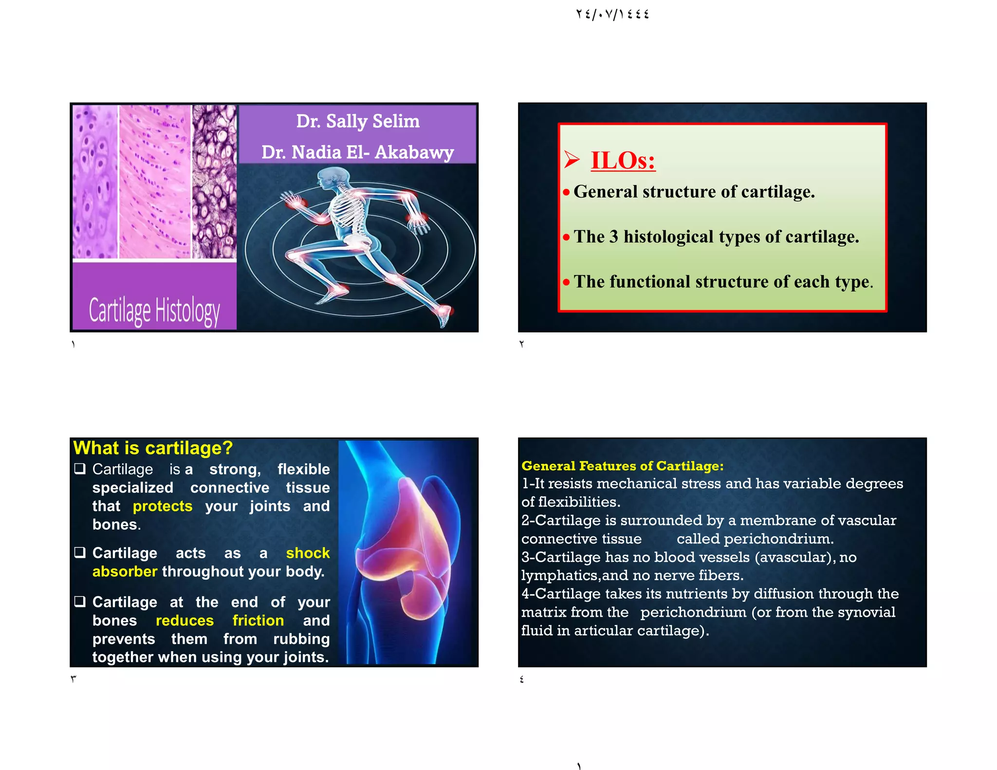
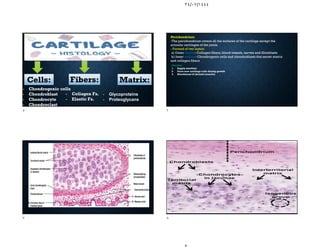
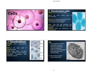
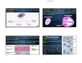
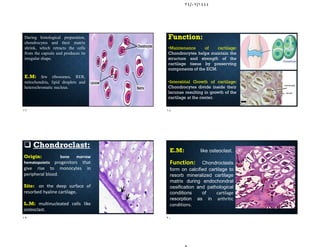
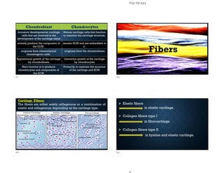
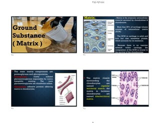
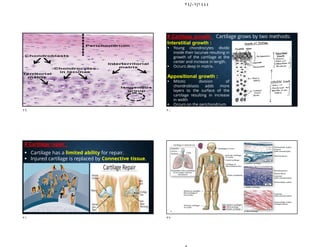
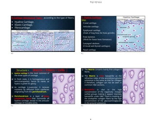
SlideShare a Scribd company logo
٢٤/٠٧/١٤٤٤
Dr. Sally Selim
Dr. Nadia El- Akabawy
 ILOs:
 General structure of cartilage.
 The 3 histological types of cartilage.
 The functional structure of each type.
What is cartilage?
 Cartilage is a strong, flexible
specialized connective tissue
that protects your joints and
bones.
 Cartilage acts as a shock
absorber throughout your body.
 Cartilage at the end of your
bones reduces friction and
prevents them from rubbing
together when using your joints.
General Features of Cartilage:
1-It resists mechanical stress and has variable degrees
of flexibilities.
2-Cartilage is surrounded by a membrane of vascular
connective tissue called perichondrium.
3-Cartilage has no blood vessels (avascular), no
lymphatics,and no nerve fibers.
4-Cartilage takes its nutrients by diffusion through the
matrix from the perichondrium (or from the synovial
fluid in articular cartilage).
١ ٢
٣ ٤
٢٤/٠٧/١٤٤٤
- Chondrogenic cells
- Chondroblast
- Chondrocyte
- Chondroclast
Matrix:
Fibers:
Cells:
- Collagen Fs.
- Elastic Fs.
- Glycoproteins
- Proteoglycans
Perichondrium:
-The perichondrium covers all the surfaces of the cartilage except the
articular cartilages of the joints.
- Formed of two layers:
a) Outer fibrous:Collagen fibers,blood vessels, nerves and fibroblasts
b) Inner cellular: Chondrogenic cells and chondroblasts that secret matrix
and collagen fibers
Function:
1. Supply nutrition
2. Form new cartilage cells during growth
3. Attachment of skeletal muscles
٥ ٦
٧ ٨
٢٤/٠٧/١٤٤٤
Cells
 Chondrogenic cells:
Origin: bone marrow derived MSCs.
Site: in the inner cellular layer of
perichondrium.
L.M: They are spindle-shaped narrow cells.
Nucleus: oval and pale basophilic.
The cytoplasm: light basophilic.
E.M: They have free ribosomes, few
mitochondria, some RER and small Golgi
apparatus.
Function: divide and differentiate into
chondroblasts.
 Chondroblast:
Origin: from chondrogenic
cells.
Site: in the inner side of
perichondrium.
L.M: elliptic shape, the long
axis is parallel to the surface,
basophilic cytoplasm with
vesicular nucleus.
EM
1. The cytoplasm is rich in
organelles of protein
synthesis (ribosomes,
rough endoplasmic
reticulum, well developed
Golgi apparatus and
mitochondria).
2. The nucleus contains
more euchromatin.
٩ ١٠
١١ ١٢
٢٤/٠٧/١٤٤٤
Function: play an important role in the formation
of cartilage.
•Development of cartilage:The primary function of the
chondroblast is the development of the cartilage.
Chondroblasts found in the inner layer of the perichondrium
mature to form chondrocytes, which form the cellular structure
of the cartilage tissue.
•Maintenance of cartilage: Chondroblast also helps maintain the
structure and strength of the cartilage tissue by producing
components of the ECM.
•Growth of cartilage: Chondroblasts carry out appositional
growth of the cartilage, i.e., thickening of cartilage tissue.
 Chondrocytes:
Origin: from chondroblast.
Site: in the cartilage matrix within
lacunae.
L.M: large rounded cells with dark
nucleus and pale basophilic cytoplasm.
Chondrocytes may also appear in
isogenous groups of up to eight cells.
Mitotic cell division of individual
chondral cells leads to cellular
grouping.
١٣ ١٤
١٥ ١٦
٢٤/٠٧/١٤٤٤
During histological preparation,
chondrocytes and their matrix
shrink, which retracts the cells
from the capsule and produces its
irregular shape.
E.M: few ribosomes, RER,
mitochondria, lipid droplets and
heterochromatic nucleus.
Function:
•Maintenance of cartilage:
Chondrocytes helps maintain the
structure and strength of the
cartilage tissue by preserving
components of the ECM.
•Interstitial Growth of cartilage:
Chondrocytes divide inside their
lacunae resulting in growth of the
cartilage at the center.
 Chondroclast:
Origin: bone marrow
hematopoietic progenitors that
give rise to monocytes in
peripheral blood.
Site: on the deep surface of
resorbed hyaline cartilage.
L.M: multinucleated cells like
osteoclast.
E.M: like osteoclast.
Function: Chondroclasts
form on calcified cartilage to
resorb mineralized cartilage
matrix during endochondral
ossification and pathological
conditions of cartilage
resorption as in arthritic
conditions.
١٧ ١٨
١٩ ٢٠
٢٤/٠٧/١٤٤٤
Chondroblast Chondrocytes
Immature developmental cartilage
cells that are involved in the
development of the cartilage tissue
Mature cartilage cells that function
to maintain the cartilage structure.
actively produce the component of
the ECM.
secrete ECM and are embedded in
it.
originate from mesenchymal
chondrogenic cells.
originate from the chondroblasts.
Appositional growth of the cartilage
by chondroblasts
Interstitial growth of the cartilage
by chondrocytes
Main function is to produce
chondrocytes and components of
the ECM
Primarily, to maintain the structure
of the cartilage and ECM
Fibers
Cartilage Fibers:
The fibers are either solely collagenous or a combination of
elastic and collagenous, depending on the cartilage type.
 Elastic fibers
in elastic cartilage.
 Collagen fibers type I
in fibrocartilage.
 Collagen fibers type II
in hyaline and elastic cartilage.
٢١ ٢٢
٢٣ ٢٤
٢٤/٠٧/١٤٤٤
Ground
Substance
( Matrix )
Matrix: - Matrix is the avascular extracellular
material secreted by chondroblast &
chondrocyte.
- More than 95% of cartilage volume
consists of extracellular matrix
(ECM).
- The ECM in cartilage is solid and
firm but also somewhat pliable,
which accounts for its resilience.
- Because there is no vascular
network within cartilage, the
composition of the ECM is crucial to
the survival of the chondrocytes.
 The main matrix components are
proteoglycans and glycoproteins;
- Proteoglycans: mostly sulfated
glycosaminoglycans responsible for matrix
basophilic staining. The main
glycosaminoglycans are chondroitin sulfate.
- Glycoprotein: adhesive proteins adhering
matrix to chondrocytes.
 The matrix closely
surrounding the
chondrocytes are
named capsular
territorial matrix, the
matrix in between
chondrocytes are
named interterritorial
matrix.
٢٥ ٢٦
٢٧ ٢٨
٢٤/٠٧/١٤٤٤
Interstitial growth :
 Young chondrocytes divide
inside their lacunae resulting in
growth of the cartilage at the
center and increase in length.
 Occurs deep in matrix.
Appositional growth :
 Mitotic division of
chondroblasts adds more
layers to the surface of the
cartilage resulting in increase
in width
 Occurs on the perichondrium.
# Cartilage growth: Cartilage grows by two methods:
# Cartilage repair :
 Cartilage has a limited ability for repair.
 Injured cartilage is replaced by Connective tissue.
٢٩ ٣٠
٣١ ٣٢
٢٤/٠٧/١٤٤٤
# Cartilage Histological Types : according to the type of fibers;
 Hyaline Cartilage.
 Elastic Cartilage.
 Fibrocartilage.
1- Hyaline Cartilage:
Site:
- Costal cartilage.
- Articular cartilage.
- Epiphyseal cartilage
(Ends of long bone for bone growth).
- Fetal skeleton
(Mold for future bone formation).
- Laryngeal skeleton
(Cricoid and thyroid cartilages).
- Nasal cartilage.
Structure = Matrix + Fibers + Cells
 Hyaline cartilage is the most common of
the three types of cartilage.
 In fresh state, it is homogeneous and
semi-transparent, hence its name, as
hyalos is greek for glassy.
 As cartilage is avascular, it receives
nutrients by diffusion from capillaries
originating from the perichondrium.
 Perichondrium is a layer of dense
connective tissue that surrounds all
hyaline cartilage, except in the articular
cartilage of movable joints.
 The Matrix contains mainly fine collagen II
fibrils.
 The Matrix is clear basophilic as the
refractive index of the amorphous ground
substance is the same as that for collagen
II therefore, collagen can not be seen in
common H&E sections.
 Basophilia is due to the high
concentration of the negatively- charged
sulfate groups making it appear intensely
basophilic under H&E. There is masking of
the acidophilic collagen fibers by the high
concentration of the glycosaminoglycans
in the ground substance.
٣٣ ٣٤
٣٥ ٣٦
٢٤/٠٧/١٤٤٤
Hyaline Cartilage
Cells:
- Chondrogenic cells:
Found mainly in the inner
chondrogenic layer of the
perichondrium.
- Chondroblasts:
Found superficially under
the perichondrium.
- Chondrocytes:
They are located in tiny
spaces within the cartilage
matrix known as lacunae.
2- Elastic Cartilage:
Site:
- Auricle and Auditory tube.
- Cuneiform Cartilage.
- Epiglottis and External auditory meatus.
Structure = Matrix +
Fibers + Cells
 Elastic cartilage is resilient, easily
returning to its original shape after
bending or distortion so, it present in
areas where elastic recoil is needed.
 In fresh state, it is yellow in color.
 The Matrix is rich in dense,
interwoven network of elastic fibers
embedded in a small amount of
amorphous extracellular ground
substance plus collagen II fibrils.
 Elastic fibers stain pink in
Hematoxylin and Eosin, black in
Verhoeff's Van Gieson stain, and
brown in Orcein.
3- Fibrocartilage:
Site:
- Symphysis pubis.
- Annulus fibrosis of
intervertebral discs.
- At points of tendons’
attachment to bone.
- Tempo- mandibular joint.
٣٧ ٣٨
٣٩ ٤٠
٢٤/٠٧/١٤٤٤
 In fresh state, fibrocartilage
is white in color.
 Fibrocartilage is the
strongest and least flexible
of the three types.
 Its primary role is to provide
reinforcement at weight-
bearing areas, It acts more
like a shock absorber.
Structure = Matrix + Fibers + Cells  Fibrocartilage has a dense arrangement of type I collagen fibers that
are arranged in an orderly manner.
 Numerous chondrocytes are located within their lacunae and are
spaced between the acidophilic regular collagen bundles.
 No perichondrium.
# Clinical note:
Disc prolapse;
- Disk herniation occurs
when the fibrocartilage
surrounding the
intervertebral disk
ruptures, releasing the
nucleus pulposus.
- The resultant pressure on
spinal nerve roots may
cause pain and damage the
surrounding nerves.
R
BE
M
E
M
E
R
٤١ ٤٢
٤٣ ٤٤
٢٤/٠٧/١٤٤٤
Articular cartilage lacks perichondrium and it receives nutrient
from the synovial fluid
The matrix is more basophilic near the chondrocyte.
Collagen type II in a basophilic matrix, but the collagen is not
visible with the L.M.
Chondrocytes divide and form small groups called isogenous
group.
found in:
- Respiratory tract - Articular cartilage
Hyaline cartilage
Elastin stain stains elastic fibers black
Collagen type II is also present.
Many elastic fibers in the matrix and in perichondrium.
More chondrocytes and less matrix than hyaline cartilage.
found in:
- External ear - Epiglottis
Elastic cartilage
No perichondrium
Collagen type I present in parallel acidophilic
bundles. (Note: Collagen type I is stronger than type II)
More fibers and less chondrocytes than that in hyaline
and elastic cartilage.
Chondrocytes arise from fibroblasts and form parallel rows.
found in: - Intervertebral discs
Fibrocartilage
All types have chondrocytes in lacunae.
All types have avascular matrix.
All types have perichondrium-->>
EXCEPT: Fibrocartilage/ Articular cartilage
Similarities between types of cartilage
٤٥ ٤٦
٤٧ ٤٨
٢٤/٠٧/١٤٤٤
٤٩

More Related Content

Similar to Cartilage presentation pdf.pdf

CARTILAGES.pptx
CARTILAGES.pptxCARTILAGES.pptx
CARTILAGES.pptx
Dr Durgadevi Boopathi
 
د حاتم البيطار استشاري وجراح الفم والاسنان 01005684344 اتصل للحجز بالعيادة ...
 د حاتم البيطار استشاري وجراح الفم والاسنان 01005684344 اتصل للحجز بالعيادة  ... د حاتم البيطار استشاري وجراح الفم والاسنان 01005684344 اتصل للحجز بالعيادة  ...
د حاتم البيطار استشاري وجراح الفم والاسنان 01005684344 اتصل للحجز بالعيادة ...
د حاتم البيطار
 
Connective tissue ppt
Connective tissue pptConnective tissue ppt
Connective tissue ppt
Prof Vijayraddi
 
Skeletal Physiology
Skeletal PhysiologySkeletal Physiology
Skeletal Physiologytd0297bn
 
Connective tissue
Connective tissueConnective tissue
Connective tissue
Yash Prajapati
 
6. SPECIALISED CONNECTIVE TISSUE.pptx
6. SPECIALISED CONNECTIVE TISSUE.pptx6. SPECIALISED CONNECTIVE TISSUE.pptx
6. SPECIALISED CONNECTIVE TISSUE.pptx
FranciKaySichu
 
SEM2 MUSK BOOK 2024.pdfًًًًًًًًًًًًًًًًً
SEM2 MUSK BOOK 2024.pdfًًًًًًًًًًًًًًًًًSEM2 MUSK BOOK 2024.pdfًًًًًًًًًًًًًًًًً
SEM2 MUSK BOOK 2024.pdfًًًًًًًًًًًًًًًًً
EA S
 
Cartilage.pptx
Cartilage.pptxCartilage.pptx
Cartilage.pptx
IMRANKHAN706639
 
Periodontal ligament dr. sherif hassan
Periodontal ligament dr. sherif hassan Periodontal ligament dr. sherif hassan
Periodontal ligament dr. sherif hassan
sherifsayed65
 
Histologia Tema 16 tejido cartilaginoso
Histologia Tema 16 tejido cartilaginosoHistologia Tema 16 tejido cartilaginoso
Histologia Tema 16 tejido cartilaginoso
ulamedicina2012
 
Cartilage (1)
Cartilage  (1)Cartilage  (1)
Cartilage (1)
Zainab&Sons
 
Animal Tissues
Animal TissuesAnimal Tissues
Animal Tissues
Biswarup Majumder
 
LP 1-cartilage ppt.pptx
LP 1-cartilage ppt.pptxLP 1-cartilage ppt.pptx
LP 1-cartilage ppt.pptx
TenzinNamgayNidrup
 
periodontal ligament dr. sherif hassan
  periodontal ligament dr. sherif hassan  periodontal ligament dr. sherif hassan
periodontal ligament dr. sherif hassan
sherifsayed65
 
OSCe.pdf in Babylon university College o
OSCe.pdf in Babylon university College oOSCe.pdf in Babylon university College o
OSCe.pdf in Babylon university College o
ham793melakthabit
 
Histology 2 connective_tissue
Histology 2 connective_tissueHistology 2 connective_tissue
Histology 2 connective_tissueTyler Wall
 
A and P Mod#2 pages 50 -56
A and P Mod#2 pages 50 -56A and P Mod#2 pages 50 -56
A and P Mod#2 pages 50 -56Kelley Crawford
 
Connective tissues
Connective tissues Connective tissues

Similar to Cartilage presentation pdf.pdf (20)

CARTILAGES.pptx
CARTILAGES.pptxCARTILAGES.pptx
CARTILAGES.pptx
 
د حاتم البيطار استشاري وجراح الفم والاسنان 01005684344 اتصل للحجز بالعيادة ...
 د حاتم البيطار استشاري وجراح الفم والاسنان 01005684344 اتصل للحجز بالعيادة  ... د حاتم البيطار استشاري وجراح الفم والاسنان 01005684344 اتصل للحجز بالعيادة  ...
د حاتم البيطار استشاري وجراح الفم والاسنان 01005684344 اتصل للحجز بالعيادة ...
 
Connective tissue ppt
Connective tissue pptConnective tissue ppt
Connective tissue ppt
 
Skeletal Physiology
Skeletal PhysiologySkeletal Physiology
Skeletal Physiology
 
Connective tissue
Connective tissueConnective tissue
Connective tissue
 
6. SPECIALISED CONNECTIVE TISSUE.pptx
6. SPECIALISED CONNECTIVE TISSUE.pptx6. SPECIALISED CONNECTIVE TISSUE.pptx
6. SPECIALISED CONNECTIVE TISSUE.pptx
 
SEM2 MUSK BOOK 2024.pdfًًًًًًًًًًًًًًًًً
SEM2 MUSK BOOK 2024.pdfًًًًًًًًًًًًًًًًًSEM2 MUSK BOOK 2024.pdfًًًًًًًًًًًًًًًًً
SEM2 MUSK BOOK 2024.pdfًًًًًًًًًًًًًًًًً
 
Cartilage
CartilageCartilage
Cartilage
 
Cartilage.pptx
Cartilage.pptxCartilage.pptx
Cartilage.pptx
 
Periodontal ligament dr. sherif hassan
Periodontal ligament dr. sherif hassan Periodontal ligament dr. sherif hassan
Periodontal ligament dr. sherif hassan
 
Histologia Tema 16 tejido cartilaginoso
Histologia Tema 16 tejido cartilaginosoHistologia Tema 16 tejido cartilaginoso
Histologia Tema 16 tejido cartilaginoso
 
Cartilage (1)
Cartilage  (1)Cartilage  (1)
Cartilage (1)
 
Animal Tissues
Animal TissuesAnimal Tissues
Animal Tissues
 
LP 1-cartilage ppt.pptx
LP 1-cartilage ppt.pptxLP 1-cartilage ppt.pptx
LP 1-cartilage ppt.pptx
 
periodontal ligament dr. sherif hassan
  periodontal ligament dr. sherif hassan  periodontal ligament dr. sherif hassan
periodontal ligament dr. sherif hassan
 
OSCe.pdf in Babylon university College o
OSCe.pdf in Babylon university College oOSCe.pdf in Babylon university College o
OSCe.pdf in Babylon university College o
 
Section 2,chapter 5
Section 2,chapter 5Section 2,chapter 5
Section 2,chapter 5
 
Histology 2 connective_tissue
Histology 2 connective_tissueHistology 2 connective_tissue
Histology 2 connective_tissue
 
A and P Mod#2 pages 50 -56
A and P Mod#2 pages 50 -56A and P Mod#2 pages 50 -56
A and P Mod#2 pages 50 -56
 
Connective tissues
Connective tissues Connective tissues
Connective tissues
 

Recently uploaded

Phone Us ❤85270-49040❤ #ℂall #gIRLS In Surat By Surat @ℂall @Girls Hotel With...
Phone Us ❤85270-49040❤ #ℂall #gIRLS In Surat By Surat @ℂall @Girls Hotel With...Phone Us ❤85270-49040❤ #ℂall #gIRLS In Surat By Surat @ℂall @Girls Hotel With...
Phone Us ❤85270-49040❤ #ℂall #gIRLS In Surat By Surat @ℂall @Girls Hotel With...
Savita Shen $i11
 
Tom Selleck Health: A Comprehensive Look at the Iconic Actor’s Wellness Journey
Tom Selleck Health: A Comprehensive Look at the Iconic Actor’s Wellness JourneyTom Selleck Health: A Comprehensive Look at the Iconic Actor’s Wellness Journey
Tom Selleck Health: A Comprehensive Look at the Iconic Actor’s Wellness Journey
greendigital
 
basicmodesofventilation2022-220313203758.pdf
basicmodesofventilation2022-220313203758.pdfbasicmodesofventilation2022-220313203758.pdf
basicmodesofventilation2022-220313203758.pdf
aljamhori teaching hospital
 
ACUTE SCROTUM.....pdf. ACUTE SCROTAL CONDITIOND
ACUTE SCROTUM.....pdf. ACUTE SCROTAL CONDITIONDACUTE SCROTUM.....pdf. ACUTE SCROTAL CONDITIOND
ACUTE SCROTUM.....pdf. ACUTE SCROTAL CONDITIOND
DR SETH JOTHAM
 
TEST BANK for Operations Management, 14th Edition by William J. Stevenson, Ve...
TEST BANK for Operations Management, 14th Edition by William J. Stevenson, Ve...TEST BANK for Operations Management, 14th Edition by William J. Stevenson, Ve...
TEST BANK for Operations Management, 14th Edition by William J. Stevenson, Ve...
kevinkariuki227
 
Hemodialysis: Chapter 3, Dialysis Water Unit - Dr.Gawad
Hemodialysis: Chapter 3, Dialysis Water Unit - Dr.GawadHemodialysis: Chapter 3, Dialysis Water Unit - Dr.Gawad
Hemodialysis: Chapter 3, Dialysis Water Unit - Dr.Gawad
NephroTube - Dr.Gawad
 
Cervical & Brachial Plexus By Dr. RIG.pptx
Cervical & Brachial Plexus By Dr. RIG.pptxCervical & Brachial Plexus By Dr. RIG.pptx
Cervical & Brachial Plexus By Dr. RIG.pptx
Dr. Rabia Inam Gandapore
 
Surat @ℂall @Girls ꧁❤8527049040❤꧂@ℂall @Girls Service Vip Top Model Safe
Surat @ℂall @Girls ꧁❤8527049040❤꧂@ℂall @Girls Service Vip Top Model SafeSurat @ℂall @Girls ꧁❤8527049040❤꧂@ℂall @Girls Service Vip Top Model Safe
Surat @ℂall @Girls ꧁❤8527049040❤꧂@ℂall @Girls Service Vip Top Model Safe
Savita Shen $i11
 
Knee anatomy and clinical tests 2024.pdf
Knee anatomy and clinical tests 2024.pdfKnee anatomy and clinical tests 2024.pdf
Knee anatomy and clinical tests 2024.pdf
vimalpl1234
 
Prix Galien International 2024 Forum Program
Prix Galien International 2024 Forum ProgramPrix Galien International 2024 Forum Program
Prix Galien International 2024 Forum Program
Levi Shapiro
 
Ozempic: Preoperative Management of Patients on GLP-1 Receptor Agonists
Ozempic: Preoperative Management of Patients on GLP-1 Receptor Agonists  Ozempic: Preoperative Management of Patients on GLP-1 Receptor Agonists
Ozempic: Preoperative Management of Patients on GLP-1 Receptor Agonists
Saeid Safari
 
Charaka Samhita Sutra sthana Chapter 15 Upakalpaniyaadhyaya
Charaka Samhita Sutra sthana Chapter 15 UpakalpaniyaadhyayaCharaka Samhita Sutra sthana Chapter 15 Upakalpaniyaadhyaya
Charaka Samhita Sutra sthana Chapter 15 Upakalpaniyaadhyaya
Dr KHALID B.M
 
HOT NEW PRODUCT! BIG SALES FAST SHIPPING NOW FROM CHINA!! EU KU DB BK substit...
HOT NEW PRODUCT! BIG SALES FAST SHIPPING NOW FROM CHINA!! EU KU DB BK substit...HOT NEW PRODUCT! BIG SALES FAST SHIPPING NOW FROM CHINA!! EU KU DB BK substit...
HOT NEW PRODUCT! BIG SALES FAST SHIPPING NOW FROM CHINA!! EU KU DB BK substit...
GL Anaacs
 
Are There Any Natural Remedies To Treat Syphilis.pdf
Are There Any Natural Remedies To Treat Syphilis.pdfAre There Any Natural Remedies To Treat Syphilis.pdf
Are There Any Natural Remedies To Treat Syphilis.pdf
Little Cross Family Clinic
 
New Drug Discovery and Development .....
New Drug Discovery and Development .....New Drug Discovery and Development .....
New Drug Discovery and Development .....
NEHA GUPTA
 
How STIs Influence the Development of Pelvic Inflammatory Disease.pptx
How STIs Influence the Development of Pelvic Inflammatory Disease.pptxHow STIs Influence the Development of Pelvic Inflammatory Disease.pptx
How STIs Influence the Development of Pelvic Inflammatory Disease.pptx
FFragrant
 
Flu Vaccine Alert in Bangalore Karnataka
Flu Vaccine Alert in Bangalore KarnatakaFlu Vaccine Alert in Bangalore Karnataka
Flu Vaccine Alert in Bangalore Karnataka
addon Scans
 
micro teaching on communication m.sc nursing.pdf
micro teaching on communication m.sc nursing.pdfmicro teaching on communication m.sc nursing.pdf
micro teaching on communication m.sc nursing.pdf
Anurag Sharma
 
BENIGN PROSTATIC HYPERPLASIA.BPH. BPHpdf
BENIGN PROSTATIC HYPERPLASIA.BPH. BPHpdfBENIGN PROSTATIC HYPERPLASIA.BPH. BPHpdf
BENIGN PROSTATIC HYPERPLASIA.BPH. BPHpdf
DR SETH JOTHAM
 
Physiology of Chemical Sensation of smell.pdf
Physiology of Chemical Sensation of smell.pdfPhysiology of Chemical Sensation of smell.pdf
Physiology of Chemical Sensation of smell.pdf
MedicoseAcademics
 

Recently uploaded (20)

Phone Us ❤85270-49040❤ #ℂall #gIRLS In Surat By Surat @ℂall @Girls Hotel With...
Phone Us ❤85270-49040❤ #ℂall #gIRLS In Surat By Surat @ℂall @Girls Hotel With...Phone Us ❤85270-49040❤ #ℂall #gIRLS In Surat By Surat @ℂall @Girls Hotel With...
Phone Us ❤85270-49040❤ #ℂall #gIRLS In Surat By Surat @ℂall @Girls Hotel With...
 
Tom Selleck Health: A Comprehensive Look at the Iconic Actor’s Wellness Journey
Tom Selleck Health: A Comprehensive Look at the Iconic Actor’s Wellness JourneyTom Selleck Health: A Comprehensive Look at the Iconic Actor’s Wellness Journey
Tom Selleck Health: A Comprehensive Look at the Iconic Actor’s Wellness Journey
 
basicmodesofventilation2022-220313203758.pdf
basicmodesofventilation2022-220313203758.pdfbasicmodesofventilation2022-220313203758.pdf
basicmodesofventilation2022-220313203758.pdf
 
ACUTE SCROTUM.....pdf. ACUTE SCROTAL CONDITIOND
ACUTE SCROTUM.....pdf. ACUTE SCROTAL CONDITIONDACUTE SCROTUM.....pdf. ACUTE SCROTAL CONDITIOND
ACUTE SCROTUM.....pdf. ACUTE SCROTAL CONDITIOND
 
TEST BANK for Operations Management, 14th Edition by William J. Stevenson, Ve...
TEST BANK for Operations Management, 14th Edition by William J. Stevenson, Ve...TEST BANK for Operations Management, 14th Edition by William J. Stevenson, Ve...
TEST BANK for Operations Management, 14th Edition by William J. Stevenson, Ve...
 
Hemodialysis: Chapter 3, Dialysis Water Unit - Dr.Gawad
Hemodialysis: Chapter 3, Dialysis Water Unit - Dr.GawadHemodialysis: Chapter 3, Dialysis Water Unit - Dr.Gawad
Hemodialysis: Chapter 3, Dialysis Water Unit - Dr.Gawad
 
Cervical & Brachial Plexus By Dr. RIG.pptx
Cervical & Brachial Plexus By Dr. RIG.pptxCervical & Brachial Plexus By Dr. RIG.pptx
Cervical & Brachial Plexus By Dr. RIG.pptx
 
Surat @ℂall @Girls ꧁❤8527049040❤꧂@ℂall @Girls Service Vip Top Model Safe
Surat @ℂall @Girls ꧁❤8527049040❤꧂@ℂall @Girls Service Vip Top Model SafeSurat @ℂall @Girls ꧁❤8527049040❤꧂@ℂall @Girls Service Vip Top Model Safe
Surat @ℂall @Girls ꧁❤8527049040❤꧂@ℂall @Girls Service Vip Top Model Safe
 
Knee anatomy and clinical tests 2024.pdf
Knee anatomy and clinical tests 2024.pdfKnee anatomy and clinical tests 2024.pdf
Knee anatomy and clinical tests 2024.pdf
 
Prix Galien International 2024 Forum Program
Prix Galien International 2024 Forum ProgramPrix Galien International 2024 Forum Program
Prix Galien International 2024 Forum Program
 
Ozempic: Preoperative Management of Patients on GLP-1 Receptor Agonists
Ozempic: Preoperative Management of Patients on GLP-1 Receptor Agonists  Ozempic: Preoperative Management of Patients on GLP-1 Receptor Agonists
Ozempic: Preoperative Management of Patients on GLP-1 Receptor Agonists
 
Charaka Samhita Sutra sthana Chapter 15 Upakalpaniyaadhyaya
Charaka Samhita Sutra sthana Chapter 15 UpakalpaniyaadhyayaCharaka Samhita Sutra sthana Chapter 15 Upakalpaniyaadhyaya
Charaka Samhita Sutra sthana Chapter 15 Upakalpaniyaadhyaya
 
HOT NEW PRODUCT! BIG SALES FAST SHIPPING NOW FROM CHINA!! EU KU DB BK substit...
HOT NEW PRODUCT! BIG SALES FAST SHIPPING NOW FROM CHINA!! EU KU DB BK substit...HOT NEW PRODUCT! BIG SALES FAST SHIPPING NOW FROM CHINA!! EU KU DB BK substit...
HOT NEW PRODUCT! BIG SALES FAST SHIPPING NOW FROM CHINA!! EU KU DB BK substit...
 
Are There Any Natural Remedies To Treat Syphilis.pdf
Are There Any Natural Remedies To Treat Syphilis.pdfAre There Any Natural Remedies To Treat Syphilis.pdf
Are There Any Natural Remedies To Treat Syphilis.pdf
 
New Drug Discovery and Development .....
New Drug Discovery and Development .....New Drug Discovery and Development .....
New Drug Discovery and Development .....
 
How STIs Influence the Development of Pelvic Inflammatory Disease.pptx
How STIs Influence the Development of Pelvic Inflammatory Disease.pptxHow STIs Influence the Development of Pelvic Inflammatory Disease.pptx
How STIs Influence the Development of Pelvic Inflammatory Disease.pptx
 
Flu Vaccine Alert in Bangalore Karnataka
Flu Vaccine Alert in Bangalore KarnatakaFlu Vaccine Alert in Bangalore Karnataka
Flu Vaccine Alert in Bangalore Karnataka
 
micro teaching on communication m.sc nursing.pdf
micro teaching on communication m.sc nursing.pdfmicro teaching on communication m.sc nursing.pdf
micro teaching on communication m.sc nursing.pdf
 
BENIGN PROSTATIC HYPERPLASIA.BPH. BPHpdf
BENIGN PROSTATIC HYPERPLASIA.BPH. BPHpdfBENIGN PROSTATIC HYPERPLASIA.BPH. BPHpdf
BENIGN PROSTATIC HYPERPLASIA.BPH. BPHpdf
 
Physiology of Chemical Sensation of smell.pdf
Physiology of Chemical Sensation of smell.pdfPhysiology of Chemical Sensation of smell.pdf
Physiology of Chemical Sensation of smell.pdf
 

Cartilage presentation pdf.pdf

  • 1. ٢٤/٠٧/١٤٤٤ Dr. Sally Selim Dr. Nadia El- Akabawy  ILOs:  General structure of cartilage.  The 3 histological types of cartilage.  The functional structure of each type. What is cartilage?  Cartilage is a strong, flexible specialized connective tissue that protects your joints and bones.  Cartilage acts as a shock absorber throughout your body.  Cartilage at the end of your bones reduces friction and prevents them from rubbing together when using your joints. General Features of Cartilage: 1-It resists mechanical stress and has variable degrees of flexibilities. 2-Cartilage is surrounded by a membrane of vascular connective tissue called perichondrium. 3-Cartilage has no blood vessels (avascular), no lymphatics,and no nerve fibers. 4-Cartilage takes its nutrients by diffusion through the matrix from the perichondrium (or from the synovial fluid in articular cartilage). ١ ٢ ٣ ٤
  • 2. ٢٤/٠٧/١٤٤٤ - Chondrogenic cells - Chondroblast - Chondrocyte - Chondroclast Matrix: Fibers: Cells: - Collagen Fs. - Elastic Fs. - Glycoproteins - Proteoglycans Perichondrium: -The perichondrium covers all the surfaces of the cartilage except the articular cartilages of the joints. - Formed of two layers: a) Outer fibrous:Collagen fibers,blood vessels, nerves and fibroblasts b) Inner cellular: Chondrogenic cells and chondroblasts that secret matrix and collagen fibers Function: 1. Supply nutrition 2. Form new cartilage cells during growth 3. Attachment of skeletal muscles ٥ ٦ ٧ ٨
  • 3. ٢٤/٠٧/١٤٤٤ Cells  Chondrogenic cells: Origin: bone marrow derived MSCs. Site: in the inner cellular layer of perichondrium. L.M: They are spindle-shaped narrow cells. Nucleus: oval and pale basophilic. The cytoplasm: light basophilic. E.M: They have free ribosomes, few mitochondria, some RER and small Golgi apparatus. Function: divide and differentiate into chondroblasts.  Chondroblast: Origin: from chondrogenic cells. Site: in the inner side of perichondrium. L.M: elliptic shape, the long axis is parallel to the surface, basophilic cytoplasm with vesicular nucleus. EM 1. The cytoplasm is rich in organelles of protein synthesis (ribosomes, rough endoplasmic reticulum, well developed Golgi apparatus and mitochondria). 2. The nucleus contains more euchromatin. ٩ ١٠ ١١ ١٢
  • 4. ٢٤/٠٧/١٤٤٤ Function: play an important role in the formation of cartilage. •Development of cartilage:The primary function of the chondroblast is the development of the cartilage. Chondroblasts found in the inner layer of the perichondrium mature to form chondrocytes, which form the cellular structure of the cartilage tissue. •Maintenance of cartilage: Chondroblast also helps maintain the structure and strength of the cartilage tissue by producing components of the ECM. •Growth of cartilage: Chondroblasts carry out appositional growth of the cartilage, i.e., thickening of cartilage tissue.  Chondrocytes: Origin: from chondroblast. Site: in the cartilage matrix within lacunae. L.M: large rounded cells with dark nucleus and pale basophilic cytoplasm. Chondrocytes may also appear in isogenous groups of up to eight cells. Mitotic cell division of individual chondral cells leads to cellular grouping. ١٣ ١٤ ١٥ ١٦
  • 5. ٢٤/٠٧/١٤٤٤ During histological preparation, chondrocytes and their matrix shrink, which retracts the cells from the capsule and produces its irregular shape. E.M: few ribosomes, RER, mitochondria, lipid droplets and heterochromatic nucleus. Function: •Maintenance of cartilage: Chondrocytes helps maintain the structure and strength of the cartilage tissue by preserving components of the ECM. •Interstitial Growth of cartilage: Chondrocytes divide inside their lacunae resulting in growth of the cartilage at the center.  Chondroclast: Origin: bone marrow hematopoietic progenitors that give rise to monocytes in peripheral blood. Site: on the deep surface of resorbed hyaline cartilage. L.M: multinucleated cells like osteoclast. E.M: like osteoclast. Function: Chondroclasts form on calcified cartilage to resorb mineralized cartilage matrix during endochondral ossification and pathological conditions of cartilage resorption as in arthritic conditions. ١٧ ١٨ ١٩ ٢٠
  • 6. ٢٤/٠٧/١٤٤٤ Chondroblast Chondrocytes Immature developmental cartilage cells that are involved in the development of the cartilage tissue Mature cartilage cells that function to maintain the cartilage structure. actively produce the component of the ECM. secrete ECM and are embedded in it. originate from mesenchymal chondrogenic cells. originate from the chondroblasts. Appositional growth of the cartilage by chondroblasts Interstitial growth of the cartilage by chondrocytes Main function is to produce chondrocytes and components of the ECM Primarily, to maintain the structure of the cartilage and ECM Fibers Cartilage Fibers: The fibers are either solely collagenous or a combination of elastic and collagenous, depending on the cartilage type.  Elastic fibers in elastic cartilage.  Collagen fibers type I in fibrocartilage.  Collagen fibers type II in hyaline and elastic cartilage. ٢١ ٢٢ ٢٣ ٢٤
  • 7. ٢٤/٠٧/١٤٤٤ Ground Substance ( Matrix ) Matrix: - Matrix is the avascular extracellular material secreted by chondroblast & chondrocyte. - More than 95% of cartilage volume consists of extracellular matrix (ECM). - The ECM in cartilage is solid and firm but also somewhat pliable, which accounts for its resilience. - Because there is no vascular network within cartilage, the composition of the ECM is crucial to the survival of the chondrocytes.  The main matrix components are proteoglycans and glycoproteins; - Proteoglycans: mostly sulfated glycosaminoglycans responsible for matrix basophilic staining. The main glycosaminoglycans are chondroitin sulfate. - Glycoprotein: adhesive proteins adhering matrix to chondrocytes.  The matrix closely surrounding the chondrocytes are named capsular territorial matrix, the matrix in between chondrocytes are named interterritorial matrix. ٢٥ ٢٦ ٢٧ ٢٨
  • 8. ٢٤/٠٧/١٤٤٤ Interstitial growth :  Young chondrocytes divide inside their lacunae resulting in growth of the cartilage at the center and increase in length.  Occurs deep in matrix. Appositional growth :  Mitotic division of chondroblasts adds more layers to the surface of the cartilage resulting in increase in width  Occurs on the perichondrium. # Cartilage growth: Cartilage grows by two methods: # Cartilage repair :  Cartilage has a limited ability for repair.  Injured cartilage is replaced by Connective tissue. ٢٩ ٣٠ ٣١ ٣٢
  • 9. ٢٤/٠٧/١٤٤٤ # Cartilage Histological Types : according to the type of fibers;  Hyaline Cartilage.  Elastic Cartilage.  Fibrocartilage. 1- Hyaline Cartilage: Site: - Costal cartilage. - Articular cartilage. - Epiphyseal cartilage (Ends of long bone for bone growth). - Fetal skeleton (Mold for future bone formation). - Laryngeal skeleton (Cricoid and thyroid cartilages). - Nasal cartilage. Structure = Matrix + Fibers + Cells  Hyaline cartilage is the most common of the three types of cartilage.  In fresh state, it is homogeneous and semi-transparent, hence its name, as hyalos is greek for glassy.  As cartilage is avascular, it receives nutrients by diffusion from capillaries originating from the perichondrium.  Perichondrium is a layer of dense connective tissue that surrounds all hyaline cartilage, except in the articular cartilage of movable joints.  The Matrix contains mainly fine collagen II fibrils.  The Matrix is clear basophilic as the refractive index of the amorphous ground substance is the same as that for collagen II therefore, collagen can not be seen in common H&E sections.  Basophilia is due to the high concentration of the negatively- charged sulfate groups making it appear intensely basophilic under H&E. There is masking of the acidophilic collagen fibers by the high concentration of the glycosaminoglycans in the ground substance. ٣٣ ٣٤ ٣٥ ٣٦
  • 10. ٢٤/٠٧/١٤٤٤ Hyaline Cartilage Cells: - Chondrogenic cells: Found mainly in the inner chondrogenic layer of the perichondrium. - Chondroblasts: Found superficially under the perichondrium. - Chondrocytes: They are located in tiny spaces within the cartilage matrix known as lacunae. 2- Elastic Cartilage: Site: - Auricle and Auditory tube. - Cuneiform Cartilage. - Epiglottis and External auditory meatus. Structure = Matrix + Fibers + Cells  Elastic cartilage is resilient, easily returning to its original shape after bending or distortion so, it present in areas where elastic recoil is needed.  In fresh state, it is yellow in color.  The Matrix is rich in dense, interwoven network of elastic fibers embedded in a small amount of amorphous extracellular ground substance plus collagen II fibrils.  Elastic fibers stain pink in Hematoxylin and Eosin, black in Verhoeff's Van Gieson stain, and brown in Orcein. 3- Fibrocartilage: Site: - Symphysis pubis. - Annulus fibrosis of intervertebral discs. - At points of tendons’ attachment to bone. - Tempo- mandibular joint. ٣٧ ٣٨ ٣٩ ٤٠
  • 11. ٢٤/٠٧/١٤٤٤  In fresh state, fibrocartilage is white in color.  Fibrocartilage is the strongest and least flexible of the three types.  Its primary role is to provide reinforcement at weight- bearing areas, It acts more like a shock absorber. Structure = Matrix + Fibers + Cells  Fibrocartilage has a dense arrangement of type I collagen fibers that are arranged in an orderly manner.  Numerous chondrocytes are located within their lacunae and are spaced between the acidophilic regular collagen bundles.  No perichondrium. # Clinical note: Disc prolapse; - Disk herniation occurs when the fibrocartilage surrounding the intervertebral disk ruptures, releasing the nucleus pulposus. - The resultant pressure on spinal nerve roots may cause pain and damage the surrounding nerves. R BE M E M E R ٤١ ٤٢ ٤٣ ٤٤
  • 12. ٢٤/٠٧/١٤٤٤ Articular cartilage lacks perichondrium and it receives nutrient from the synovial fluid The matrix is more basophilic near the chondrocyte. Collagen type II in a basophilic matrix, but the collagen is not visible with the L.M. Chondrocytes divide and form small groups called isogenous group. found in: - Respiratory tract - Articular cartilage Hyaline cartilage Elastin stain stains elastic fibers black Collagen type II is also present. Many elastic fibers in the matrix and in perichondrium. More chondrocytes and less matrix than hyaline cartilage. found in: - External ear - Epiglottis Elastic cartilage No perichondrium Collagen type I present in parallel acidophilic bundles. (Note: Collagen type I is stronger than type II) More fibers and less chondrocytes than that in hyaline and elastic cartilage. Chondrocytes arise from fibroblasts and form parallel rows. found in: - Intervertebral discs Fibrocartilage All types have chondrocytes in lacunae. All types have avascular matrix. All types have perichondrium-->> EXCEPT: Fibrocartilage/ Articular cartilage Similarities between types of cartilage ٤٥ ٤٦ ٤٧ ٤٨