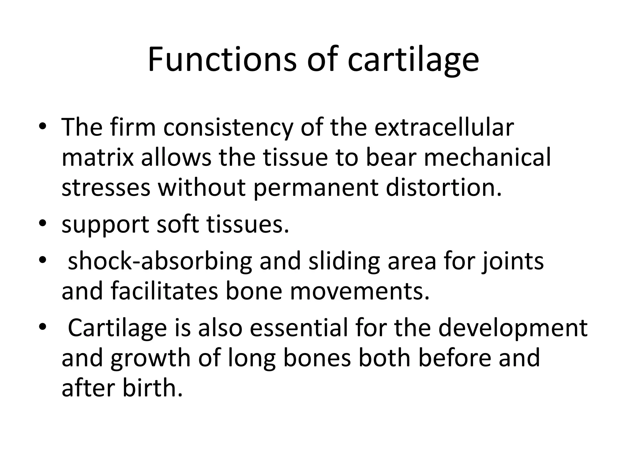
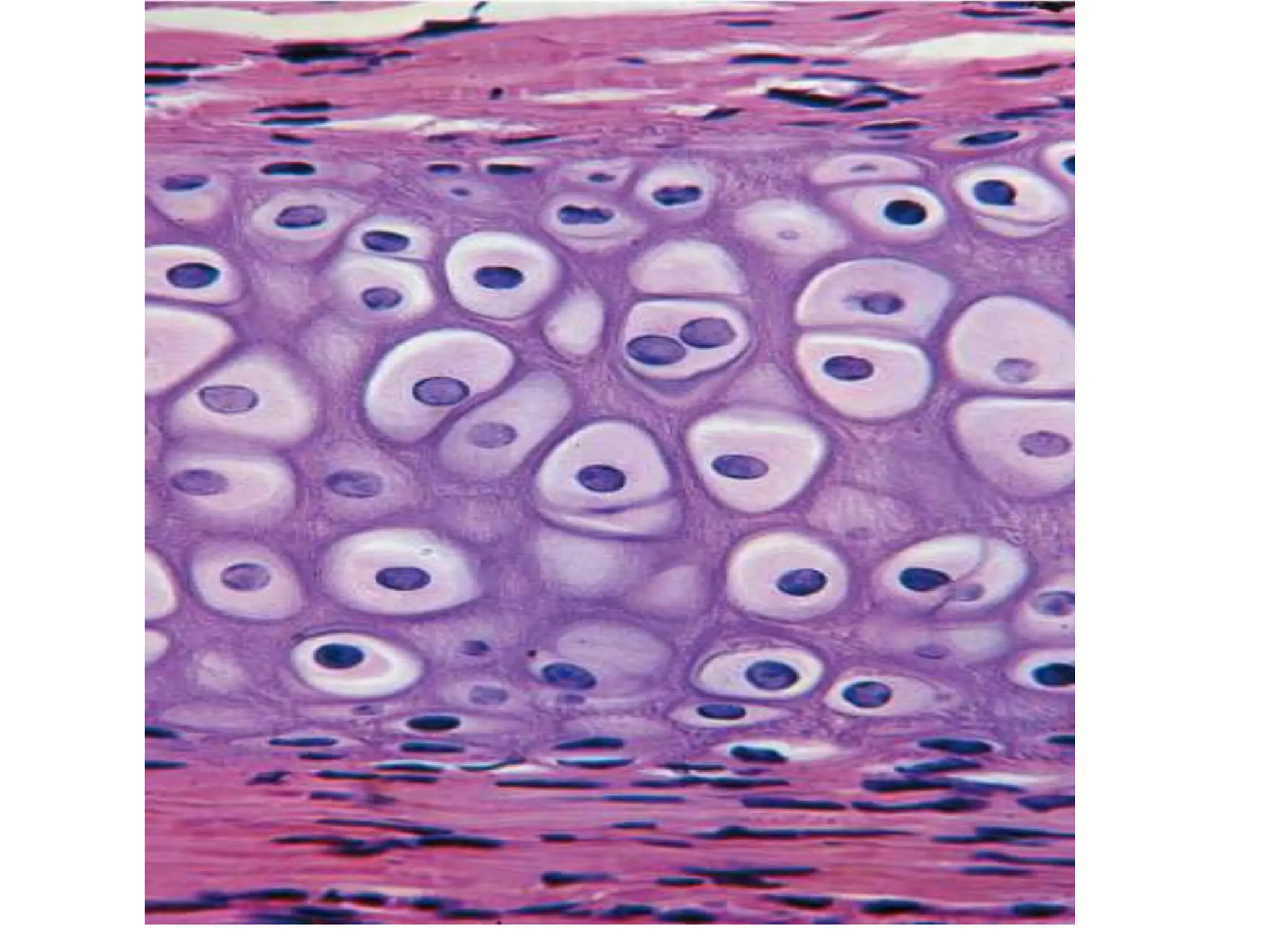
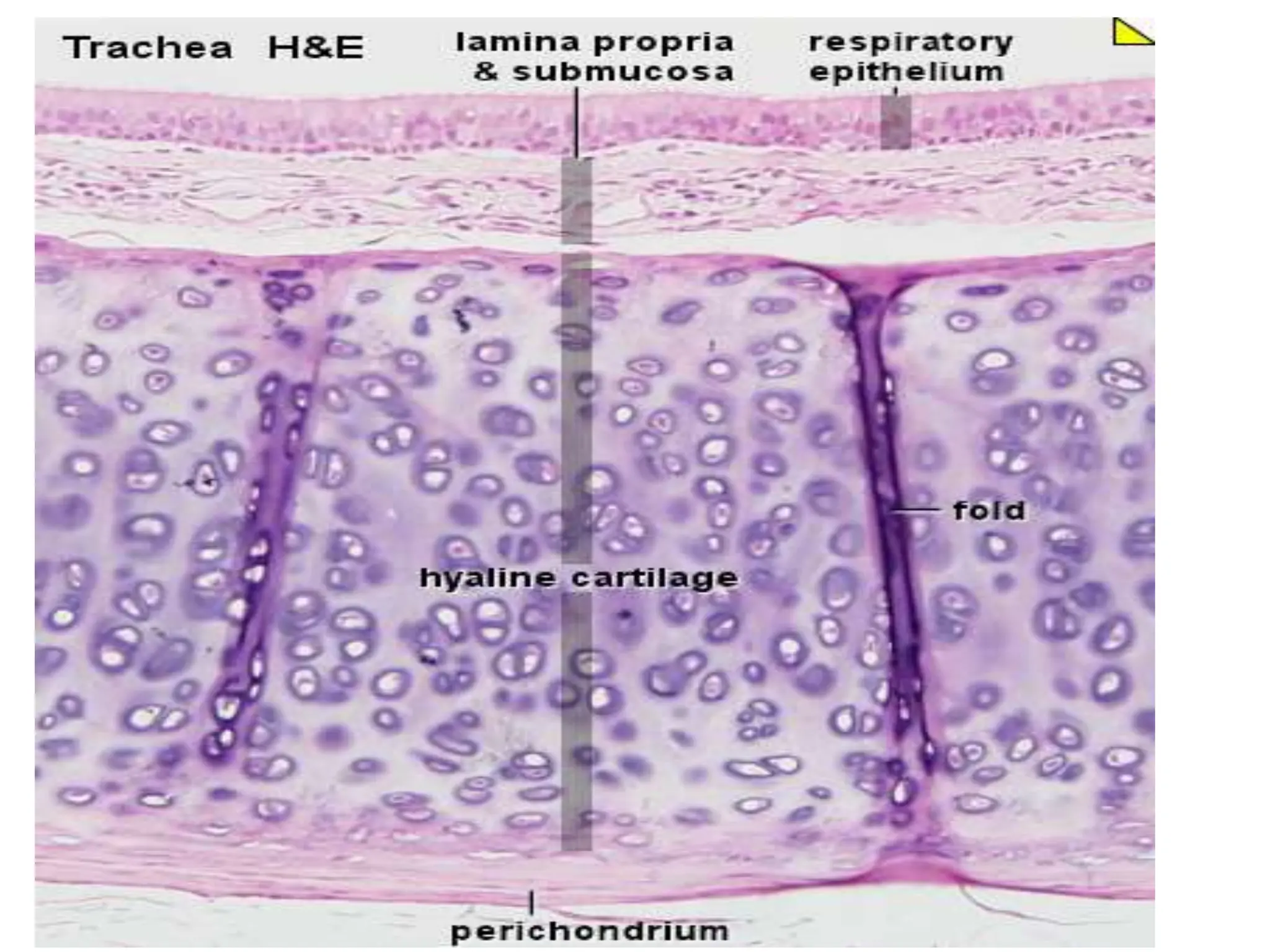
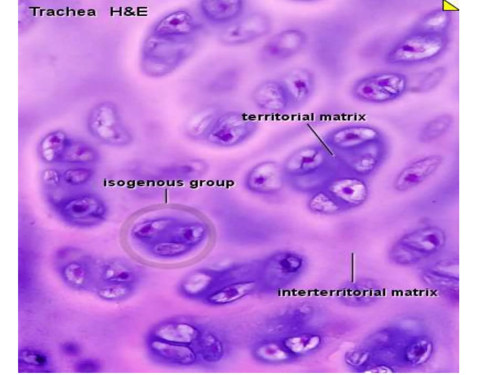
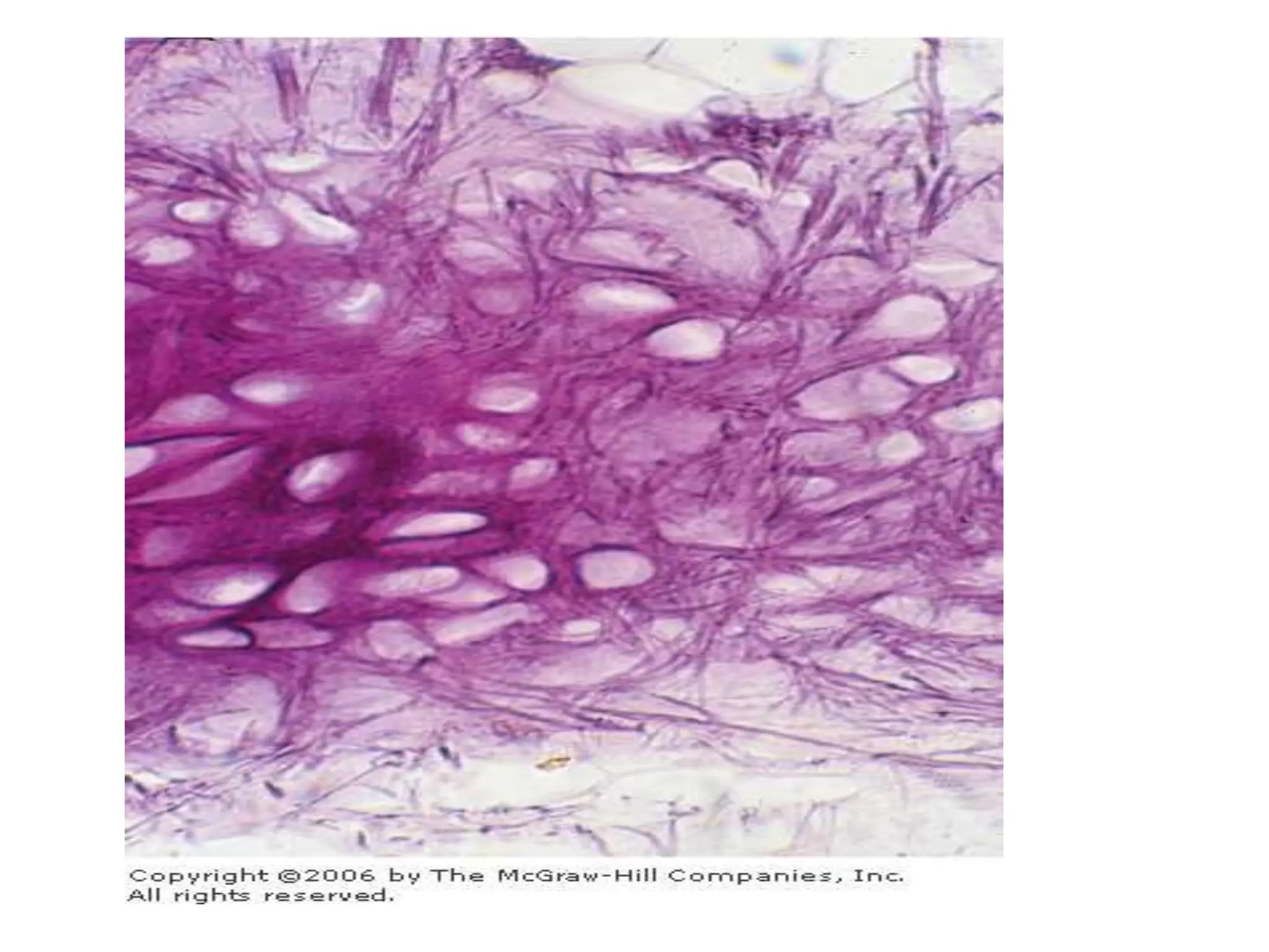
Cartilage
• Cartilage is a specialized form of connective
tissue.
• it consists of cells (chondrocytes) and
extracellular matrix.
• The chondrocytes are found in matrix cavities
called lacunae .
• They synthesize and secrete the extracellular
matrix
Functions of cartilage
• The firm consistency of the extracellular
matrix allows the tissue to bear mechanical
stresses without permanent distortion.
• support soft tissues.
• shock-absorbing and sliding area for joints
and facilitates bone movements.
• Cartilage is also essential for the development
and growth of long bones both before and
after birth.
• Cartilage is avascular and have no lymphatic
vessels or nerve endings.
• Its surrounded by dense connective tissue the
perichondrium and it gives the vascular,
lymphatic and sensory supply to cartilage .
• Articular cartilage has no perichondrium and
gets the nutrients by diffusion from the
synovial fluid.
Types
• According to the functional requirement and
the composition of extracellular matrix there
are 3 types of cartilage:
– Hyaline
– Elastic
– Fibrocartilage.
Hyaline cartilage
• The most common type.
• Is found in the articular surfaces of the
movable joints, in the walls of larger
respiratory passages (nose, larynx, trachea,
bronchi), in the ventral ends of ribs, and in the
epiphyseal plate, where it is responsible for
the longitudinal growth of bone.
• The matrix contains collagen fibrils type 2
mainly and macromolecules.
• At the periphery of hyaline cartilage, young
chondrocytes have an elliptic shape, with the
long axis parallel to the surface. Farther in,
they are round and may appear in groups of
up to eight cells originating from mitotic
divisions of a single chondrocyte. These
groups are called isogenous.
Growth of cartilage
• interstitial growth;
resulting from the mitotic division of preexisting
chondrocytes, it occurs in the epiphyseal plates
of long bones and within articular cartilage.
• appositional growth;
Chondroblasts of the perichondrium proliferate
and become chondrocytes once they have
surrounded themselves with cartilaginous
matrix and are incorporated into the existing
cartilage
Elastic cartilage
• occurs in the epiglottic cartilage, the
corniculate and cuneiform cartilage of the
larynx, the cartilage of the external ear and
the auditory tube.
• corresponds histologically to hyaline cartilage,
but, in addition, elastic cartilage contains a
dense network of delicately branched elastic
fibres.
Fibrocartilage
• it’s a transition between dense connective
tissue and hyaline cartilage.
• Chondrocytes may lie singly or in pairs, but
most often they form short rows between
dense bundles of collagen fibres.
• collagen fibers type I is dominant in fibrous
cartilage.
It is found in intervertebral disks, in attachments
of certain ligaments to the cartilaginous surface
of bones, and in the symphysis pubis.
Histology lecture ..Cartilage tissue.pptx

Histology lecture ..Cartilage tissue.pptx

  • 1.
  • 2.
    • Cartilage isa specialized form of connective tissue. • it consists of cells (chondrocytes) and extracellular matrix.
  • 3.
    • The chondrocytesare found in matrix cavities called lacunae . • They synthesize and secrete the extracellular matrix
  • 4.
    Functions of cartilage •The firm consistency of the extracellular matrix allows the tissue to bear mechanical stresses without permanent distortion. • support soft tissues. • shock-absorbing and sliding area for joints and facilitates bone movements. • Cartilage is also essential for the development and growth of long bones both before and after birth.
  • 5.
    • Cartilage isavascular and have no lymphatic vessels or nerve endings. • Its surrounded by dense connective tissue the perichondrium and it gives the vascular, lymphatic and sensory supply to cartilage . • Articular cartilage has no perichondrium and gets the nutrients by diffusion from the synovial fluid.
  • 6.
    Types • According tothe functional requirement and the composition of extracellular matrix there are 3 types of cartilage: – Hyaline – Elastic – Fibrocartilage.
  • 7.
    Hyaline cartilage • Themost common type. • Is found in the articular surfaces of the movable joints, in the walls of larger respiratory passages (nose, larynx, trachea, bronchi), in the ventral ends of ribs, and in the epiphyseal plate, where it is responsible for the longitudinal growth of bone. • The matrix contains collagen fibrils type 2 mainly and macromolecules.
  • 9.
    • At theperiphery of hyaline cartilage, young chondrocytes have an elliptic shape, with the long axis parallel to the surface. Farther in, they are round and may appear in groups of up to eight cells originating from mitotic divisions of a single chondrocyte. These groups are called isogenous.
  • 12.
    Growth of cartilage •interstitial growth; resulting from the mitotic division of preexisting chondrocytes, it occurs in the epiphyseal plates of long bones and within articular cartilage.
  • 13.
    • appositional growth; Chondroblastsof the perichondrium proliferate and become chondrocytes once they have surrounded themselves with cartilaginous matrix and are incorporated into the existing cartilage
  • 14.
    Elastic cartilage • occursin the epiglottic cartilage, the corniculate and cuneiform cartilage of the larynx, the cartilage of the external ear and the auditory tube. • corresponds histologically to hyaline cartilage, but, in addition, elastic cartilage contains a dense network of delicately branched elastic fibres.
  • 17.
    Fibrocartilage • it’s atransition between dense connective tissue and hyaline cartilage. • Chondrocytes may lie singly or in pairs, but most often they form short rows between dense bundles of collagen fibres. • collagen fibers type I is dominant in fibrous cartilage.
  • 19.
    It is foundin intervertebral disks, in attachments of certain ligaments to the cartilaginous surface of bones, and in the symphysis pubis.

Editor's Notes

  • #13  In the epiphyseal plates, interstitial growth is important in increasing the length of long bones and in providing a cartilage model for endochondral bone formation (see Chapter 8: Bone). In articular cartilage, as the cells and matrix near the articulating surface are gradually worn away, the cartilage must be replaced from within, since there is no perichondrium