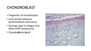
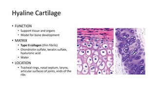
Cartilage
MCT 101
mmulipilwa
Cartilage
Cartilage is a specialized form of firm and resilient connective tissue
that can bear stresses without permanent distortion.
It serves as a precursor or model for the embryonic development and
subsequent growth of many long bones.
Structure of the Cartilage
• It consists of cells
• chondroblasts
• chondrocytes
• extracelluar matrix, consisting of fibers and ground substance
(hyaluranan, proteoglycans, glycoproteins).
Cartilage composition
• Cells
Chondrocytes: located within matrix; lacunae
Chondroblasts: located at periphery; secrete extracellular matrix
• Fibers
Collagen and elastic fibers
• Ground substance
Rich in glycosaminoglycans and proteoglycans
CHONDROBLAST
• Progenitor of chondrocytes
• Lines border between
perichondrium and matrix
• Secretes type II collagen and
other ECM components
• Chondroblasts build
CHONDROCYTE
• Mature cartilage cell
• Reside in a space called the
lacuna
• Clear areas = Golgi and lipid
droplets
Cells of the cartilage
• Chondrocytes completely fill
their lacunae
• RER and euchromatic nuclei
• Synthetically active, secrete
matrix
MATRIX
• Provides the rigidity, elasticity, &
resilience
• FIBERS
• Collagenous and elastic
• GROUND SUBSTANCE
• Glycosaminoglycans (chondroitin
sulfates, keratin sulfate, hyaluronic acid)
• Proteoglycans: GAGs + core protein
• Water
• Basophilic
• Territorial matrix – high conc of
sulfated proteoglycans
CARTILAGE GROWTH
• Appositional
• Increasing in WIDTH;
chondroblasts deposit matrix on
surface of pre-existing cartilage
• Interstitial
• Increasing in LENGTH;
chondrocytes divide and secrete
matrix from w/in lacunae
Cartilage composition
Perichondrium
• Perichondrium is a layer of dense irregular connective tissue.
• It is essential for the growth and maintenance of cartilage.
• Dense irregularly arranged connective tissue (type I collagen)
• Ensheaths the cartilage
• Houses the blood vessels that nourish chondrocytes
Features of Cartilage
• Avascular;
• Nutrients diffusion from perichondrium or synovial fluid;
• No innervation;
• No lymphatic vessels;
• Chondrocytes have low metabolic activity
TYPES OF CARTILAGE
• HYALINE
• ELASTIC
• FIBROUS
Types of Cartilage
• Hyaline cartilage: most common, much type II collagen
• Elastic cartilage: elastic fibers
• Fibrocatilage: dense network of type I collagen
Hyaline Cartilage
• FUNCTION
• Support tissue and organs
• Model for bone development
• MATRIX
• Type II collagen (thin fibrils)
• Chondroitin sulfate, keratin sulfate,
hyaluronic acid
• Water
• LOCATION
• Tracheal rings, nasal septum, larynx,
articular surfaces of joints, ends of the
ribs
Hyaline Cartilage
• Chondrocytes present in groups
of 4 to 8 cells
• Cell nests ( isogenous
aggregates)
• Teritorial matrix lacunar capsule
:- deeply stained matrix
• interstitial matrix
ELASTIC CARTILAGE
• FUNCTION
• Support with flexibility
• MATRIX
• Normal components of hyaline matrix
plus ELASTIC fibers
• LOCATION
• External ear, external auditory canal,
epiglottis, laryngeal cartilage (corniculate
and cuneiform)
• STAINS
• Elastic fibers stain BLACK with Weigert
stain
Elastic Cartilage
• Yellow cartilage
• Covered by perichondrium
NOTE: Elastic cartilage is located
in sites associated with
production and or reception of
sound.
Fibrocartilage
• FUNCTION
• Support with great tensile strength
• MATRIX
• Type I collagen - Oriented parallel
to stress plane
• LOCATION
• Intervertebral disks, pubic
symphysis
Fibrocartilage
• White fibrocartilage
• Looks like dense fibrous tissue
• Chondrocytes occur singly in the
lacunar
• No distinct surrounding
perichondrium
NB; Fibrocartilage is associated
with structures in the midline of
the body.
Applied Anatomy
• Hyaline cartilage is susceptible to calcification during aging
• Oesteoarthritis, a chronic condition during aging affect hyaline
cartilage in joints
• Cartilage inability to full repair due to its avascularity
• Tumours of the cartilage
Chondromas
chondrosarcomas

Cartilage_[Autosaved].pptx

  • 1.
  • 2.
    Cartilage Cartilage is aspecialized form of firm and resilient connective tissue that can bear stresses without permanent distortion. It serves as a precursor or model for the embryonic development and subsequent growth of many long bones.
  • 3.
    Structure of theCartilage • It consists of cells • chondroblasts • chondrocytes • extracelluar matrix, consisting of fibers and ground substance (hyaluranan, proteoglycans, glycoproteins).
  • 4.
    Cartilage composition • Cells Chondrocytes:located within matrix; lacunae Chondroblasts: located at periphery; secrete extracellular matrix • Fibers Collagen and elastic fibers • Ground substance Rich in glycosaminoglycans and proteoglycans
  • 6.
    CHONDROBLAST • Progenitor ofchondrocytes • Lines border between perichondrium and matrix • Secretes type II collagen and other ECM components • Chondroblasts build
  • 7.
    CHONDROCYTE • Mature cartilagecell • Reside in a space called the lacuna • Clear areas = Golgi and lipid droplets
  • 8.
    Cells of thecartilage • Chondrocytes completely fill their lacunae • RER and euchromatic nuclei • Synthetically active, secrete matrix
  • 9.
    MATRIX • Provides therigidity, elasticity, & resilience • FIBERS • Collagenous and elastic • GROUND SUBSTANCE • Glycosaminoglycans (chondroitin sulfates, keratin sulfate, hyaluronic acid) • Proteoglycans: GAGs + core protein • Water • Basophilic • Territorial matrix – high conc of sulfated proteoglycans
  • 10.
    CARTILAGE GROWTH • Appositional •Increasing in WIDTH; chondroblasts deposit matrix on surface of pre-existing cartilage • Interstitial • Increasing in LENGTH; chondrocytes divide and secrete matrix from w/in lacunae
  • 11.
    Cartilage composition Perichondrium • Perichondriumis a layer of dense irregular connective tissue. • It is essential for the growth and maintenance of cartilage. • Dense irregularly arranged connective tissue (type I collagen) • Ensheaths the cartilage • Houses the blood vessels that nourish chondrocytes
  • 12.
    Features of Cartilage •Avascular; • Nutrients diffusion from perichondrium or synovial fluid; • No innervation; • No lymphatic vessels; • Chondrocytes have low metabolic activity
  • 13.
    TYPES OF CARTILAGE •HYALINE • ELASTIC • FIBROUS
  • 14.
    Types of Cartilage •Hyaline cartilage: most common, much type II collagen • Elastic cartilage: elastic fibers • Fibrocatilage: dense network of type I collagen
  • 15.
    Hyaline Cartilage • FUNCTION •Support tissue and organs • Model for bone development • MATRIX • Type II collagen (thin fibrils) • Chondroitin sulfate, keratin sulfate, hyaluronic acid • Water • LOCATION • Tracheal rings, nasal septum, larynx, articular surfaces of joints, ends of the ribs
  • 16.
    Hyaline Cartilage • Chondrocytespresent in groups of 4 to 8 cells • Cell nests ( isogenous aggregates) • Teritorial matrix lacunar capsule :- deeply stained matrix • interstitial matrix
  • 18.
    ELASTIC CARTILAGE • FUNCTION •Support with flexibility • MATRIX • Normal components of hyaline matrix plus ELASTIC fibers • LOCATION • External ear, external auditory canal, epiglottis, laryngeal cartilage (corniculate and cuneiform) • STAINS • Elastic fibers stain BLACK with Weigert stain
  • 19.
    Elastic Cartilage • Yellowcartilage • Covered by perichondrium NOTE: Elastic cartilage is located in sites associated with production and or reception of sound.
  • 21.
    Fibrocartilage • FUNCTION • Supportwith great tensile strength • MATRIX • Type I collagen - Oriented parallel to stress plane • LOCATION • Intervertebral disks, pubic symphysis
  • 22.
    Fibrocartilage • White fibrocartilage •Looks like dense fibrous tissue • Chondrocytes occur singly in the lacunar • No distinct surrounding perichondrium NB; Fibrocartilage is associated with structures in the midline of the body.
  • 24.
    Applied Anatomy • Hyalinecartilage is susceptible to calcification during aging • Oesteoarthritis, a chronic condition during aging affect hyaline cartilage in joints • Cartilage inability to full repair due to its avascularity • Tumours of the cartilage Chondromas chondrosarcomas