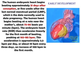
SlideShare a Scribd company logo
Cardiovascular System Denise Chan "Matters of the Heart"  Andrew McCaskill
The  circulatory system  (scientifically known as the  cardiovascular system ) is an organ system that  moves substances to and from cells ; it can also help  stabilize body temperature and pH (part of homeostasis).   The main components of the circulatory system are the  heart , the  blood , and the  blood vessels .
The human embryonic heart begins beating approximately  21 days after conception , or five weeks after the last normal menstrual period (LMP), which is the date normally used to date pregnancy. The human heart begins beating at a rate near the mother’s, about  75-80  beats per minute (bpm). The embryonic heart rate (EHR) then accelerates linearly for the first month of beating, peaking at  165-185 bpm . This acceleration is approximately 3.3 bpm per day, or about 10 bpm every three days, an increase of 100 bpm in the first month. Early development  Patrick J. Lynch, medical illustrator; C. Carl Jaffe, MD, cardiologist
[object Object],[object Object],[object Object],[object Object],[object Object],[object Object],[object Object],[object Object],[object Object],They are roughly grouped as  arterial  and  venous , determined by whether the blood in it is flowing  toward  or  away from  the heart. The term "arterial blood" is used to indicate blood high in oxygen. Types: There are various kinds of blood vessels
Anatomy All blood vessels have the same basic structure. The inner lining is the  endothelium  and is surrounded by  connective tissue . Around this there is a layer of  vascular smooth muscle , which is highly developed in arteries. Finally, there is a further layer of connective tissue known as the  adventitia , which contains nerves that supply the muscular layer, as well as nutrient capillaries in the larger blood vessels. The hollow internal cavity in which the blood flows is called the  lumen . Capillaries consist of little more than a layer of endothelium and occasional connective tissue. Laid end to end, the blood vessels in an average human body will stretch approximately 62,000 miles.
Arteries  are muscular blood vessels that carry blood  away  from the heart. Its proper functioning is responsible for the  delivery of oxygen and nutrients to all cells, maintenance of optimum pH, and the mobility of the elements, proteins and cells of the immune system.  In developed countries, the two leading causes of death, myocardial infarction and stroke each may directly result from an arterial system that has been slowly and progressively compromised by years of deterioration.
Blood pressure The arterial system is the higher-pressure portion of the circulatory system. Arterial pressure varies between the peak pressure during heart contraction, called the  systolic pressure , and the minimum, or  diastolic pressure  between contractions, when the heart rests between cycles. This pressure variation within the artery produces the pulse which is observable in any artery, and reflects heart activity. To withstand and adapt to the pressures within, arteries are surrounded by varying thicknesses of  smooth muscle  which have extensive elastic and inelastic connective tissues. The pulse pressure, i.e. Systolic vs. Diastolic difference, is determined primarily by the amount of blood ejected by each heart beat versus the volume and elasticity of the major arteries. Over time, elevated arterial blood sugar, cholesterol, and pressure, smoking, and other factors are all involved in  damaging both the endothelium and walls of the arteries , resulting in atherosclerosis or Diabetes Mellitus.
Pulmonary arteries The pulmonary arteries carry  deoxygenated  blood that has just returned from the body to the lungs, where carbon dioxide is exchanged for oxygen. Systemic arteries Systemic arteries  deliver blood to the arterioles, and then to the capillaries, where nutrients and gases are exchanged. The Aorta The aorta is the root systemic artery. It  receives blood  directly from the  left ventricle  of the heart via the aortic valve. As the aorta branches, and these arteries branch in turn, they become successively smaller in diameter, down to the arteriole. The arterioles supply capillaries which in turn empty into venules. Arterioles Arterioles,  the smallest of the true arteries, help regulate blood pressure and deliver blood to capillaries. Types of Arteries
Structure The walls of capillaries are composed of only a  single layer of cells , the  endothelium . This layer is so thin that molecules such as oxygen, water and lipids can pass through them by  diffusion  and enter the tissues.  Waste products  such as carbon dioxide and urea can diffuse back into the blood to be carried away for removal from the body. Capillaries  are the  smallest of a body's blood vessels,  measuring 5-10 μm. They  connect arterioles and venules , and they are the blood vessels that most  closely interact with tissues.
[object Object],Function: Veins serve to  return blood from organs to the heart.  The de-oxygenated blood is taken by veins to the  right atrium  of the heart. In pulmonary circulation the pulmonary veins return oxygenated blood from the lungs to the left atrium, which empties into the left ventricle, completing the cycle of blood circulation.
Most veins have  one-way valves  called  venous valves  to prevent  backflow  caused by gravity. They also have a thick  collagen outer layer , which helps maintain blood pressure and stop blood pooling. The hollow internal cavity in which the blood flows is called the  lumen . Veins are surrounded by helical bands of smooth muscles which help maintain blood flow to the right atrium.  Fainting can be caused by failure of the skeletal-muscular pump.  Long periods of standing can result in blood pooling in the legs , with blood pressure too low to ascend to the heart. In these cases the smooth muscles surrounding the veins become slack and fill with blood, absorbing a large portion of the total blood volume,  keeping blood away from the brain and causing unconsciousness.
Notable veins and vein systems The pulmonary veins carry  oxygenated  blood from the lungs to the heart.  ***The  superior and inferior vena cavae carry  deoxygenated  blood from the upper and lower systemic circulations, respectively. ***A portal venous system is a series of veins or venules that  directly connect two capillary beds.  An example of such a system includes the hepatic portal vein. List of important named veins Pulmonary veins Portal vein  Superior vena cava  Inferior vena cava  Femoral vein  Jugular veins
William Harvey  (April 1, 1578 – June 3, 1657) was an English medical doctor, who is credited with being the first to correctly describe, in exact detail, the properties of blood being pumped around the body by the heart.  “ I profess both to learn and to teach anatomy, not from books but from dissections; not from positions of philosophers but from the fabric of nature.”
Having circulated through the body, all the relatively  de-oxygenated blood  collects in the venous system which coalesces into two major veins: the  superior vena cava (roughly speaking from areas above the heart) and the inferior vena cava (roughly speaking from areas below the heart ). These two great vessels empty into the  right atrium  of the heart. The right atrium is the larger of the two atria, although both receive the same amount of blood. The blood is then pumped through the  tricuspid valve , or right atrioventricular valve , into the right ventricle . From the right ventricle, blood is pumped through the  pulmonary semi-lunar valve  into the  pulmonary artery . This blood enters the two pulmonary arteries (one for each lung) and travels through the lungs, where it is  oxygenated  and then flows into the pulmonary veins.  PULMONARY CIRCUIT:
This oxygenated blood then enters the  left atrium , which pumps it through the  bicuspid valve , also called the mitral or left atrioventricular valve, into the  left ventricle . The left ventricle is thicker and more muscular than the right ventricle because it pumps blood at a higher pressure. From the left ventricle, blood is pumped through the  aortic semi-lunar valve  into the aorta, a massive and thick-walled artery. The aorta arches and gives off major arteries to the upper body before piercing the diaphragm in order to supply the lower parts of the body with its various branches. Once the blood enters the peripheral tissues oxygen and nutrients are extracted from it and  carbon dioxide and wastes added , and it will again be collected in the veins and the  process will be repeated .  http://www.smm.org/heart/heart/pumping.htm Systemic Circuit:
Heart Circulation: In Conclusion! PULMONARY CIRCUIT:  (De-Oxygenated Blood) From Body    Superior/Inferior Vena Cava     Right Atria    via Tricuspid Valve    Right Ventricle     via Pulmonary Valve     Pulmonary Arteries    To Lungs (L&R) Systemic Circuit:  (Oxygenated Blood)  From Lungs    Pulmonary Vein    Left Atria    via Bicuspid Valve    Left Ventricle    via Aortic Semi-Lunar Valve    Aorta    to Body
The  sinoatrial node  (abbreviated  SA node ) is the  impulse generating (pacemaker) tissue located in the right atrium of the heart.  It is a group of cells positioned on the wall of the right atrium, near the entrance of the superior vena cava.  Although all of the heart's cells possess the ability to generate the electrical impulses (or action potentials), the sinoatrial node is what normally  initiates it , sometimes called the  primary pacemaker..   Cells in the SA node will naturally discharge (create action potentials) at  about 70-80 times/minute .  The atrioventricular node (abbreviated AV node) is an area of specialized tissue between the atria and the ventricles of the heart, which conducts the normal electrical impulse from the atria to the ventricles.
Acute  myocardial infarction  ( AMI  or  MI ), commonly known as a  heart attack , is a disease state that occurs when the  blood supply to a part of the heart is interrupted.  The resulting  ischemia  or oxygen shortage causes  damage and potential death of heart tissue.  It is a medical emergency, and the leading cause of death for both men and women all over the world. Important risk factors are   a previous history of vascular disease such as atherosclerotic coronary heart disease and/or angina a previous heart attack or stroke any previous episodes of abnormal heart rhythms or syncope older age—especially men over 40 and women over 50 smoking excessive alcohol consumption the abuse of certain illicit drugs high triglyceride levels high LDL ("Low-density lipoprotein") and low HDL ("High density lipoprotein") diabetes high blood pressureD obesity and chronically high levels of stress in certain persons.
The term  myocardial infarction  is derived from  myocardium  (the heart muscle) and  infarction  (tissue death due to oxygen starvation). The phrase "heart attack" is sometimes used incorrectly to describe sudden cardiac death, which may or may not be the result of acute myocardial infarction. Classical symptoms of acute myocardial infarction include  chest pain, shortness of breath, nausea, vomiting, palpitations, sweating, and anxiety or a feeling of impending doom.  Patients frequently feel suddenly ill.  Women often experience different symptoms than men. The most common symptoms of MI in women include shortness of breath, weakness, and fatigue. Approximately one third of all myocardial infarctions are silent, without chest pain or other symptoms.
The  jugular veins  are veins that  bring deoxygenated blood from the head back to the heart via the superior vena cava. The are two sets of jugular veins: external and internal. The  internal jugular runs  with the common carotid artery and vagus nerve inside the carotid sheath.  It provides venous drainage for the contents of the skull.  The  external jugular  runs superficially to sternocleidomastoid.  Both connect to the brachiocephalic veins, the external jugular joining more laterally than the internal. The brachiocephalic veins then join the subclavian veins from both sides then join to form the superior vena cava. The Jugular: The Main Brain Draining Vein
In human anatomy, the  common carotid artery  is an artery that  supplies the head and neck; it divides in the neck to form the external and internal carotid arteries. The common carotid artery is a paired structure, meaning that there are two in the body, one for each half.  The left and right common carotid arteries follow the same course with the exception of their origin.  The right common carotid originates in the neck from the brachiocephalic trunk. The left arises from the aortic arch in the thoracic region.
Renal Artery The  renal arteries  normally arise off the side of the abdominal aorta and supply the  kidneys with blood . The renal arteries carry a large portion of total blood flow to the kidneys.  Up to a third of total cardiac output can pass through the renal arteries to be filtered by the kidneys. The arterial supply of the kidneys is variable and there may be one or more renal arteries supplying each kidney. It is located above the renal vein.
The  renal veins  are veins that drain the kidney. They connect the kidney to the inferior vena cava. Because the inferior vena cava is on the right half of the body, the left renal vein is generally the longer of the two.
The hepatic portal vein, a large vein that carries blood from the digestive tract to the liver. The portal venous system is responsible for directing blood from parts of the  gastrointestinal tract to the liver.  Things absorbed in the small intestine, for example, would be taken to the liver for processing before being sent to the heart. Many drugs that are absorbed through the GI tract are  substantially metabolized by the liver before reaching general circulation.
The  subclavian artery  is a major artery of the upper thorax that mainly  supplies blood to the head and arms.  It is located below the clavicle, hence the name. There is a left subclavian and a right subclavian. The  subclavian veins  are two large veins, one on either side of the body. Its diameter is approximately that of a man's small finger. Each  subclavian drains the extremities and upper thorax.
The  axillary artery  is a large blood vessel that  conveys oxygenated blood to the lateral aspect of the thorax, the axilla (armpit) and the upper limb.  Its origin is the subclavian artery. The  axillary vein  is a large blood vessel that  conveys blood from the lateral aspect of the thorax, axilla (armpit) and upper limb toward the heart.  There is one axillary vein on each side of the body.
The  common iliac arteries  are two large arteries that originate from the aorta and supply all lower extremities. There are two divisions: the external iliac artery and internal iliac artery.  The  common iliac veins  are formed by the external iliac veins and internal iliac veins and together, in the abdomen, form the inferior vena cava. They drain blood from the pelvis and lower limbs. Iliac artery and vein
The  femoral vein  is a blood vessel that accompanies the femoral artery. The femoral vein returns de-oxygenated blood to the inferior vena cavae.  In anatomy, the  femoral artery  is a large artery in the muscles of the thigh that supplies the lower extremities with needed oxygen.
The  pulmonary arteries  carry blood from the heart to the lungs. They are the only arteries (other than umbilical arteries in the fetus) that carry deoxygenated blood. Pulmonary hypertension occurs alone and as a consequence of a number of lung diseases. It can be a consequence of heart disease.
Patrick J. Lynch, medical illustrator; C. Carl Jaffe, MD, cardiologist.  http://creativecommons.org/licenses/by/2.5/   Pulmonary Arteries Pulmonary Veins
200 million RBSs are produced daily by the hematopoetic tissue in bone marrow About the same number of RBCs are broken down by the spleen and converted into bilirubin which (along with bile) help break down fats.
Multiple Allele Traits Traits that are controlled by more than two alleles. Blood type in humans is controlled by three alleles: A, B, and O www.biologycorner.com/bio2/specialgenetics.html Blood Transfusions Blood can only be transferred to a body of a person who's immune system will "recognize" the blood. A and B are  antigens  on the blood that will be recognized. If the antigen is unfamiliar to the body, your body will  attack  and destroy the transfused blood as if it were a hostile invader (which can cause death). O is like a blank, it has no antigens.  O  is called the  universal donor  because a person can receive a transfusion from O blood without having an immune response AB  is the  universal acceptor , because a person with AB blood has both the A and B antigens already in the body, A and B blood can be transfused to the person (as well as O) and the body will recognize it and not attack.  Phenotype Genotype A AA or AO B BB or BO AB AB only O OO only
Conclusion: Circulatory System Blood Clotting Cascade The circulatory system, just like any other system, is a complex, interrelated, system with multiple parts that function as a whole.  The circulatory system exhibits “irreducible complexity” in the highest degree.  The heart without the complex arterial system would be useless.  The arterial system alone would be useless without the venous system (and its many valves) to return the blood to the heart.  The complexity of the heart alone is enough to astound anyone, let alone the system of vessels that accompany the passage of blood to every cell of the body.  Considering the relatedness to every other system of the body makes the circulatory system the most essential system of the human body.

More Related Content

What's hot

In b.sc nursing important question for psychology by S.Lakshmanan psychologist
In b.sc nursing important question for psychology by S.Lakshmanan psychologistIn b.sc nursing important question for psychology by S.Lakshmanan psychologist
In b.sc nursing important question for psychology by S.Lakshmanan psychologist
LAKSHMANAN S
 
Respiratory system by_monalisa
Respiratory system by_monalisaRespiratory system by_monalisa
Respiratory system by_monalisaAnu Radha G
 
POSITIONS
POSITIONSPOSITIONS
POSITIONS
Arifa T N
 
History of nursing in india.
History of nursing in india.History of nursing in india.
History of nursing in india.
Arsi University, Asella, Ethiopia
 
The Axial Skeleton & Appendicular Skeleton
The Axial Skeleton & Appendicular SkeletonThe Axial Skeleton & Appendicular Skeleton
The Axial Skeleton & Appendicular SkeletonLinda Langevoort
 
Cardiovascular physiology
Cardiovascular physiologyCardiovascular physiology
Cardiovascular physiology
Jesse Spurr
 
Anatomy and-physiology-of-the-cardiovascular-system-medical-surgical-nursing-ppt
Anatomy and-physiology-of-the-cardiovascular-system-medical-surgical-nursing-pptAnatomy and-physiology-of-the-cardiovascular-system-medical-surgical-nursing-ppt
Anatomy and-physiology-of-the-cardiovascular-system-medical-surgical-nursing-pptShineshy
 
Coronary circulation
Coronary circulationCoronary circulation
Coronary circulationTehmina Naru
 
The Cardiac Cycle
The Cardiac CycleThe Cardiac Cycle
The Cardiac Cycle
Christabelle Cécile
 
DIFFERENT PULSE SITES (pulse -introduction & images)
DIFFERENT  PULSE SITES (pulse -introduction & images)DIFFERENT  PULSE SITES (pulse -introduction & images)
DIFFERENT PULSE SITES (pulse -introduction & images)
martinshaji
 
Physiology of respiration
Physiology of respirationPhysiology of respiration
Physiology of respiration
Dr.RMLIMS lucknow
 
The digestive system
The digestive systemThe digestive system
The digestive system
Sathish Rajamani
 
Muskuloskeletal system for 1st year bsc nursing
Muskuloskeletal system for 1st year bsc nursingMuskuloskeletal system for 1st year bsc nursing
Muskuloskeletal system for 1st year bsc nursing
RGCN
 
Introduction of CVS
Introduction of CVSIntroduction of CVS
Introduction of CVS
Dr. Vibhash Kumar Vaidya
 
Lecture 21 respiratory system
Lecture 21    respiratory systemLecture 21    respiratory system
Lecture 21 respiratory systemShakir Ellahi
 
joints
jointsjoints
Respiratory system
 Respiratory system Respiratory system
Respiratory system
Amani Riyadh
 

What's hot (20)

In b.sc nursing important question for psychology by S.Lakshmanan psychologist
In b.sc nursing important question for psychology by S.Lakshmanan psychologistIn b.sc nursing important question for psychology by S.Lakshmanan psychologist
In b.sc nursing important question for psychology by S.Lakshmanan psychologist
 
Respiratory system by_monalisa
Respiratory system by_monalisaRespiratory system by_monalisa
Respiratory system by_monalisa
 
History of nursing
History of nursingHistory of nursing
History of nursing
 
POSITIONS
POSITIONSPOSITIONS
POSITIONS
 
History of nursing in india.
History of nursing in india.History of nursing in india.
History of nursing in india.
 
The Axial Skeleton & Appendicular Skeleton
The Axial Skeleton & Appendicular SkeletonThe Axial Skeleton & Appendicular Skeleton
The Axial Skeleton & Appendicular Skeleton
 
Cardiovascular physiology
Cardiovascular physiologyCardiovascular physiology
Cardiovascular physiology
 
Anatomy and-physiology-of-the-cardiovascular-system-medical-surgical-nursing-ppt
Anatomy and-physiology-of-the-cardiovascular-system-medical-surgical-nursing-pptAnatomy and-physiology-of-the-cardiovascular-system-medical-surgical-nursing-ppt
Anatomy and-physiology-of-the-cardiovascular-system-medical-surgical-nursing-ppt
 
Coronary circulation
Coronary circulationCoronary circulation
Coronary circulation
 
The Cardiac Cycle
The Cardiac CycleThe Cardiac Cycle
The Cardiac Cycle
 
DIFFERENT PULSE SITES (pulse -introduction & images)
DIFFERENT  PULSE SITES (pulse -introduction & images)DIFFERENT  PULSE SITES (pulse -introduction & images)
DIFFERENT PULSE SITES (pulse -introduction & images)
 
Physiology of respiration
Physiology of respirationPhysiology of respiration
Physiology of respiration
 
The digestive system
The digestive systemThe digestive system
The digestive system
 
Muskuloskeletal system for 1st year bsc nursing
Muskuloskeletal system for 1st year bsc nursingMuskuloskeletal system for 1st year bsc nursing
Muskuloskeletal system for 1st year bsc nursing
 
Introduction of CVS
Introduction of CVSIntroduction of CVS
Introduction of CVS
 
Joint movement
Joint movementJoint movement
Joint movement
 
Lecture 21 respiratory system
Lecture 21    respiratory systemLecture 21    respiratory system
Lecture 21 respiratory system
 
Physiology of heart
Physiology of heartPhysiology of heart
Physiology of heart
 
joints
jointsjoints
joints
 
Respiratory system
 Respiratory system Respiratory system
Respiratory system
 

Viewers also liked

Circulatory System
Circulatory  SystemCirculatory  System
Circulatory System
Jeana Maeve Yandog
 
The circulatory system
The circulatory systemThe circulatory system
The circulatory system
Mike Tamas
 
Circulatory system slide show
Circulatory system slide showCirculatory system slide show
Circulatory system slide show
Tracy Kuhn
 
Circulatory System 19.1-19.2
Circulatory System 19.1-19.2Circulatory System 19.1-19.2
Circulatory System 19.1-19.2
hld80789
 
1st science 12 describes the function of each respiratory organ
1st science 12 describes the function of each respiratory organ1st science 12 describes the function of each respiratory organ
1st science 12 describes the function of each respiratory organ
Shirley Sison
 
Function of each part of the circulatory system (LESSON 2)
Function of each part of the circulatory system (LESSON 2)Function of each part of the circulatory system (LESSON 2)
Function of each part of the circulatory system (LESSON 2)
Jenil Urianza-Moises
 
Cardiovascular diagnostic procedures
Cardiovascular diagnostic proceduresCardiovascular diagnostic procedures
Cardiovascular diagnostic procedureslfowlkes28
 
Chapter 15 Cardiovascular System
Chapter 15   Cardiovascular SystemChapter 15   Cardiovascular System
Chapter 15 Cardiovascular System
1957Hamlet
 

Viewers also liked (10)

Circulatory system
Circulatory systemCirculatory system
Circulatory system
 
Circulatory System
Circulatory  SystemCirculatory  System
Circulatory System
 
The circulatory system
The circulatory systemThe circulatory system
The circulatory system
 
Circulatory system slide show
Circulatory system slide showCirculatory system slide show
Circulatory system slide show
 
Circulatory System 19.1-19.2
Circulatory System 19.1-19.2Circulatory System 19.1-19.2
Circulatory System 19.1-19.2
 
1st science 12 describes the function of each respiratory organ
1st science 12 describes the function of each respiratory organ1st science 12 describes the function of each respiratory organ
1st science 12 describes the function of each respiratory organ
 
Function of each part of the circulatory system (LESSON 2)
Function of each part of the circulatory system (LESSON 2)Function of each part of the circulatory system (LESSON 2)
Function of each part of the circulatory system (LESSON 2)
 
Cardiovascular diagnostic procedures
Cardiovascular diagnostic proceduresCardiovascular diagnostic procedures
Cardiovascular diagnostic procedures
 
Chapter 15 Cardiovascular System
Chapter 15   Cardiovascular SystemChapter 15   Cardiovascular System
Chapter 15 Cardiovascular System
 
Urinary system
Urinary systemUrinary system
Urinary system
 

Similar to Cardiovascular system

circulatory system mammals.ppt
circulatory system mammals.pptcirculatory system mammals.ppt
circulatory system mammals.ppt
ssuser880f82
 
Cardiovascular System
Cardiovascular SystemCardiovascular System
Cardiovascular System000 07
 
Tiesha miller and jillian mc clennen circulatory final
Tiesha miller and jillian mc clennen  circulatory finalTiesha miller and jillian mc clennen  circulatory final
Tiesha miller and jillian mc clennen circulatory final
Trmillerwidener
 
Tiesha miller and jillian mc clennen circulatory final
Tiesha miller and jillian mc clennen  circulatory finalTiesha miller and jillian mc clennen  circulatory final
Tiesha miller and jillian mc clennen circulatory finalTrmillerwidener
 
Cape biology unit 2 -_circulatory_system_in_humans_and_exercise
Cape biology unit 2 -_circulatory_system_in_humans_and_exerciseCape biology unit 2 -_circulatory_system_in_humans_and_exercise
Cape biology unit 2 -_circulatory_system_in_humans_and_exercise
Hilton Ritch
 
Circulatory System.ppt
Circulatory System.pptCirculatory System.ppt
Circulatory System.pptShama
 
Circulatory System - Heart_ST.ppt
Circulatory System - Heart_ST.pptCirculatory System - Heart_ST.ppt
Circulatory System - Heart_ST.pptShama
 
Case study pregnancy induced hypertension
Case study pregnancy induced hypertensionCase study pregnancy induced hypertension
Case study pregnancy induced hypertensionkiarratot
 
Cardiovascular System Part 1
Cardiovascular System Part 1Cardiovascular System Part 1
Cardiovascular System Part 1
melecia megan castillo
 
Cardiovascular system
Cardiovascular systemCardiovascular system
Cardiovascular system
malkeetsingh104
 
CARDIOVASCULAR SYSTEM.pptx
CARDIOVASCULAR SYSTEM.pptxCARDIOVASCULAR SYSTEM.pptx
CARDIOVASCULAR SYSTEM.pptx
Sweety gupta
 
cardiovascular-system-1216560905497953-8 (1).pptx
cardiovascular-system-1216560905497953-8 (1).pptxcardiovascular-system-1216560905497953-8 (1).pptx
cardiovascular-system-1216560905497953-8 (1).pptx
safuraqazi
 
Circulatory-System-STEM.ppt
Circulatory-System-STEM.pptCirculatory-System-STEM.ppt
Circulatory-System-STEM.ppt
KeithJelianSocajel
 
Biology Project [Circulatory System] Vijay Raja Std Vii Navdeep With Sound
Biology Project [Circulatory System] Vijay Raja Std Vii Navdeep With SoundBiology Project [Circulatory System] Vijay Raja Std Vii Navdeep With Sound
Biology Project [Circulatory System] Vijay Raja Std Vii Navdeep With Sound
vijayaswathy
 
Cardiovascular System
Cardiovascular SystemCardiovascular System
Cardiovascular Systemxtrm nurse
 
Cardiovascular System
Cardiovascular SystemCardiovascular System
Cardiovascular Systemxtrm nurse
 
Circulatory system
Circulatory systemCirculatory system
Circulatory system
Ghantali Mitra Mandal
 
CARDIOVASCULAR SYSTEM
CARDIOVASCULAR SYSTEMCARDIOVASCULAR SYSTEM
CARDIOVASCULAR SYSTEM
sandeep chandakavate
 
Organ system for internal transport (circulatory system)
Organ system for internal transport (circulatory system)Organ system for internal transport (circulatory system)
Organ system for internal transport (circulatory system)Cesar Rentino
 
Blood circulation
Blood circulationBlood circulation
Blood circulation
kkumargupta2003
 

Similar to Cardiovascular system (20)

circulatory system mammals.ppt
circulatory system mammals.pptcirculatory system mammals.ppt
circulatory system mammals.ppt
 
Cardiovascular System
Cardiovascular SystemCardiovascular System
Cardiovascular System
 
Tiesha miller and jillian mc clennen circulatory final
Tiesha miller and jillian mc clennen  circulatory finalTiesha miller and jillian mc clennen  circulatory final
Tiesha miller and jillian mc clennen circulatory final
 
Tiesha miller and jillian mc clennen circulatory final
Tiesha miller and jillian mc clennen  circulatory finalTiesha miller and jillian mc clennen  circulatory final
Tiesha miller and jillian mc clennen circulatory final
 
Cape biology unit 2 -_circulatory_system_in_humans_and_exercise
Cape biology unit 2 -_circulatory_system_in_humans_and_exerciseCape biology unit 2 -_circulatory_system_in_humans_and_exercise
Cape biology unit 2 -_circulatory_system_in_humans_and_exercise
 
Circulatory System.ppt
Circulatory System.pptCirculatory System.ppt
Circulatory System.ppt
 
Circulatory System - Heart_ST.ppt
Circulatory System - Heart_ST.pptCirculatory System - Heart_ST.ppt
Circulatory System - Heart_ST.ppt
 
Case study pregnancy induced hypertension
Case study pregnancy induced hypertensionCase study pregnancy induced hypertension
Case study pregnancy induced hypertension
 
Cardiovascular System Part 1
Cardiovascular System Part 1Cardiovascular System Part 1
Cardiovascular System Part 1
 
Cardiovascular system
Cardiovascular systemCardiovascular system
Cardiovascular system
 
CARDIOVASCULAR SYSTEM.pptx
CARDIOVASCULAR SYSTEM.pptxCARDIOVASCULAR SYSTEM.pptx
CARDIOVASCULAR SYSTEM.pptx
 
cardiovascular-system-1216560905497953-8 (1).pptx
cardiovascular-system-1216560905497953-8 (1).pptxcardiovascular-system-1216560905497953-8 (1).pptx
cardiovascular-system-1216560905497953-8 (1).pptx
 
Circulatory-System-STEM.ppt
Circulatory-System-STEM.pptCirculatory-System-STEM.ppt
Circulatory-System-STEM.ppt
 
Biology Project [Circulatory System] Vijay Raja Std Vii Navdeep With Sound
Biology Project [Circulatory System] Vijay Raja Std Vii Navdeep With SoundBiology Project [Circulatory System] Vijay Raja Std Vii Navdeep With Sound
Biology Project [Circulatory System] Vijay Raja Std Vii Navdeep With Sound
 
Cardiovascular System
Cardiovascular SystemCardiovascular System
Cardiovascular System
 
Cardiovascular System
Cardiovascular SystemCardiovascular System
Cardiovascular System
 
Circulatory system
Circulatory systemCirculatory system
Circulatory system
 
CARDIOVASCULAR SYSTEM
CARDIOVASCULAR SYSTEMCARDIOVASCULAR SYSTEM
CARDIOVASCULAR SYSTEM
 
Organ system for internal transport (circulatory system)
Organ system for internal transport (circulatory system)Organ system for internal transport (circulatory system)
Organ system for internal transport (circulatory system)
 
Blood circulation
Blood circulationBlood circulation
Blood circulation
 

More from Andrew McCaskill (20)

Human reproduction and development
Human reproduction and developmentHuman reproduction and development
Human reproduction and development
 
Digestive system
Digestive systemDigestive system
Digestive system
 
Respiratory syst
Respiratory systRespiratory syst
Respiratory syst
 
37 plantnutrition text
37 plantnutrition text37 plantnutrition text
37 plantnutrition text
 
53 communities text
53 communities text53 communities text
53 communities text
 
48 nervous text
48 nervous text48 nervous text
48 nervous text
 
41 animalnutrition text
41  animalnutrition text41  animalnutrition text
41 animalnutrition text
 
51 behavior text
51 behavior text51 behavior text
51 behavior text
 
46 reproduction text
46 reproduction text46 reproduction text
46 reproduction text
 
44 excretion text
44 excretion text44 excretion text
44 excretion text
 
40 animalform&function text
40 animalform&function text40 animalform&function text
40 animalform&function text
 
32 animaldiversity text
32  animaldiversity text32  animaldiversity text
32 animaldiversity text
 
35 plantstructure text
35  plantstructure text35  plantstructure text
35 plantstructure text
 
28 protists
28  protists28  protists
28 protists
 
29 plants ii text
29  plants ii text29  plants ii text
29 plants ii text
 
31 fungi text
31  fungi text31  fungi text
31 fungi text
 
24 originofspecies text
24  originofspecies text24  originofspecies text
24 originofspecies text
 
27 prokaryotes text
27  prokaryotes text27  prokaryotes text
27 prokaryotes text
 
23 evolutionofpopulations text
23  evolutionofpopulations text23  evolutionofpopulations text
23 evolutionofpopulations text
 
22 darwin text
22  darwin text22  darwin text
22 darwin text
 

Recently uploaded

NVBDCP.pptx Nation vector borne disease control program
NVBDCP.pptx Nation vector borne disease control programNVBDCP.pptx Nation vector borne disease control program
NVBDCP.pptx Nation vector borne disease control program
Sapna Thakur
 
Tests for analysis of different pharmaceutical.pptx
Tests for analysis of different pharmaceutical.pptxTests for analysis of different pharmaceutical.pptx
Tests for analysis of different pharmaceutical.pptx
taiba qazi
 
Ozempic: Preoperative Management of Patients on GLP-1 Receptor Agonists
Ozempic: Preoperative Management of Patients on GLP-1 Receptor Agonists  Ozempic: Preoperative Management of Patients on GLP-1 Receptor Agonists
Ozempic: Preoperative Management of Patients on GLP-1 Receptor Agonists
Saeid Safari
 
#cALL# #gIRLS# In Dehradun ꧁❤8107221448❤꧂#cALL# #gIRLS# Service In Dehradun W...
#cALL# #gIRLS# In Dehradun ꧁❤8107221448❤꧂#cALL# #gIRLS# Service In Dehradun W...#cALL# #gIRLS# In Dehradun ꧁❤8107221448❤꧂#cALL# #gIRLS# Service In Dehradun W...
#cALL# #gIRLS# In Dehradun ꧁❤8107221448❤꧂#cALL# #gIRLS# Service In Dehradun W...
chandankumarsmartiso
 
The Electrocardiogram - Physiologic Principles
The Electrocardiogram - Physiologic PrinciplesThe Electrocardiogram - Physiologic Principles
The Electrocardiogram - Physiologic Principles
MedicoseAcademics
 
Non-respiratory Functions of the Lungs.pdf
Non-respiratory Functions of the Lungs.pdfNon-respiratory Functions of the Lungs.pdf
Non-respiratory Functions of the Lungs.pdf
MedicoseAcademics
 
DISSERTATION on NEW DRUG DISCOVERY AND DEVELOPMENT STAGES OF DRUG DISCOVERY
DISSERTATION on NEW DRUG DISCOVERY AND DEVELOPMENT STAGES OF DRUG DISCOVERYDISSERTATION on NEW DRUG DISCOVERY AND DEVELOPMENT STAGES OF DRUG DISCOVERY
DISSERTATION on NEW DRUG DISCOVERY AND DEVELOPMENT STAGES OF DRUG DISCOVERY
NEHA GUPTA
 
Aortic Association CBL Pilot April 19 – 20 Bern
Aortic Association CBL Pilot April 19 – 20 BernAortic Association CBL Pilot April 19 – 20 Bern
Aortic Association CBL Pilot April 19 – 20 Bern
suvadeepdas911
 
Identification and nursing management of congenital malformations .pptx
Identification and nursing management of congenital malformations .pptxIdentification and nursing management of congenital malformations .pptx
Identification and nursing management of congenital malformations .pptx
MGM SCHOOL/COLLEGE OF NURSING
 
Dehradun #ℂall #gIRLS Oyo Hotel 8107221448 #ℂall #gIRL in Dehradun
Dehradun #ℂall #gIRLS Oyo Hotel 8107221448 #ℂall #gIRL in DehradunDehradun #ℂall #gIRLS Oyo Hotel 8107221448 #ℂall #gIRL in Dehradun
Dehradun #ℂall #gIRLS Oyo Hotel 8107221448 #ℂall #gIRL in Dehradun
chandankumarsmartiso
 
Cardiac Assessment for B.sc Nursing Student.pdf
Cardiac Assessment for B.sc Nursing Student.pdfCardiac Assessment for B.sc Nursing Student.pdf
Cardiac Assessment for B.sc Nursing Student.pdf
shivalingatalekar1
 
Superficial & Deep Fascia of the NECK.pptx
Superficial & Deep Fascia of the NECK.pptxSuperficial & Deep Fascia of the NECK.pptx
Superficial & Deep Fascia of the NECK.pptx
Dr. Rabia Inam Gandapore
 
Physiology of Special Chemical Sensation of Taste
Physiology of Special Chemical Sensation of TastePhysiology of Special Chemical Sensation of Taste
Physiology of Special Chemical Sensation of Taste
MedicoseAcademics
 
ABDOMINAL TRAUMA in pediatrics part one.
ABDOMINAL TRAUMA in pediatrics part one.ABDOMINAL TRAUMA in pediatrics part one.
ABDOMINAL TRAUMA in pediatrics part one.
drhasanrajab
 
Sex determination from mandible pelvis and skull
Sex determination from mandible pelvis and skullSex determination from mandible pelvis and skull
Sex determination from mandible pelvis and skull
ShashankRoodkee
 
How STIs Influence the Development of Pelvic Inflammatory Disease.pptx
How STIs Influence the Development of Pelvic Inflammatory Disease.pptxHow STIs Influence the Development of Pelvic Inflammatory Disease.pptx
How STIs Influence the Development of Pelvic Inflammatory Disease.pptx
FFragrant
 
Integrating Ayurveda into Parkinson’s Management: A Holistic Approach
Integrating Ayurveda into Parkinson’s Management: A Holistic ApproachIntegrating Ayurveda into Parkinson’s Management: A Holistic Approach
Integrating Ayurveda into Parkinson’s Management: A Holistic Approach
Ayurveda ForAll
 
Best Ayurvedic medicine for Gas and Indigestion
Best Ayurvedic medicine for Gas and IndigestionBest Ayurvedic medicine for Gas and Indigestion
Best Ayurvedic medicine for Gas and Indigestion
Swastik Ayurveda
 
Novas diretrizes da OMS para os cuidados perinatais de mais qualidade
Novas diretrizes da OMS para os cuidados perinatais de mais qualidadeNovas diretrizes da OMS para os cuidados perinatais de mais qualidade
Novas diretrizes da OMS para os cuidados perinatais de mais qualidade
Prof. Marcus Renato de Carvalho
 
Ketone bodies and metabolism-biochemistry
Ketone bodies and metabolism-biochemistryKetone bodies and metabolism-biochemistry
Ketone bodies and metabolism-biochemistry
Dhayanithi C
 

Recently uploaded (20)

NVBDCP.pptx Nation vector borne disease control program
NVBDCP.pptx Nation vector borne disease control programNVBDCP.pptx Nation vector borne disease control program
NVBDCP.pptx Nation vector borne disease control program
 
Tests for analysis of different pharmaceutical.pptx
Tests for analysis of different pharmaceutical.pptxTests for analysis of different pharmaceutical.pptx
Tests for analysis of different pharmaceutical.pptx
 
Ozempic: Preoperative Management of Patients on GLP-1 Receptor Agonists
Ozempic: Preoperative Management of Patients on GLP-1 Receptor Agonists  Ozempic: Preoperative Management of Patients on GLP-1 Receptor Agonists
Ozempic: Preoperative Management of Patients on GLP-1 Receptor Agonists
 
#cALL# #gIRLS# In Dehradun ꧁❤8107221448❤꧂#cALL# #gIRLS# Service In Dehradun W...
#cALL# #gIRLS# In Dehradun ꧁❤8107221448❤꧂#cALL# #gIRLS# Service In Dehradun W...#cALL# #gIRLS# In Dehradun ꧁❤8107221448❤꧂#cALL# #gIRLS# Service In Dehradun W...
#cALL# #gIRLS# In Dehradun ꧁❤8107221448❤꧂#cALL# #gIRLS# Service In Dehradun W...
 
The Electrocardiogram - Physiologic Principles
The Electrocardiogram - Physiologic PrinciplesThe Electrocardiogram - Physiologic Principles
The Electrocardiogram - Physiologic Principles
 
Non-respiratory Functions of the Lungs.pdf
Non-respiratory Functions of the Lungs.pdfNon-respiratory Functions of the Lungs.pdf
Non-respiratory Functions of the Lungs.pdf
 
DISSERTATION on NEW DRUG DISCOVERY AND DEVELOPMENT STAGES OF DRUG DISCOVERY
DISSERTATION on NEW DRUG DISCOVERY AND DEVELOPMENT STAGES OF DRUG DISCOVERYDISSERTATION on NEW DRUG DISCOVERY AND DEVELOPMENT STAGES OF DRUG DISCOVERY
DISSERTATION on NEW DRUG DISCOVERY AND DEVELOPMENT STAGES OF DRUG DISCOVERY
 
Aortic Association CBL Pilot April 19 – 20 Bern
Aortic Association CBL Pilot April 19 – 20 BernAortic Association CBL Pilot April 19 – 20 Bern
Aortic Association CBL Pilot April 19 – 20 Bern
 
Identification and nursing management of congenital malformations .pptx
Identification and nursing management of congenital malformations .pptxIdentification and nursing management of congenital malformations .pptx
Identification and nursing management of congenital malformations .pptx
 
Dehradun #ℂall #gIRLS Oyo Hotel 8107221448 #ℂall #gIRL in Dehradun
Dehradun #ℂall #gIRLS Oyo Hotel 8107221448 #ℂall #gIRL in DehradunDehradun #ℂall #gIRLS Oyo Hotel 8107221448 #ℂall #gIRL in Dehradun
Dehradun #ℂall #gIRLS Oyo Hotel 8107221448 #ℂall #gIRL in Dehradun
 
Cardiac Assessment for B.sc Nursing Student.pdf
Cardiac Assessment for B.sc Nursing Student.pdfCardiac Assessment for B.sc Nursing Student.pdf
Cardiac Assessment for B.sc Nursing Student.pdf
 
Superficial & Deep Fascia of the NECK.pptx
Superficial & Deep Fascia of the NECK.pptxSuperficial & Deep Fascia of the NECK.pptx
Superficial & Deep Fascia of the NECK.pptx
 
Physiology of Special Chemical Sensation of Taste
Physiology of Special Chemical Sensation of TastePhysiology of Special Chemical Sensation of Taste
Physiology of Special Chemical Sensation of Taste
 
ABDOMINAL TRAUMA in pediatrics part one.
ABDOMINAL TRAUMA in pediatrics part one.ABDOMINAL TRAUMA in pediatrics part one.
ABDOMINAL TRAUMA in pediatrics part one.
 
Sex determination from mandible pelvis and skull
Sex determination from mandible pelvis and skullSex determination from mandible pelvis and skull
Sex determination from mandible pelvis and skull
 
How STIs Influence the Development of Pelvic Inflammatory Disease.pptx
How STIs Influence the Development of Pelvic Inflammatory Disease.pptxHow STIs Influence the Development of Pelvic Inflammatory Disease.pptx
How STIs Influence the Development of Pelvic Inflammatory Disease.pptx
 
Integrating Ayurveda into Parkinson’s Management: A Holistic Approach
Integrating Ayurveda into Parkinson’s Management: A Holistic ApproachIntegrating Ayurveda into Parkinson’s Management: A Holistic Approach
Integrating Ayurveda into Parkinson’s Management: A Holistic Approach
 
Best Ayurvedic medicine for Gas and Indigestion
Best Ayurvedic medicine for Gas and IndigestionBest Ayurvedic medicine for Gas and Indigestion
Best Ayurvedic medicine for Gas and Indigestion
 
Novas diretrizes da OMS para os cuidados perinatais de mais qualidade
Novas diretrizes da OMS para os cuidados perinatais de mais qualidadeNovas diretrizes da OMS para os cuidados perinatais de mais qualidade
Novas diretrizes da OMS para os cuidados perinatais de mais qualidade
 
Ketone bodies and metabolism-biochemistry
Ketone bodies and metabolism-biochemistryKetone bodies and metabolism-biochemistry
Ketone bodies and metabolism-biochemistry
 

Cardiovascular system

  • 1. Cardiovascular System Denise Chan "Matters of the Heart" Andrew McCaskill
  • 2. The circulatory system (scientifically known as the cardiovascular system ) is an organ system that moves substances to and from cells ; it can also help stabilize body temperature and pH (part of homeostasis). The main components of the circulatory system are the heart , the blood , and the blood vessels .
  • 3. The human embryonic heart begins beating approximately 21 days after conception , or five weeks after the last normal menstrual period (LMP), which is the date normally used to date pregnancy. The human heart begins beating at a rate near the mother’s, about 75-80 beats per minute (bpm). The embryonic heart rate (EHR) then accelerates linearly for the first month of beating, peaking at 165-185 bpm . This acceleration is approximately 3.3 bpm per day, or about 10 bpm every three days, an increase of 100 bpm in the first month. Early development Patrick J. Lynch, medical illustrator; C. Carl Jaffe, MD, cardiologist
  • 4.
  • 5. Anatomy All blood vessels have the same basic structure. The inner lining is the endothelium and is surrounded by connective tissue . Around this there is a layer of vascular smooth muscle , which is highly developed in arteries. Finally, there is a further layer of connective tissue known as the adventitia , which contains nerves that supply the muscular layer, as well as nutrient capillaries in the larger blood vessels. The hollow internal cavity in which the blood flows is called the lumen . Capillaries consist of little more than a layer of endothelium and occasional connective tissue. Laid end to end, the blood vessels in an average human body will stretch approximately 62,000 miles.
  • 6. Arteries are muscular blood vessels that carry blood away from the heart. Its proper functioning is responsible for the delivery of oxygen and nutrients to all cells, maintenance of optimum pH, and the mobility of the elements, proteins and cells of the immune system. In developed countries, the two leading causes of death, myocardial infarction and stroke each may directly result from an arterial system that has been slowly and progressively compromised by years of deterioration.
  • 7. Blood pressure The arterial system is the higher-pressure portion of the circulatory system. Arterial pressure varies between the peak pressure during heart contraction, called the systolic pressure , and the minimum, or diastolic pressure between contractions, when the heart rests between cycles. This pressure variation within the artery produces the pulse which is observable in any artery, and reflects heart activity. To withstand and adapt to the pressures within, arteries are surrounded by varying thicknesses of smooth muscle which have extensive elastic and inelastic connective tissues. The pulse pressure, i.e. Systolic vs. Diastolic difference, is determined primarily by the amount of blood ejected by each heart beat versus the volume and elasticity of the major arteries. Over time, elevated arterial blood sugar, cholesterol, and pressure, smoking, and other factors are all involved in damaging both the endothelium and walls of the arteries , resulting in atherosclerosis or Diabetes Mellitus.
  • 8. Pulmonary arteries The pulmonary arteries carry deoxygenated blood that has just returned from the body to the lungs, where carbon dioxide is exchanged for oxygen. Systemic arteries Systemic arteries deliver blood to the arterioles, and then to the capillaries, where nutrients and gases are exchanged. The Aorta The aorta is the root systemic artery. It receives blood directly from the left ventricle of the heart via the aortic valve. As the aorta branches, and these arteries branch in turn, they become successively smaller in diameter, down to the arteriole. The arterioles supply capillaries which in turn empty into venules. Arterioles Arterioles, the smallest of the true arteries, help regulate blood pressure and deliver blood to capillaries. Types of Arteries
  • 9. Structure The walls of capillaries are composed of only a single layer of cells , the endothelium . This layer is so thin that molecules such as oxygen, water and lipids can pass through them by diffusion and enter the tissues. Waste products such as carbon dioxide and urea can diffuse back into the blood to be carried away for removal from the body. Capillaries are the smallest of a body's blood vessels, measuring 5-10 μm. They connect arterioles and venules , and they are the blood vessels that most closely interact with tissues.
  • 10.
  • 11. Most veins have one-way valves called venous valves to prevent backflow caused by gravity. They also have a thick collagen outer layer , which helps maintain blood pressure and stop blood pooling. The hollow internal cavity in which the blood flows is called the lumen . Veins are surrounded by helical bands of smooth muscles which help maintain blood flow to the right atrium. Fainting can be caused by failure of the skeletal-muscular pump. Long periods of standing can result in blood pooling in the legs , with blood pressure too low to ascend to the heart. In these cases the smooth muscles surrounding the veins become slack and fill with blood, absorbing a large portion of the total blood volume, keeping blood away from the brain and causing unconsciousness.
  • 12. Notable veins and vein systems The pulmonary veins carry oxygenated blood from the lungs to the heart. ***The superior and inferior vena cavae carry deoxygenated blood from the upper and lower systemic circulations, respectively. ***A portal venous system is a series of veins or venules that directly connect two capillary beds. An example of such a system includes the hepatic portal vein. List of important named veins Pulmonary veins Portal vein Superior vena cava Inferior vena cava Femoral vein Jugular veins
  • 13. William Harvey (April 1, 1578 – June 3, 1657) was an English medical doctor, who is credited with being the first to correctly describe, in exact detail, the properties of blood being pumped around the body by the heart. “ I profess both to learn and to teach anatomy, not from books but from dissections; not from positions of philosophers but from the fabric of nature.”
  • 14. Having circulated through the body, all the relatively de-oxygenated blood collects in the venous system which coalesces into two major veins: the superior vena cava (roughly speaking from areas above the heart) and the inferior vena cava (roughly speaking from areas below the heart ). These two great vessels empty into the right atrium of the heart. The right atrium is the larger of the two atria, although both receive the same amount of blood. The blood is then pumped through the tricuspid valve , or right atrioventricular valve , into the right ventricle . From the right ventricle, blood is pumped through the pulmonary semi-lunar valve into the pulmonary artery . This blood enters the two pulmonary arteries (one for each lung) and travels through the lungs, where it is oxygenated and then flows into the pulmonary veins. PULMONARY CIRCUIT:
  • 15. This oxygenated blood then enters the left atrium , which pumps it through the bicuspid valve , also called the mitral or left atrioventricular valve, into the left ventricle . The left ventricle is thicker and more muscular than the right ventricle because it pumps blood at a higher pressure. From the left ventricle, blood is pumped through the aortic semi-lunar valve into the aorta, a massive and thick-walled artery. The aorta arches and gives off major arteries to the upper body before piercing the diaphragm in order to supply the lower parts of the body with its various branches. Once the blood enters the peripheral tissues oxygen and nutrients are extracted from it and carbon dioxide and wastes added , and it will again be collected in the veins and the process will be repeated . http://www.smm.org/heart/heart/pumping.htm Systemic Circuit:
  • 16. Heart Circulation: In Conclusion! PULMONARY CIRCUIT: (De-Oxygenated Blood) From Body  Superior/Inferior Vena Cava  Right Atria  via Tricuspid Valve  Right Ventricle  via Pulmonary Valve  Pulmonary Arteries  To Lungs (L&R) Systemic Circuit: (Oxygenated Blood) From Lungs  Pulmonary Vein  Left Atria  via Bicuspid Valve  Left Ventricle  via Aortic Semi-Lunar Valve  Aorta  to Body
  • 17. The sinoatrial node (abbreviated SA node ) is the impulse generating (pacemaker) tissue located in the right atrium of the heart. It is a group of cells positioned on the wall of the right atrium, near the entrance of the superior vena cava. Although all of the heart's cells possess the ability to generate the electrical impulses (or action potentials), the sinoatrial node is what normally initiates it , sometimes called the primary pacemaker.. Cells in the SA node will naturally discharge (create action potentials) at about 70-80 times/minute . The atrioventricular node (abbreviated AV node) is an area of specialized tissue between the atria and the ventricles of the heart, which conducts the normal electrical impulse from the atria to the ventricles.
  • 18. Acute myocardial infarction ( AMI or MI ), commonly known as a heart attack , is a disease state that occurs when the blood supply to a part of the heart is interrupted. The resulting ischemia or oxygen shortage causes damage and potential death of heart tissue. It is a medical emergency, and the leading cause of death for both men and women all over the world. Important risk factors are a previous history of vascular disease such as atherosclerotic coronary heart disease and/or angina a previous heart attack or stroke any previous episodes of abnormal heart rhythms or syncope older age—especially men over 40 and women over 50 smoking excessive alcohol consumption the abuse of certain illicit drugs high triglyceride levels high LDL ("Low-density lipoprotein") and low HDL ("High density lipoprotein") diabetes high blood pressureD obesity and chronically high levels of stress in certain persons.
  • 19. The term myocardial infarction is derived from myocardium (the heart muscle) and infarction (tissue death due to oxygen starvation). The phrase "heart attack" is sometimes used incorrectly to describe sudden cardiac death, which may or may not be the result of acute myocardial infarction. Classical symptoms of acute myocardial infarction include chest pain, shortness of breath, nausea, vomiting, palpitations, sweating, and anxiety or a feeling of impending doom. Patients frequently feel suddenly ill. Women often experience different symptoms than men. The most common symptoms of MI in women include shortness of breath, weakness, and fatigue. Approximately one third of all myocardial infarctions are silent, without chest pain or other symptoms.
  • 20. The jugular veins are veins that bring deoxygenated blood from the head back to the heart via the superior vena cava. The are two sets of jugular veins: external and internal. The internal jugular runs with the common carotid artery and vagus nerve inside the carotid sheath. It provides venous drainage for the contents of the skull. The external jugular runs superficially to sternocleidomastoid. Both connect to the brachiocephalic veins, the external jugular joining more laterally than the internal. The brachiocephalic veins then join the subclavian veins from both sides then join to form the superior vena cava. The Jugular: The Main Brain Draining Vein
  • 21. In human anatomy, the common carotid artery is an artery that supplies the head and neck; it divides in the neck to form the external and internal carotid arteries. The common carotid artery is a paired structure, meaning that there are two in the body, one for each half. The left and right common carotid arteries follow the same course with the exception of their origin. The right common carotid originates in the neck from the brachiocephalic trunk. The left arises from the aortic arch in the thoracic region.
  • 22. Renal Artery The renal arteries normally arise off the side of the abdominal aorta and supply the kidneys with blood . The renal arteries carry a large portion of total blood flow to the kidneys. Up to a third of total cardiac output can pass through the renal arteries to be filtered by the kidneys. The arterial supply of the kidneys is variable and there may be one or more renal arteries supplying each kidney. It is located above the renal vein.
  • 23. The renal veins are veins that drain the kidney. They connect the kidney to the inferior vena cava. Because the inferior vena cava is on the right half of the body, the left renal vein is generally the longer of the two.
  • 24. The hepatic portal vein, a large vein that carries blood from the digestive tract to the liver. The portal venous system is responsible for directing blood from parts of the gastrointestinal tract to the liver. Things absorbed in the small intestine, for example, would be taken to the liver for processing before being sent to the heart. Many drugs that are absorbed through the GI tract are substantially metabolized by the liver before reaching general circulation.
  • 25. The subclavian artery is a major artery of the upper thorax that mainly supplies blood to the head and arms. It is located below the clavicle, hence the name. There is a left subclavian and a right subclavian. The subclavian veins are two large veins, one on either side of the body. Its diameter is approximately that of a man's small finger. Each subclavian drains the extremities and upper thorax.
  • 26. The axillary artery is a large blood vessel that conveys oxygenated blood to the lateral aspect of the thorax, the axilla (armpit) and the upper limb. Its origin is the subclavian artery. The axillary vein is a large blood vessel that conveys blood from the lateral aspect of the thorax, axilla (armpit) and upper limb toward the heart. There is one axillary vein on each side of the body.
  • 27. The common iliac arteries are two large arteries that originate from the aorta and supply all lower extremities. There are two divisions: the external iliac artery and internal iliac artery. The common iliac veins are formed by the external iliac veins and internal iliac veins and together, in the abdomen, form the inferior vena cava. They drain blood from the pelvis and lower limbs. Iliac artery and vein
  • 28. The femoral vein is a blood vessel that accompanies the femoral artery. The femoral vein returns de-oxygenated blood to the inferior vena cavae. In anatomy, the femoral artery is a large artery in the muscles of the thigh that supplies the lower extremities with needed oxygen.
  • 29. The pulmonary arteries carry blood from the heart to the lungs. They are the only arteries (other than umbilical arteries in the fetus) that carry deoxygenated blood. Pulmonary hypertension occurs alone and as a consequence of a number of lung diseases. It can be a consequence of heart disease.
  • 30. Patrick J. Lynch, medical illustrator; C. Carl Jaffe, MD, cardiologist. http://creativecommons.org/licenses/by/2.5/ Pulmonary Arteries Pulmonary Veins
  • 31. 200 million RBSs are produced daily by the hematopoetic tissue in bone marrow About the same number of RBCs are broken down by the spleen and converted into bilirubin which (along with bile) help break down fats.
  • 32. Multiple Allele Traits Traits that are controlled by more than two alleles. Blood type in humans is controlled by three alleles: A, B, and O www.biologycorner.com/bio2/specialgenetics.html Blood Transfusions Blood can only be transferred to a body of a person who's immune system will "recognize" the blood. A and B are antigens on the blood that will be recognized. If the antigen is unfamiliar to the body, your body will attack and destroy the transfused blood as if it were a hostile invader (which can cause death). O is like a blank, it has no antigens. O is called the universal donor because a person can receive a transfusion from O blood without having an immune response AB is the universal acceptor , because a person with AB blood has both the A and B antigens already in the body, A and B blood can be transfused to the person (as well as O) and the body will recognize it and not attack. Phenotype Genotype A AA or AO B BB or BO AB AB only O OO only
  • 33. Conclusion: Circulatory System Blood Clotting Cascade The circulatory system, just like any other system, is a complex, interrelated, system with multiple parts that function as a whole. The circulatory system exhibits “irreducible complexity” in the highest degree. The heart without the complex arterial system would be useless. The arterial system alone would be useless without the venous system (and its many valves) to return the blood to the heart. The complexity of the heart alone is enough to astound anyone, let alone the system of vessels that accompany the passage of blood to every cell of the body. Considering the relatedness to every other system of the body makes the circulatory system the most essential system of the human body.