Sonagachi Call Girls Services 9907093804 @24x7 High Class Babes Here Call Now
The Cardiovascular System Explained
1.
2. What is the
cardiovascular
system?
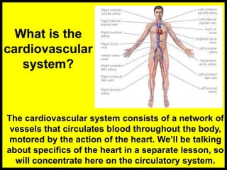
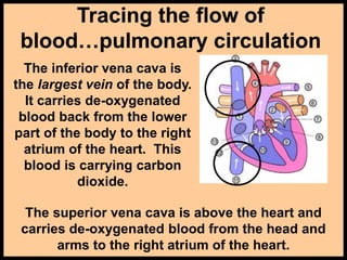
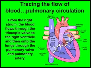
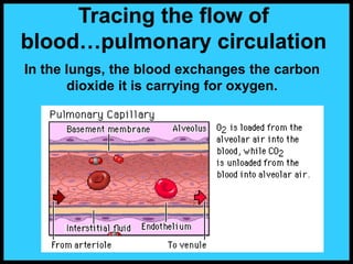
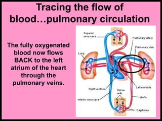
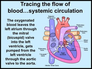
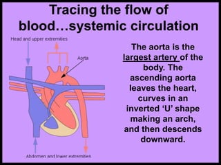
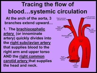
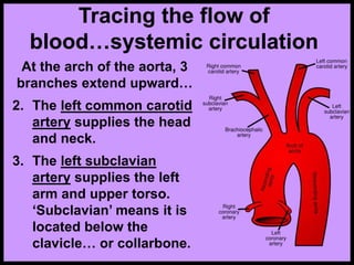
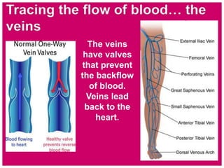
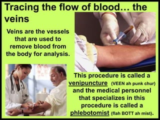
The cardiovascular system consists of a network of
vessels that circulates blood throughout the body,
motored by the action of the heart. We’ll be talking
about specifics of the heart in a separate lesson, so
will concentrate here on the circulatory system.
3. Tracing the flow of
blood…pulmonary circulation
The inferior vena cava is
the largest vein of the body.
It carries de-oxygenated
blood back from the lower
part of the body to the right
atrium of the heart. This
blood is carrying carbon
dioxide.
The superior vena cava is above the heart and
carries de-oxygenated blood from the head and
arms to the right atrium of the heart.
4. Tracing the flow of
blood…pulmonary circulation
From the right
atrium, the blood
flows through the
tricuspid valve to
the right ventricle
and then onto the
lungs through the
pulmonary valve
and pulmonary
artery.
5. In the lungs, the blood exchanges the carbon
dioxide it is carrying for oxygen.
Tracing the flow of
blood…pulmonary circulation
6. Tracing the flow of
blood…pulmonary circulation
The fully oxygenated
blood now flows
BACK to the left
atrium of the heart
through the
pulmonary veins.
7. Tracing the flow of
blood…systemic circulation
The oxygenated
blood leaves the
left atrium through
the mitral
(bicuspid) valve
into the left
ventricle, gets
pumped from the
left ventricle
through the aortic
valve to the aorta.
8. Tracing the flow of
blood…systemic circulation
The aorta is the
largest artery of the
body. The
ascending aorta
leaves the heart,
curves in an
inverted ‘U’ shape
making an arch,
and then descends
downward.
9. Tracing the flow of
blood…systemic circulation
At the arch of the aorta, 3
branches extend upward…
1. The brachiocephalic
artery (or innominate
artery) quickly divides into
the right subclavian artery
that supplies blood to the
right arm and upper torso
AND the right common
carotid artery that supplies
the head and neck.
10. Tracing the flow of
blood…systemic circulation
At the arch of the aorta, 3
branches extend upward…
2. The left common carotid
artery supplies the head
and neck.
3. The left subclavian
artery supplies the left
arm and upper torso.
‘Subclavian’ means it is
located below the
clavicle… or collarbone.
11. Tracing the flow of
blood… the arteries
Arteries are elastic tubes that carry
blood in pulsating waves. The blood
exerts pressure against the walls of
the arteries as it passes through. The
peak pressure occurs during the
heart’s contraction, and is called
systolic pressure. The minimum
pressure occurs between
contractions when the heart expands
and refills, and is called diastolic
pressure. This pressure variation
within the artery produces a pulse.
All arteries have a pulse.
12. Tracing the flow of blood… the
arteries
Common pulse sites used to
check circulation are:
1. TEMPORAL (TEM por ul) - side of
forehead
2 & 3. CAROTID (kuh RAH did) - neck
4. BRACHIAL (BRAY kee ul) - inside
the elbow
5. RADIAL - thumb side of wrist
6. FEMORAL (FEM er all) - groin
7. POPLITEAL (pah plah TEE ul) -
behind the knee
8 & 9. DORSALIS PEDIS (dor SAL us
PED iss) - upper surface of foot
13. Arterioles act like adjustable
nozzles in the circulatory system,
so they have the greatest
influence over blood pressure.
Tracing the flow of blood… the
arterioles
The arteries
branch off
into even
smaller
vessels called
arterioles,
and then to
smaller
vessels yet
called
capillaries.
14. Tracing the flow of blood… the
capillaries
The capillaries are the
smallest of the blood
vessels, and the walls
are so thin that
molecules can pass
through them. They
branch out from the
arterioles, passing
next to the organs,
intestines, and
through all the
cellular tissue.
In the cellular tissue, the
capillaries provide the
means of exchange,
through the process of
absorption.
15. Tracing the flow of blood… the
capillaries
The capillaries
branching away
from the arteries in
the abdomen pass
by the liver and
intestines, picking
up nutrients and
water.
The capillaries in the cellular tissue exchange
their oxygen, nutrients, and water… and pick up
carbon dioxide and other wastes.
The capillaries
branching away
from the arteries in
the lungs absorb
oxygen.
16. Tracing the flow of blood… the
venules
The capillaries,
now carrying
carbon dioxide
and cell wastes,
start merging
into bigger vessels
called venules (VEEN
or VEN yoo als) The
venules widen
even further,
emptying into
veins.
17. The veins
have valves
that prevent
the backflow
of blood.
Veins lead
back to the
heart.
Tracing the flow of blood… the
veins
18. Veins are the vessels
that are used to
remove blood from
the body for analysis.
Tracing the flow of blood… the
veins
This procedure is called a
venipuncture (VEEN ah punk chur)
and the medical personnel
that specializes in this
procedure is called a
phlebotomist (flah BOTT ah mist).