Physiology of heart

The document summarizes the conduction system of the heart and the cardiac cycle. It discusses the following key points: 1. The heart's conduction system originates in specialized cardiac muscle cells called autorhythmic cells, which generate electrical impulses to initiate and coordinate heart contractions. The main structures of the conduction system are the sinoatrial node, atrioventricular node, bundle of His, and Purkinje fibers. 2. The cardiac cycle involves coordinated contraction and relaxation of the atria and ventricles. It begins with depolarization of the sinoatrial node, followed by atrial contraction, ventricular depolarization through the conduction system, ventricular contraction, and finally relaxation of both chambers.

Recommended
More Related Content
What's hot
What's hot (20)
Similar to Physiology of heart
Similar to Physiology of heart (20)
More from Dr. Binu Babu Nursing Lectures Incredibly Easy
More from Dr. Binu Babu Nursing Lectures Incredibly Easy (20)
Recently uploaded
Recently uploaded (20)
Physiology of heart
- 1. Mr. Binu Babu Asst. Professor Mrs. Jincy Ealias Asst. Professor
- 2. Electrical Activity of the Heart • The heartbeat originates in a specialized cardiac conduction system. • The heart beats normally in an orderly sequence: Contraction of the atria (atrial systole) is followed by contraction of the ventricles (ventricular systole), and during diastole all four chambers are relaxed.
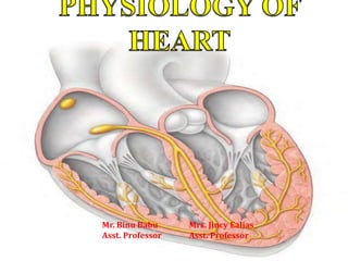
- 3. CONDUCTION SYSTEM OF THE HEART • Action potentials (electrical impulses) in the heart originate in specialized cardiac muscle cells, called autorhythmic cells. • These cells are self-excitable, able to generate an action potential without external stimulation by nerve cells. • The autorhythmic cells serve as a pacemaker to initiate the cardiac cycle (pumping cycle of the heart) and provide a conduction system to coordinate the contraction of muscle cells throughout the heart.
- 4. • The autorhythmic cells are concentrated in the structures that make up the conduction system – the sinoatrial node (SA node) – the internodal atrial pathways – the atrioventricular node (AV node) – the bundle of His and its branches – the Purkinje system.
- 5. Sino atrial node • It is a part of the wall of right atrium close to the opening of superior venacava. It generates impulses approximately at the rate of 72 times/min. SA node is called the pacemaker because it depolarizes at a faster rate than any other group of cells in the heart, and imposes that faster rate on the heart as a whole. Atrio ventricular node • It is a part of the wall of right atrium close to the atrioventricular septum and near to the tricuspid valve. It generates impulses approximately at the rate of 60 times/min.
- 6. Bundle of His • It is a thick band of muscle fibers starting from A.V Node. It runs along with intra ventricular septum. It divides into right and left bundle branch. It generates impulses approximately at the rate of 40 times/min. Purkinje Fibers • These fibers arise from the branches of bundle of His. These fibers pierce into the ventricular myocardium.
- 7. • Cardiac excitation normally begins in the sinoatrial (SA) node, located in the right atrial wall just inferior and lateral to the opening of the superior vena cava. • The spontaneous depolarization is a pacemaker potential. (The pacemaker potential is a slow, positive increase in voltage across the cell's membrane) • When the pacemaker potential reaches threshold, it triggers an action potential. Each action potential from the SA node propagates both atria via gap junctions in the intercalated discs of atrial muscle fibers and contract both atria at the same time.
- 9. • By conducting along atrial muscle fibers, the action potential reaches the atrioventricular (AV) node, located in the interatrial septum, just anterior to the opening of the coronary sinus. • From the AV node, the action potential enters the atrioventricular (AV) bundle (also known as the bundle of His). This bundle is the only site where action potentials can conduct from the atria to the ventricles.
- 11. • After propagating along the AV bundle, the action potential enters both the right and left bundle branches. • Finally, the large-diameter Purkinje fibers rapidly conduct the action potential beginning at the apex of the heart upward to the remainder of the ventricular myocardium. Then the ventricles contract, pushing the blood upward toward the semilunar valves.
- 12. Conduction Speeds in Cardiac Tissue Tissue Conduction Rate (m/s) SA node 0.05 Atrial pathways 0.1 AV node 0.05 Bundle of His 0.1 Purkinje system 0.4 Ventricular muscle 0.1
- 13. • SA node is called the pacemaker of the heart. If for any reason the SA node stops beating, the AV node, which has the next fastest rate of depolarization, would become the heart’s pacemaker. If the AV node failed, the bundle of His would take over. If it failed, the Perkinje fibers would start the heartbeat, and if they failed as well, a group of cells somewhere else in the heart would start pulsing. If failed, eventually it can no longer sustain life.
- 14. The Electrocardiogram • The body fluids are good conductors, fluctuations in potential that represent the algebraic sum of the action potentials of myocardial fibers can be recorded extracellularly. • The record of these potential fluctuations during the cardiac cycle is the electrocardiogram (ECG).
- 16. ECG Intervals. Normal Durations Intervals Average Range Events in the Heart during Interval PR intervala 0.18b 0.12–0.20 Atrial depolarization and conduction through AV node QRS duration 0.08 to 0.10 Ventricular depolarization and atrial repolarization QT interval 0.40 to 0.43 Ventricular depolarization plus ventricular repolarization ST interval (QT minus QRS) 0.32 . . . Ventricular repolarization (during T wave) aMeasured from the beginning of the P wave to the beginning of the QRS complex. bShortens as heart rate increases from average of 0.18 s at a rate of 70 beats/min to 0.14 s at a rate of 130 beats/min.
- 17. Properties of cardiac muscles • Excitability • Conduction • Contraction • Refractory period • Functional Syncytium • Auto rhythmicity • Staircase phenomenon
- 18. Excitation • It is an electrical event. Calcium ion are responsible for this event Conduction • The action potential is propagated all along the length of the muscle fiber this phenomenon is known as conduction. Contraction • It is the shortening of muscle fibres. Refractory period • It is the period during which the 2nd stimulus cannot generate a fresh action potential. It is divided in to two – Absolute refractory period (0.25 sec) – Relative refractory period (0.05 sec) it may generate an action potential.
- 19. Functional Syncytium • Cardiac muscles act as single unit this phenomenon is called Functional Syncytium. Auto rhythmicity • Cardiac muscle can generate their own impulse, this property of heart is called the auto rhythmicity Staircase phenomenon • When a series of stimuli of the same intensity are sent into the muscle after a calm period, the first few contractions of the series show a successive increase in amplitude.
- 20. Cardiac Cycle • Cardiac cycle is defined as sequence of cyclical changes taking place in the heart from one beat to the next. • A cardiac cycle consists of systole and diastole of the atria plus systole and diastole of the ventricles. • A cardiac cycle duration is 0.8 sec. Changes during Cardiac cycle 1. Mechanical changes: contraction and relaxation of atria and ventricles. 2. Electrical changes 3. Volume change inside the heart 4. Pressure change inside the chambers 5. Opening and closing of valves 6. Heart sounds
- 21. • In each cardiac cycle, the atria and ventricles alternately contract and relax, forcing blood from areas of higher pressure to areas of lower pressure. As a chamber of the heart contracts, blood pressure within it increases.
- 22. Atrial Systole • Depolarization of the SA node causes atrial depolarization which results atrial systole. • During atrial systole, both the atria contracts at the same time, where as the ventricles are relaxed. • The contraction of atrial muscles narrows the venacaval orifices and pulmonary vein orifices. • And exert a pressure on the blood atria, which forces blood to move into ventricles through the open tricuspid and mitral valves. • Atrial systole which lasts about 0.1 sec. The end of atrial systole is also the end of ventricular diastole (relaxation). • Each ventricle contains about 130 mL at the end of its relaxation period (diastole). This blood volume is called the end-diastolic volume (EDV).
- 23. Ventricular Systole • When the ventricles are contracting the atria are relaxed (atrial diastole). • Ventricular depolarization causes ventricular systole. When ventricular systole begins, pressure rises inside the ventricles and pushes blood up against the atrioventricular (AV) valves, forcing them shut. For about 0.05 seconds, both the semilunar and AV valves are closed. This is the period of isovolumetric contraction.
- 24. • When left ventricular pressure exceeds aortic pressure (80 mmHg) and right ventricular pressure exceeds pressure in the pulmonary artery (20 mmHg), both semilunar valves open. • At this point, ejection of blood from the ventricles begins. • The left ventricle ejects about 70 mL of blood into the aorta and the right ventricle ejects the same volume of blood into the pulmonary trunk. The volume remaining in each ventricle at the end of systole, about 60 mL, is the end- systolic volume (ESV). • Ventricular systole lasts about 0.3 sec. • Stroke volume (SV) is the amount of blood ejected by the left ventricle in one contraction. • SV = EDV - ESV (130-60=70 ml)
- 25. Relaxation Period (diastole) • During the relaxation period, which lasts about 0.4 sec, the atria and the ventricles are relaxed. Blood flows into the heart throughout diastole, filling the atria and ventricles. • Once the atrial muscles are relaxed venacaval orifices and pulmonary veins open and atria is filled blood. • Once the ventricular muscle is fully contracted, the ventricular pressures drop rapidly. And blood in the aorta and pulmonary trunk begins to flow backward toward the regions of lower pressure in the ventricles.
- 26. • As the ventricles continue to relax, the pressure falls quickly. When ventricular pressure drops below atrial pressure, the AV valves open, and ventricular filling begins. Blood that has been flowing into and building up in the atria during ventricular systole then rushes rapidly into the ventricles. At the end of the relaxation period, the ventricles are about three-quarters full. The P wave appears in the ECG, signaling the start of another cardiac cycle.
- 27. Cardiac Output • Cardiac output (CO) is the volume of blood ejected from the heart in each minute. • Cardiac output is expressed in litres per minute (L/min). • Cardiac output is the product of heart rate (HR) and stroke volume (SV). • CO= HR x SV • In a resting, supine man, it averages about 5 L/min (72 beats/min x 70 mL).
- 28. During exercise, venous return increases and stretches the ventricular myocardium, which in response contracts more forcefully, and results in increase stroke volume. More blood is pumped with each beat, and at the same time, the heart rate increases, causing cardiac output to increase to as much as four times the resting level, and even more for athletes.
- 29. Factors affecting cardiac output • Factors increases cardiac output – Anxiety and excitement – Eating – Exercise – High environmental temperature – Pregnancy • Factors decreases cardiac output – Sitting or standing from lying position – Rapid arrhythmias – Heart disease
- 30. Regulation of cardiac output • Changes in cardiac output can be produced by changes in heart rate or stroke volume or both.
- 31. Regulation of Heart Rate • The heart generates its own electrical impulse, which begins at the SA node. The nervous system can change the heart rate in response to environmental circumstances. In the brain, the medulla oblongata contains the cardiovascular centers: the accelerator center and the inhibitory center.
- 32. • The cardiovascular center directs appropriate output by increasing or decreasing the frequency of nerve impulses in both the sympathetic and parasympathetic branches of the ANS. • Sympathetic impulses—along sympathetic nerves from the thoracic spinal cord to the SA node, AV node, and most of the myocardium— increase heart rate and force of contraction. • Parasympathetic impulses—along the vagus nerve to the SA node, AV node, and atrial myocardium—decrease heart rate.
- 34. Medullary cardio vascular center receives information necessary for changes in the heart rate from higher brain centers (cerebral cortex, limbic system and hypothalamus) and sensory receptors such as – Proprioceptors – Chemoreceptors – Baroreceptors located in the internal carotid arteries and the aortic arch. • The baroreceptors detect changes in blood pressure. • The chemoreceptors are cells specialized to detect changes in the oxygen content of the blood (as well as changes in carbon dioxide and hydrogen ion content). • The Proprioceptors detect changes in position and movement.
- 36. • In response to the information received by the medullary cardiac center, either sympathetic impulses or parasympathetic impulses gets initiated. • Sympathetic system response Impulses in the cardiac accelerator nerves trigger the release of norepinephrine (by adrenal medulla), which binds to beta-1 (β1) receptors on cardiac muscle fibers. This results, – SA (and AV) node speeds the rate of spontaneous depolarization so that they fire impulses more rapidly and heart rate increases. – Increased contraction of atria and ventricles thus increases stroke volume.
- 37. • Parasympathetic system response, They release acetylcholine, which ↓ HR by ↓ slow inflow of Na+ and Ca++ and by ↑ the subsequent outflow of potassium (K+). – Decreased rate of spontaneous depolarization of SA (and AV) node decreases heart rate.
- 38. Factors contribute to regulation of heart rate Chemical regulation – Cardiac activity depressed by • Hypoxia • Acidosis • Alkalosis – Hormones • Catecholamines and thyroid hormones increase HR and contractility – Cations • Alterations in balance of K+, Na+ and Ca2+ alter HR and contractility
- 39. Age Gender • Female HR higher Physical fitness • Resting bradycardia Body temperature • Increase causes SA node to discharge more rapidly
- 40. Regulation of Stroke Volume • Stroke volume is determined by sympathetic stimuli making the myocardial muscle fibers contract with greater strength and parasympathetic stimuli having the opposite effect. • The factors regulate stroke volume – Preload: the degree of stretch of heart muscle before it contracts. – Contractility: the forcefulness of contraction of individual ventricular muscle fibers. – Afterload: the resistance to ejection of blood from the ventricle.
- 41. Preload • Preload is defined as the force acting to stretch the ventricular fibers at end-diastole. • An increase in myocardial muscle fiber length is associated with an increase in the force of contraction and thus increases the stroke volume and cardiac output. • Higher the preload, the higher the stroke volume will be.
- 42. Contractility • Contractility refers to the force generated by the contracting myocardium. • Increased contractility results in increased stroke volume. • Contractility is enhanced by circulating catecholamines, sympathetic neuronal activity, and certain medications • Contractility is depressed by hypoxemia, acidosis, and certain medications (eg, beta- adrenergic blocking agents)
- 43. Afterload • Afterload is the resistance to ejection of blood from the ventricle. • The ejection of blood from the heart begins when pressure in the right ventricle exceeds the pressure in the pulmonary trunk (about 20 mmHg), and when the pressure in the left ventricle exceeds the pressure in the aorta (about 80 mmHg). At that point, the higher pressure in the ventricles causes blood to push the semilunar valves open. The pressure that must be overcome before a semilunar valve can open is termed the afterload. • An increase in afterload causes stroke volume to decrease.