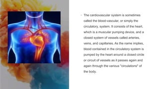
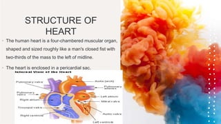
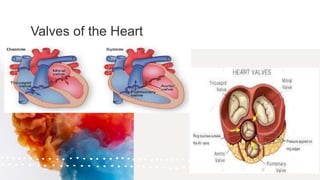
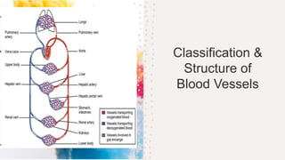
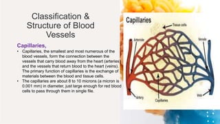
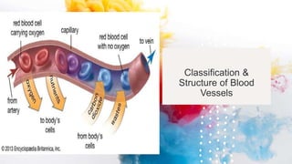
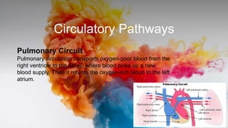
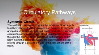
The cardiovascular system consists of the heart and blood vessels. The heart pumps blood through arteries, capillaries, and veins in a closed circulatory system. In the capillaries, nutrients and waste are exchanged. The heart has four chambers and valves that ensure blood flows in one direction through the pulmonary and systemic circuits. It beats regularly due to an intrinsic pacemaker and conduction system. Blood transports oxygen, nutrients, hormones and waste throughout the body in a continuous cycle.


![ONFH[AVN HIP] -TRIPLE REGIME -A NOVAL SURGICAL CONCEPT .pptx](https://cdn.slidesharecdn.com/ss_thumbnails/onfhavnhip2026koaconcalicutdrgokuldevdrmashraf-260210064517-213ec005-thumbnail.jpg?width=640&height=640&fit=bounds)


![CTEV [ clubfoot] DR ARUN LAL ,DR MOHAMED ASHRAF travancore medical college k...](https://cdn.slidesharecdn.com/ss_thumbnails/ctevclubfootdrarunlaldrmohamedashraftravancoremedicalcollegekollamkeralaindia-260208063247-18fc466c-thumbnail.jpg?width=640&height=640&fit=bounds)

