Downloaded 449 times
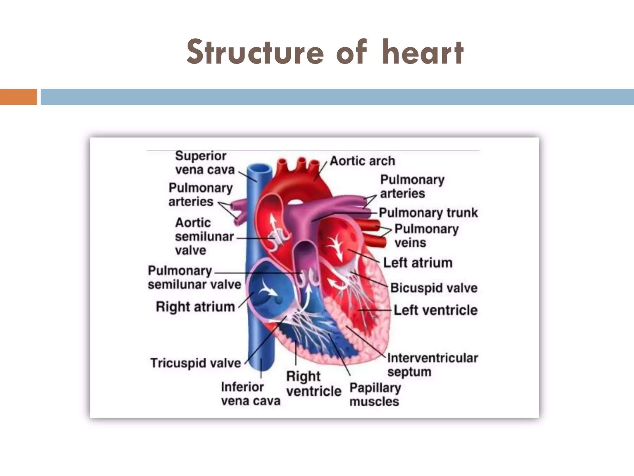


The cardiac cycle refers to the series of events in the heart from the start of one heartbeat to the next, initiated by the sino-atrial node (SAN) that generates contraction signals. It consists of four main stages: atrial diastole, atrial systole, ventricular systole, and ventricular diastole, each involving the relaxation and contraction of heart chambers to effectively circulate blood. The atrioventricular node (AVN) and Purkinje fibers play essential roles in conducting the excitation waves that coordinate these contractions.


