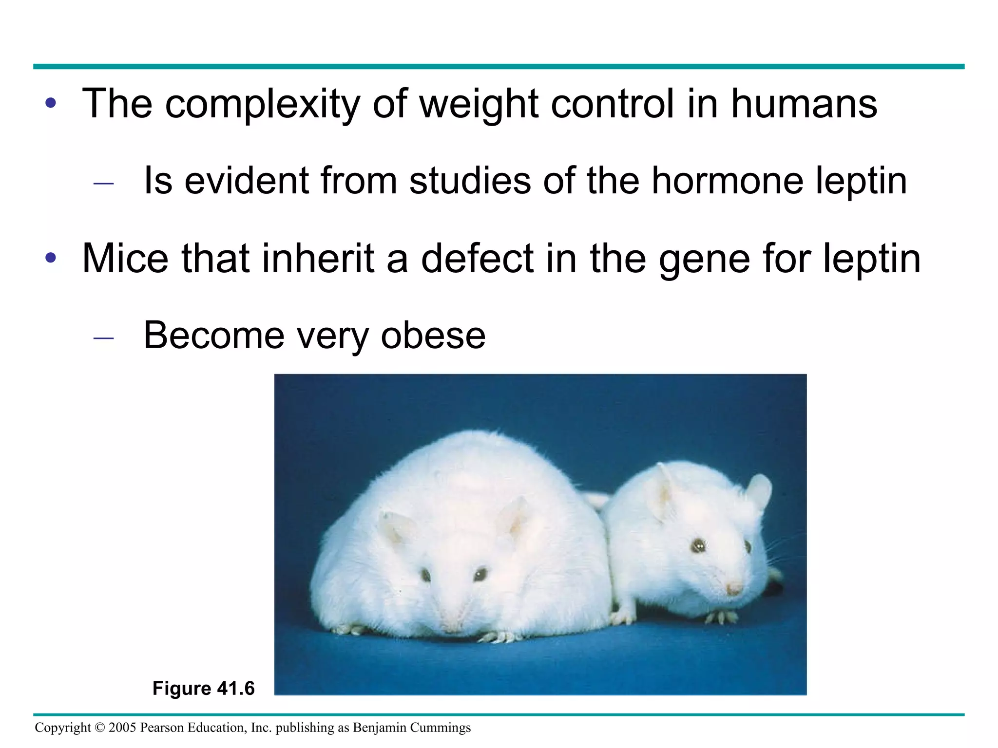
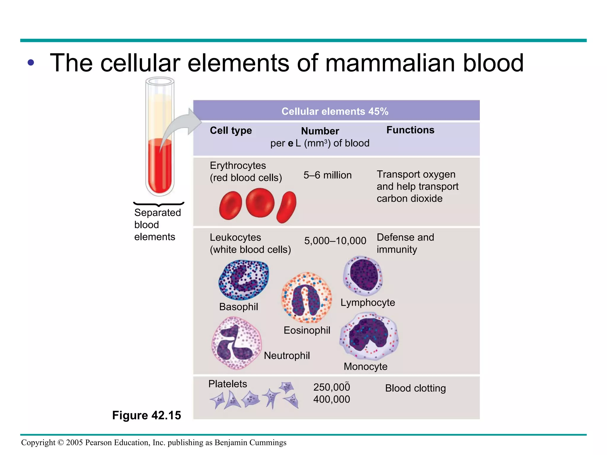
The document provides an overview of animal nutrition and digestion. It discusses that animals fall into three dietary categories - herbivores, carnivores, and omnivores. The main stages of food processing in animals are ingestion, digestion, absorption, and elimination. Digestion occurs through specialized digestive organs and glands that break down food into smaller molecules for absorption. Homeostatic mechanisms regulate nutrient intake and storage to maintain energy balance.
The liver is the central laboratory of a chicken’s body. It is essential that this organ is kept in an excellent condition in order to maintain a healthy bird. Understanding the metabolic function and causes of disruptions in liver functions helps us to provide the birds with the right feed and health treatment.
When we cut open the body of a chicken, the first organ that is most likely revealed is the liver. The message is clear. Nature wants us to examine the liver carefully before
proceeding to the other organs.The liver contains great functional reserve capacity, which is very important in domestic animals subjected to high production requirements. This organ adapts easily to different conditions by increasing the intensity of its functions.
Particularly in broilers, the liver has to cope with many challenges, including
high energy level feed, the addition of chemotherapeutics, coccidiostats
and others, whose desired metabolites must be maintained in equilibrium by hepatic homeostasis.Incidental treatments with highly hepatotoxic and nephrotoxic antibiotics
or sulfonamides pose serious risks and cause situations of difficult prognosis during a 40-45 day period in which the body acquires satisfactory muscular mass. What is the function of the liver and what might be the cause of malfunctioning?
Human digestive system structure and function
overview
Major organs
Mouth
Esophagus
Stomach
small intestine
large intestine
Acessory organs:
Liver
gall bladder
Pancreas.
Human digestive system
Major organs
Mouth
Esophagus
Stomach
small intestine
large intestine.
Acessory organs:
Liver
Gall bladder
Pancreas.
MAJOR ORGANSThe Mouth
pH: 7
The first part of the digestive system
the entry point of food.
Structures in the mouth that aids digestion
Teeth – cut, tear, crush and grind food.
Salivary glands – produce and secrete saliva into the oral cavity.
saliva
moistens the food
contains enzymes (ptyalin or salivary amylase)
begins digestion of starch into smaller polysaccharides.
Function:
Mechanical digestion.
increasing surface area for faster chemical digestion.
The Esophagus
a tube connecting the mouth to the stomach
running through the Thoracic cavity.
Location:
lies behind windpipe (Trachea).
The trachea has as an epiglottis
preventing food from entering the windpipe,
moving the food to the esophagus while swallowing.
Food travels down the esophagus, through a series of involuntary rhythmic contractions (wave-like) called peristalsis.
Function:
The lining of the esophagus secretes mucus
lubricating
to support the movement of food.
Esophageal sphincter:
bolus reaches the stomach
must pass through a muscular ringed valve called the esophageal sphincter (Cardiac Sphincter).
Function:
prevent stomach acids from back flowing into the esophagus.
Stomach
J-shaped muscular sac
Has inner folds (rugae)
Increasing surface area of the stomach.
Function:
Stomach performs mechanical digestion
HOW By churning the bolus and mixing it with the gastric juices
secreted by the lining of the stomach.
GASTRIC JUICES HCl, salts, enzymes, water and mucus)
HCL helps break down of food and kills bacteria that came along with the food.
The bolus is now called Chyme.
Enzymes in stomach:
Acidic environment
HCl secreation
kill any microbes that are found in the bolus,
creating a pH of 2.
Mucus prevents the stomach from digesting itself.
Pepsin secreation
responsible for initiating the breakdown of proteins (in )food.
hydrolyzes proteins to yield polypeptides.
pH is 2, the enzyme from the salivary glands stops breaking down carbohydrates.
Pyloric sphincter:
chyme moves from the stomach to the small intestine.
It passes through a muscular ringed sphincter called the pyloric sphincter.
stomach does not digest itselfWhy ?
Protective Mechanism:
three protective mechanisms.
First the stomach only secretes small amounts of gastric juices until food is present.
Second the secretion of mucus coats the lining of the stomach protecting it from the gastric juices.
The third mechanism is the digestive enzyme pepsin is secreted in an inactive protein c
43. Figure 41.15 IIeum of small intestine Duodenum of small intestine Appendix Cecum Ascending portion of large intestine Anus Small intestine Large intestine Rectum Liver Gall- bladder Tongue Oral cavity Pharynx Esophagus Stomach Pyloric sphincter Cardiac orifice Mouth Esophagus Salivary glands Stomach Liver Pancreas Gall- bladder Large intestines Small intestines Rectum Anus Parotid gland Sublingual gland Submandibular gland Salivary glands A schematic diagram of the human digestive system Pancreas
165. Figure 42.27 Inhaled air Exhaled air 160 0.2 O 2 CO 2 O 2 CO 2 O 2 CO 2 O 2 CO 2 O 2 CO 2 O 2 CO 2 O 2 CO 2 O 2 CO 2 40 45 40 45 100 40 104 40 104 40 120 27 CO 2 O 2 Alveolar epithelial cells Pulmonary arteries Blood entering alveolar capillaries Blood leaving tissue capillaries Blood entering tissue capillaries Blood leaving alveolar capillaries CO 2 O 2 Tissue capillaries Heart Alveolar capillaries of lung <40 >45 Tissue cells Pulmonary veins Systemic arteries Systemic veins O 2 CO 2 O 2 CO 2 Alveolar spaces 1 2 4 3
166.
167.
168.
169.
170.
171. O 2 unloaded from hemoglobin during normal metabolism O 2 reserve that can be unloaded from hemoglobin to tissues with high metabolism Tissues during exercise Tissues at rest 100 80 60 40 20 0 100 80 60 40 20 0 100 80 60 40 20 0 100 80 60 40 20 0 Lungs P O 2 (mm Hg) P O 2 (mm Hg) O 2 saturation of hemoglobin (%) O 2 saturation of hemoglobin (%) Bohr shift: Additional O 2 released from hemoglobin at lower pH (higher CO 2 concentration) pH 7.4 pH 7.2 (a) P O 2 and Hemoglobin Dissociation at 37°C and pH 7.4 (b) pH and Hemoglobin Dissociation Figure 42.29a, b
172.
173.
174. Figure 42.30 Tissue cell CO 2 Interstitial fluid CO 2 produced CO 2 transport from tissues CO 2 CO 2 Blood plasma within capillary Capillary wall H 2 O Red blood cell Hb Carbonic acid H 2 CO 3 HCO 3 – H + + Bicarbonate HCO 3 – Hemoglobin picks up CO 2 and H + HCO 3 – HCO 3 – H + + H 2 CO 3 Hb Hemoglobin releases CO 2 and H + CO 2 transport to lungs H 2 O CO 2 CO 2 CO 2 CO 2 Alveolar space in lung 2 1 3 4 5 6 7 8 9 10 11 To lungs Carbon dioxide produced by body tissues diffuses into the interstitial fluid and the plasma. Over 90% of the CO 2 diffuses into red blood cells, leaving only 7% in the plasma as dissolved CO 2 . Some CO 2 is picked up and transported by hemoglobin. However, most CO 2 reacts with water in red blood cells, forming carbonic acid (H 2 CO 3 ), a reaction catalyzed by carbonic anhydrase contained. Within red blood cells. Carbonic acid dissociates into a biocarbonate ion (HCO 3 – ) and a hydrogen ion (H + ). Hemoglobin binds most of the H + from H 2 CO 3 preventing the H + from acidifying the blood and thus preventing the Bohr shift. CO 2 diffuses into the alveolar space, from which it is expelled during exhalation. The reduction of CO 2 concentration in the plasma drives the breakdown of H 2 CO 3 Into CO 2 and water in the red blood cells (see step 9), a reversal of the reaction that occurs in the tissues (see step 4). Most of the HCO 3 – diffuse into the plasma where it is carried in the bloodstream to the lungs. In the HCO 3 – diffuse from the plasma red blood cells, combining with H + released from hemoglobin and forming H 2 CO 3 . Carbonic acid is converted back into CO 2 and water. CO 2 formed from H 2 CO 3 is unloaded from hemoglobin and diffuses into the interstitial fluid. 1 2 3 4 5 6 7 8 9 10 11
225. 2 1 3 B cell Bacterium Peptide antigen Class II MHC molecule TCR Helper T cell CD4 Activated helper T cell Clone of memory B cells Cytokines Clone of plasma cells Secreted antibody molecules Endoplasmic reticulum of plasma cell Macrophage After a macrophage engulfs and degrades a bacterium, it displays a peptide antigen complexed with a class II MHC molecule. A helper T cell that recognizes the displayed complex is activated with the aid of cytokines secreted from the macrophage, forming a clone of activated helper T cells (not shown). 1 A B cell that has taken up and degraded the same bacterium displays class II MHC–peptide antigen complexes. An activated helper T cell bearing receptors specific for the displayed antigen binds to the B cell. This interaction, with the aid of cytokines from the T cell, activates the B cell. 2 The activated B cell proliferates and differentiates into memory B cells and antibody-secreting plasma cells. The secreted antibodies are specific for the same bacterial antigen that initiated the response. 3 Figure 43.17