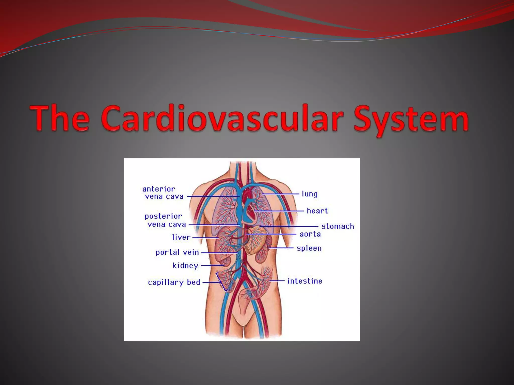
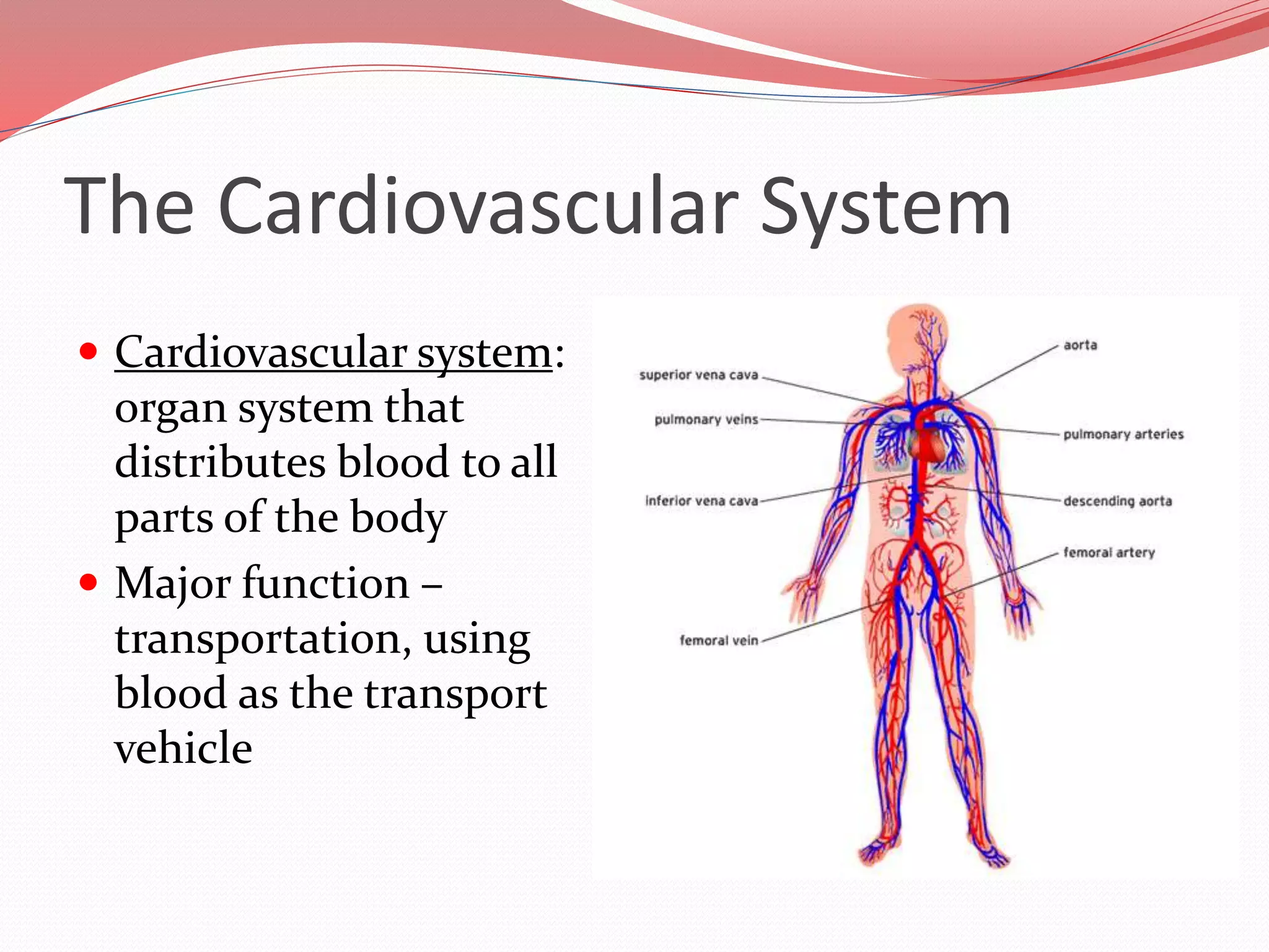
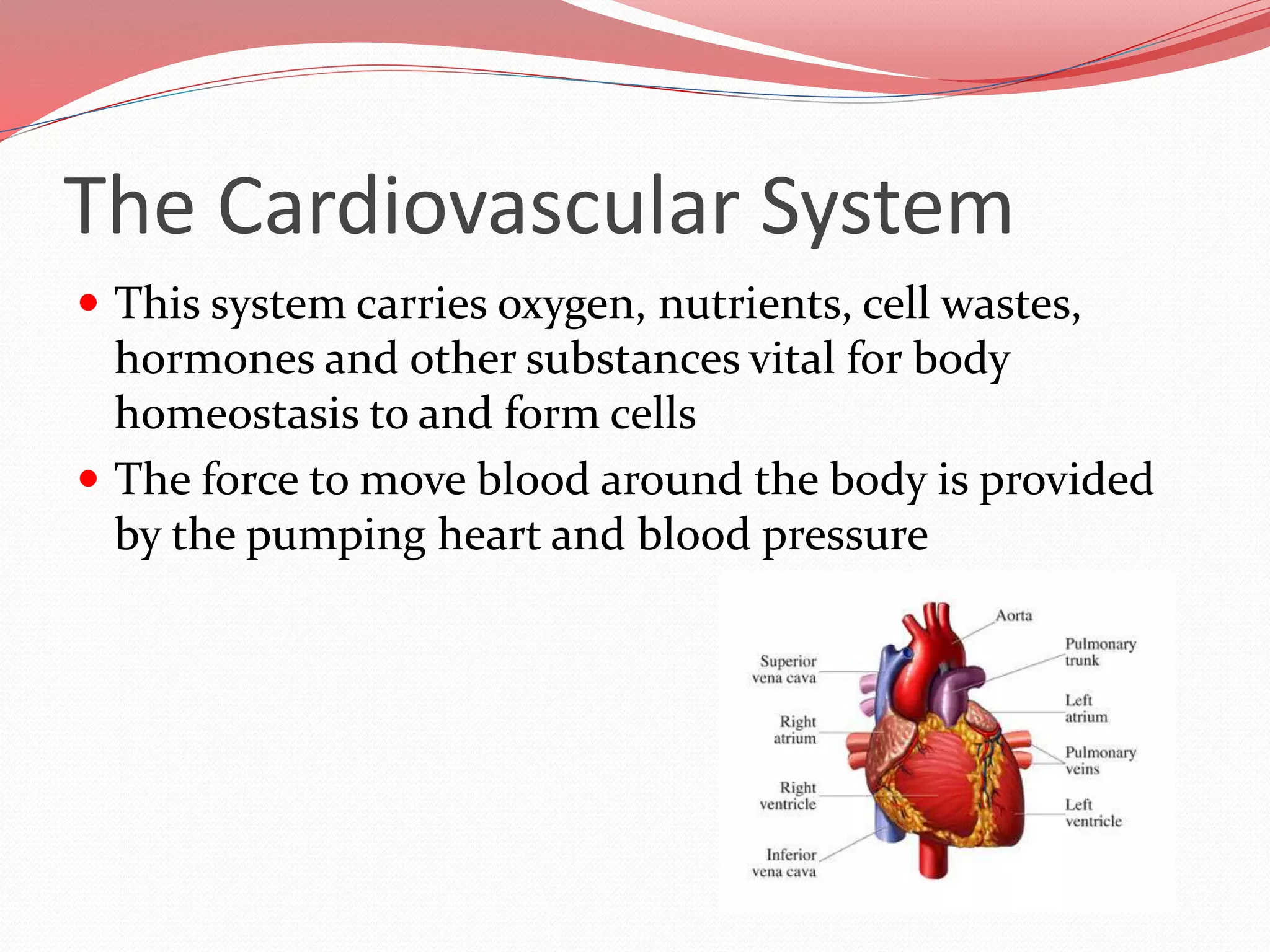
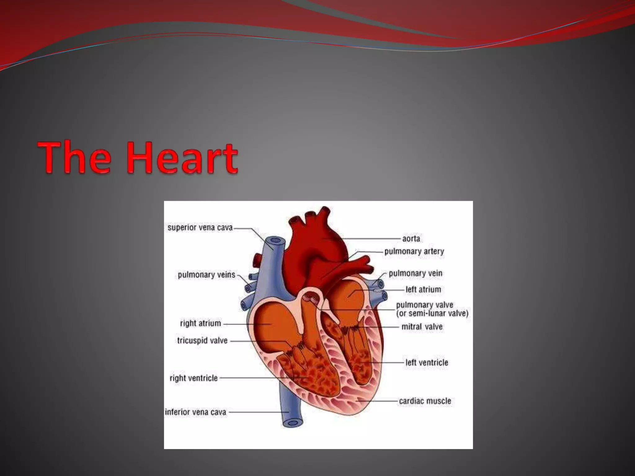
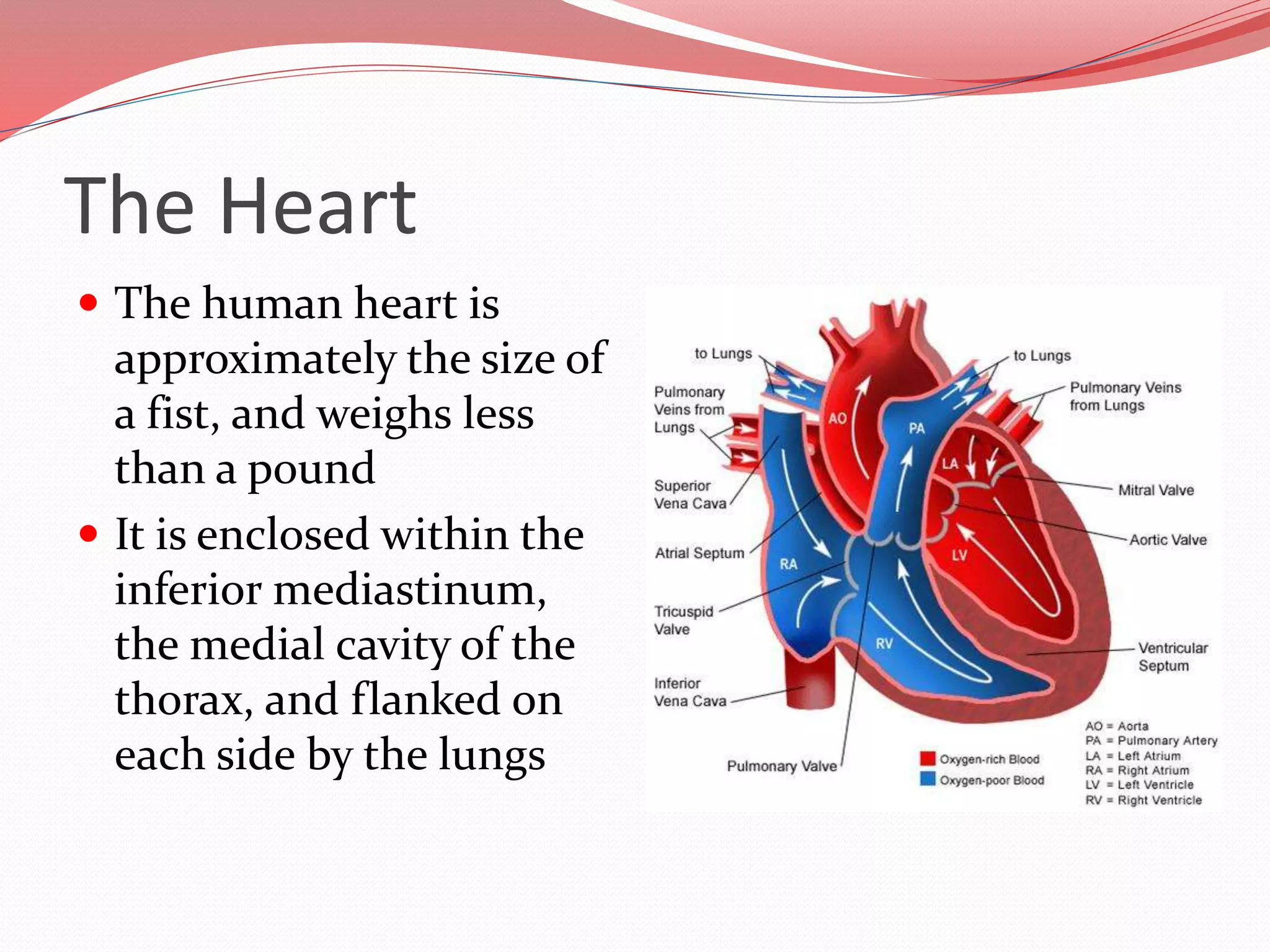
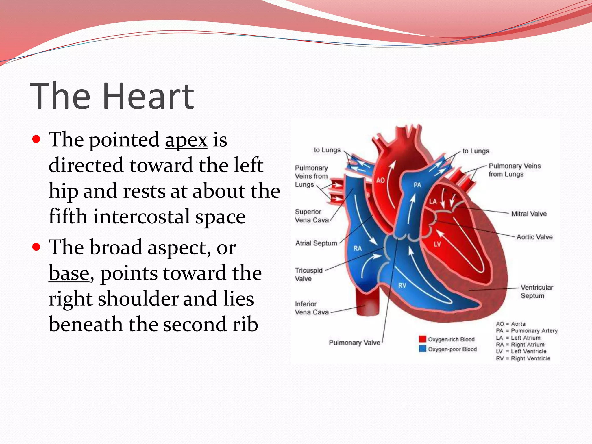
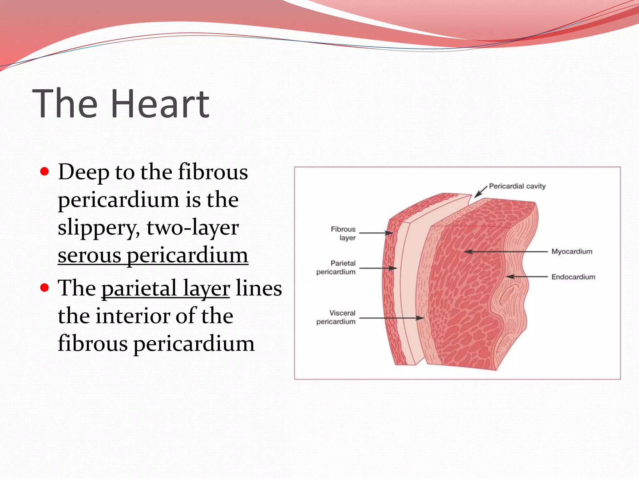
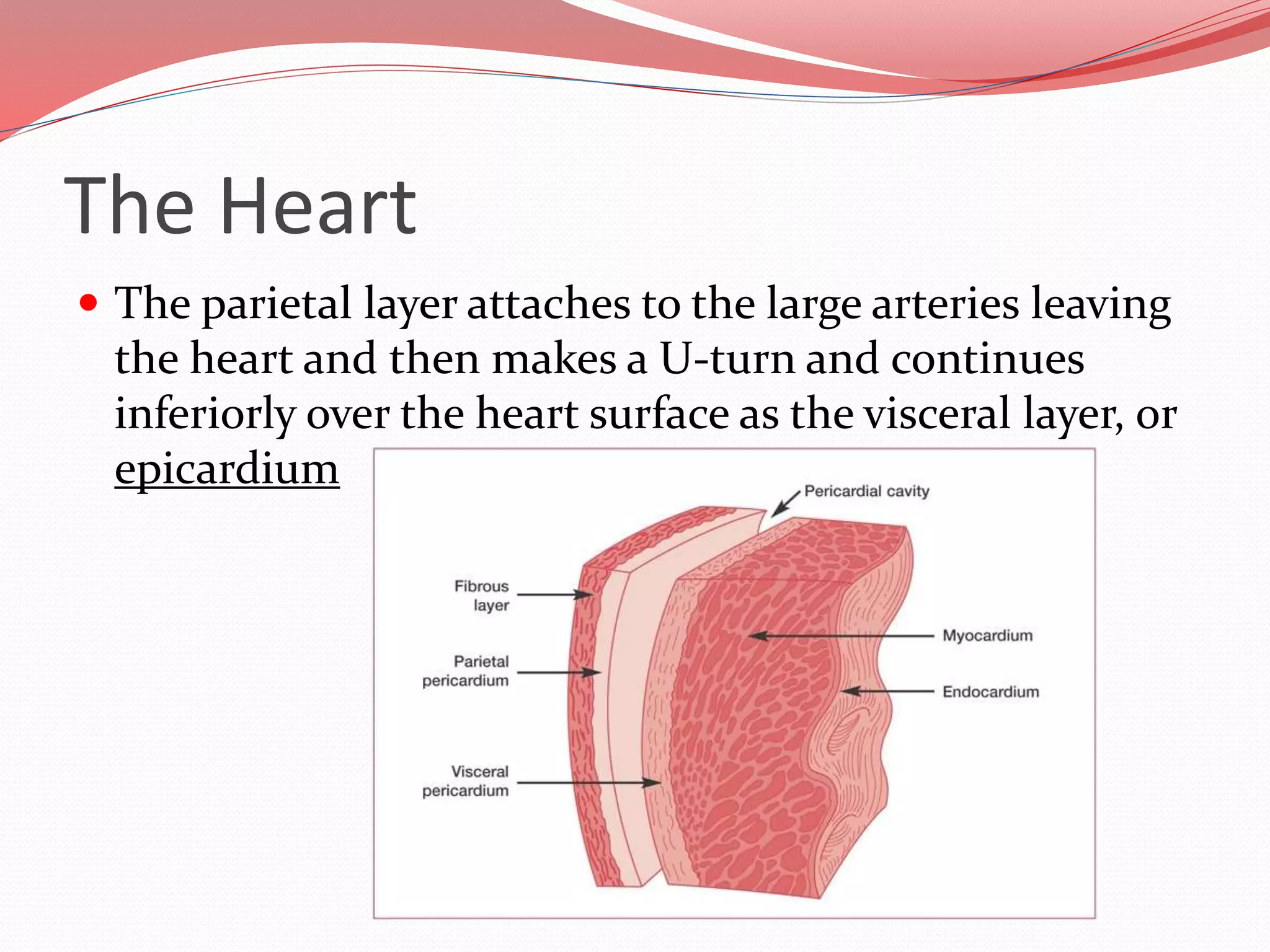
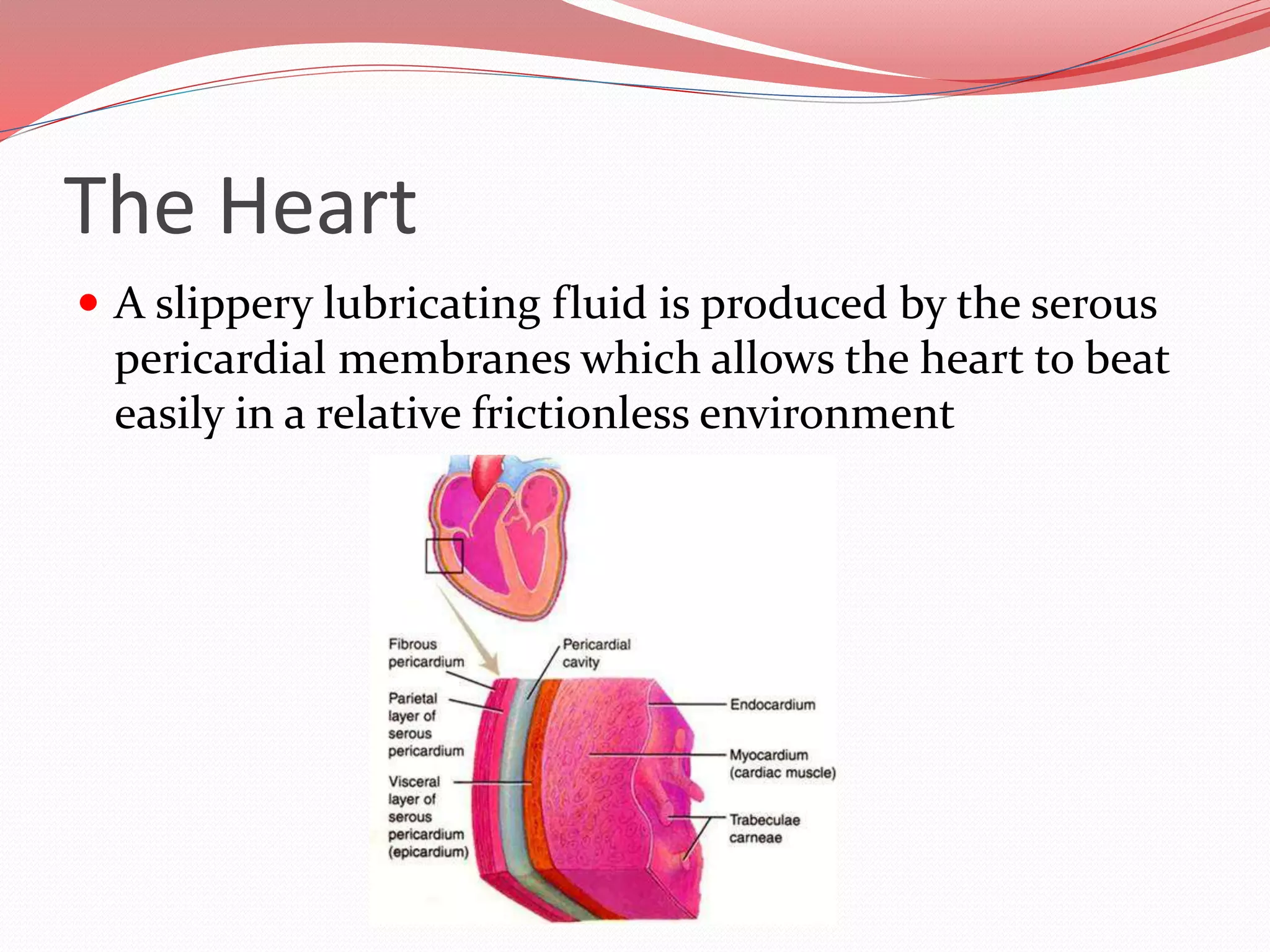
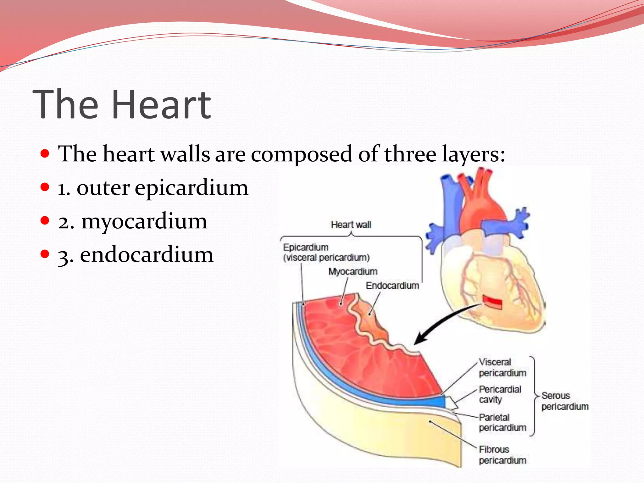
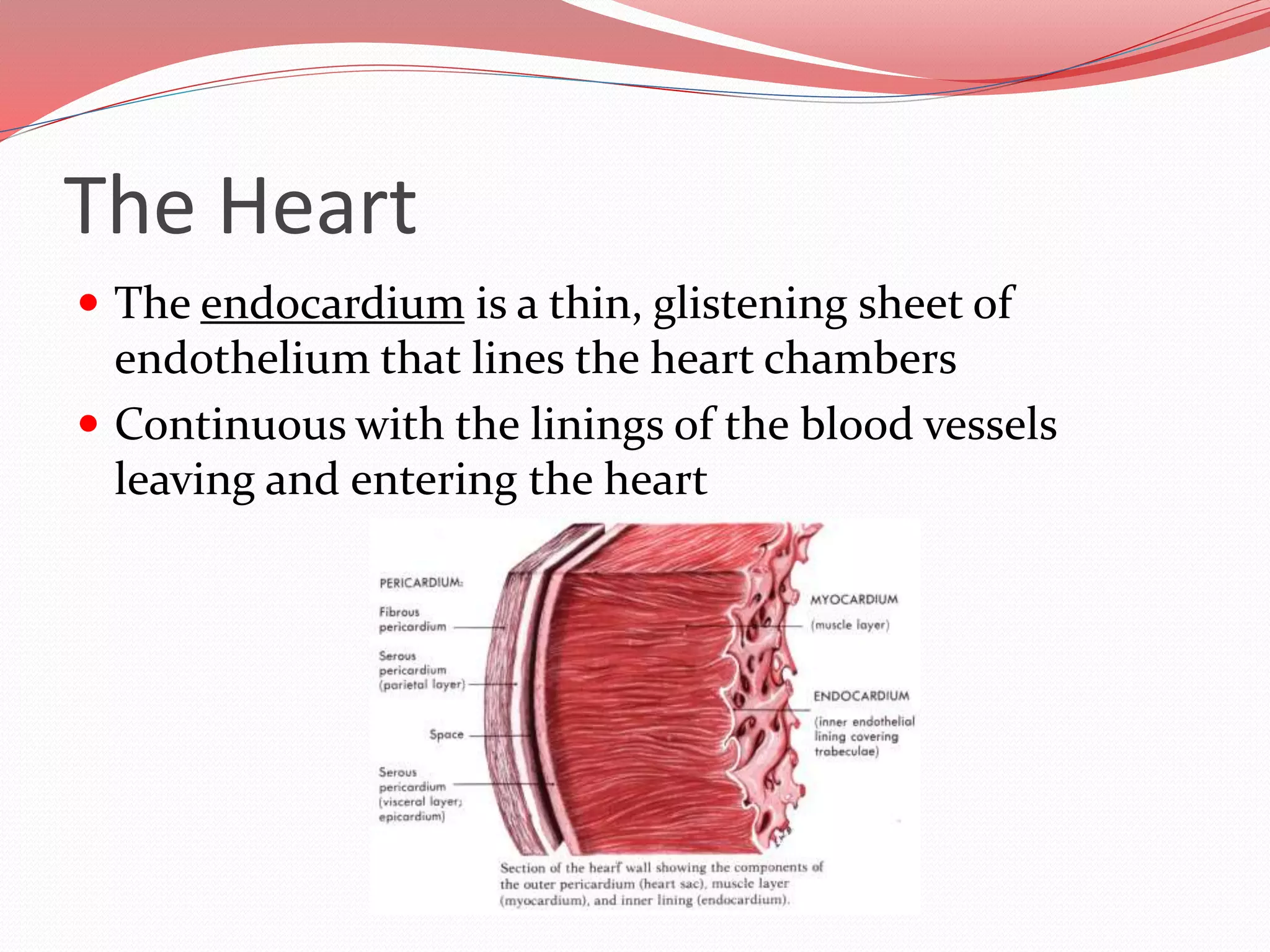
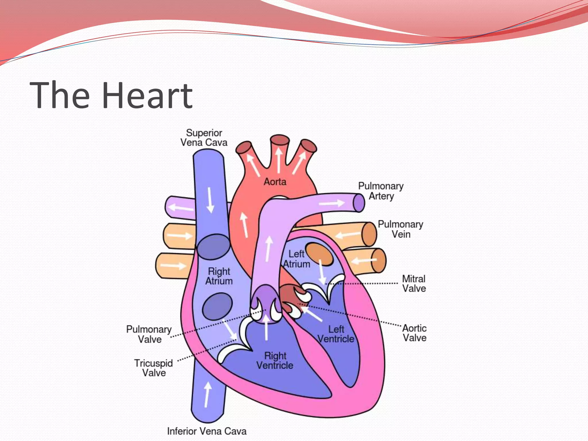
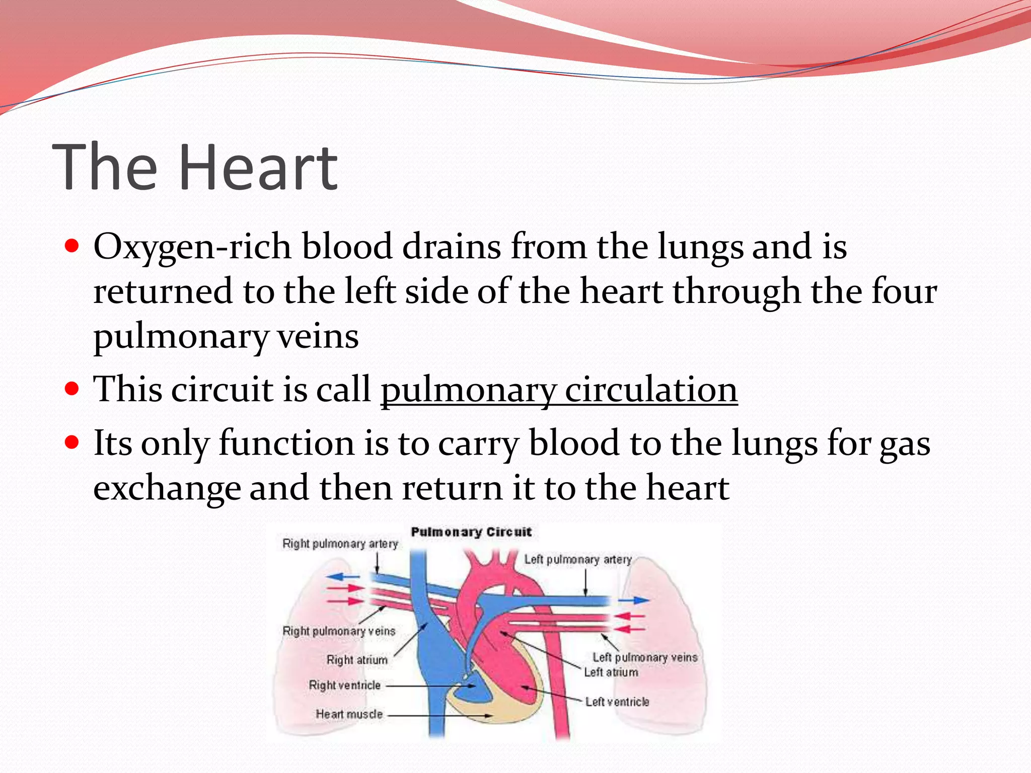
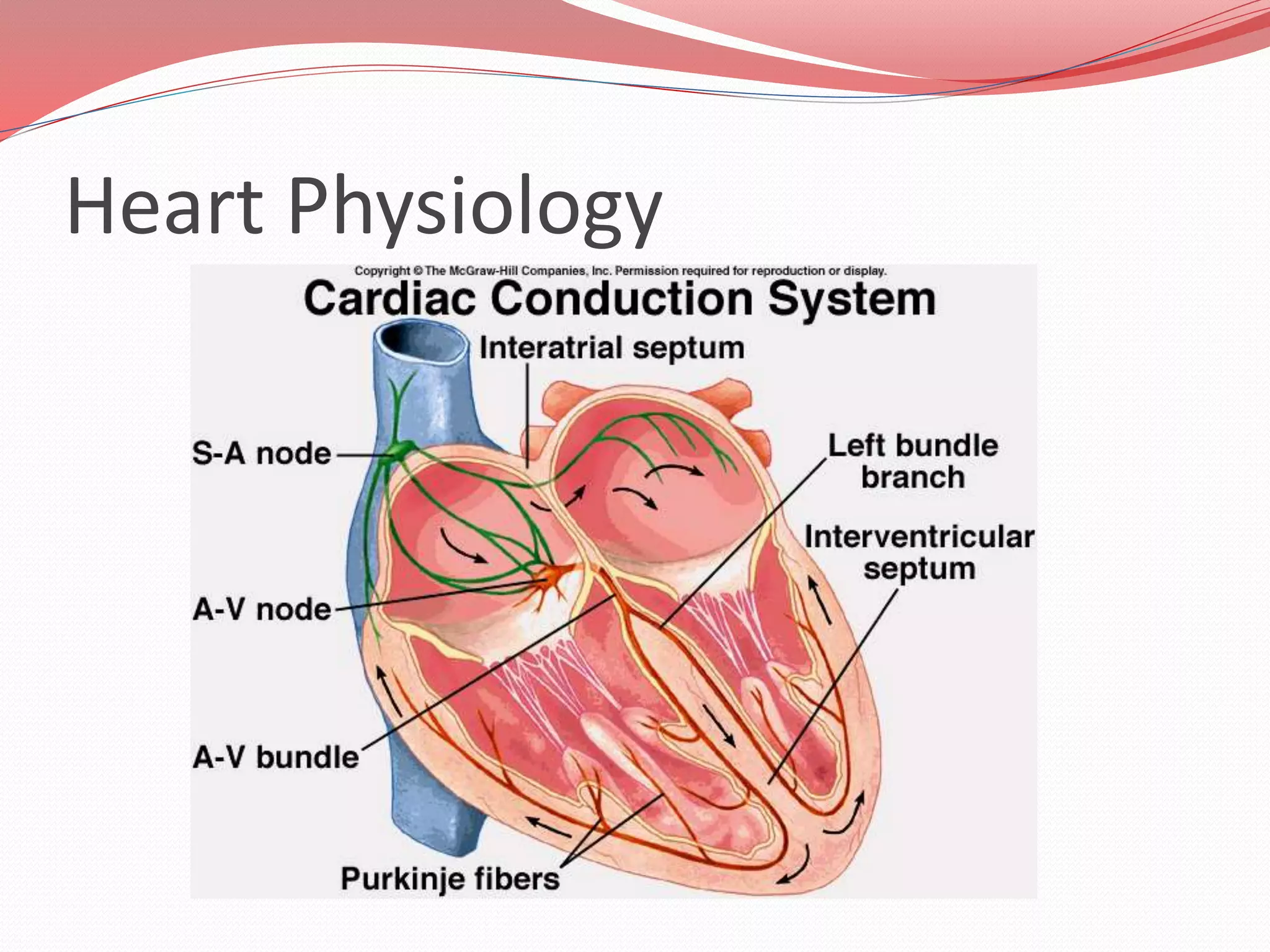
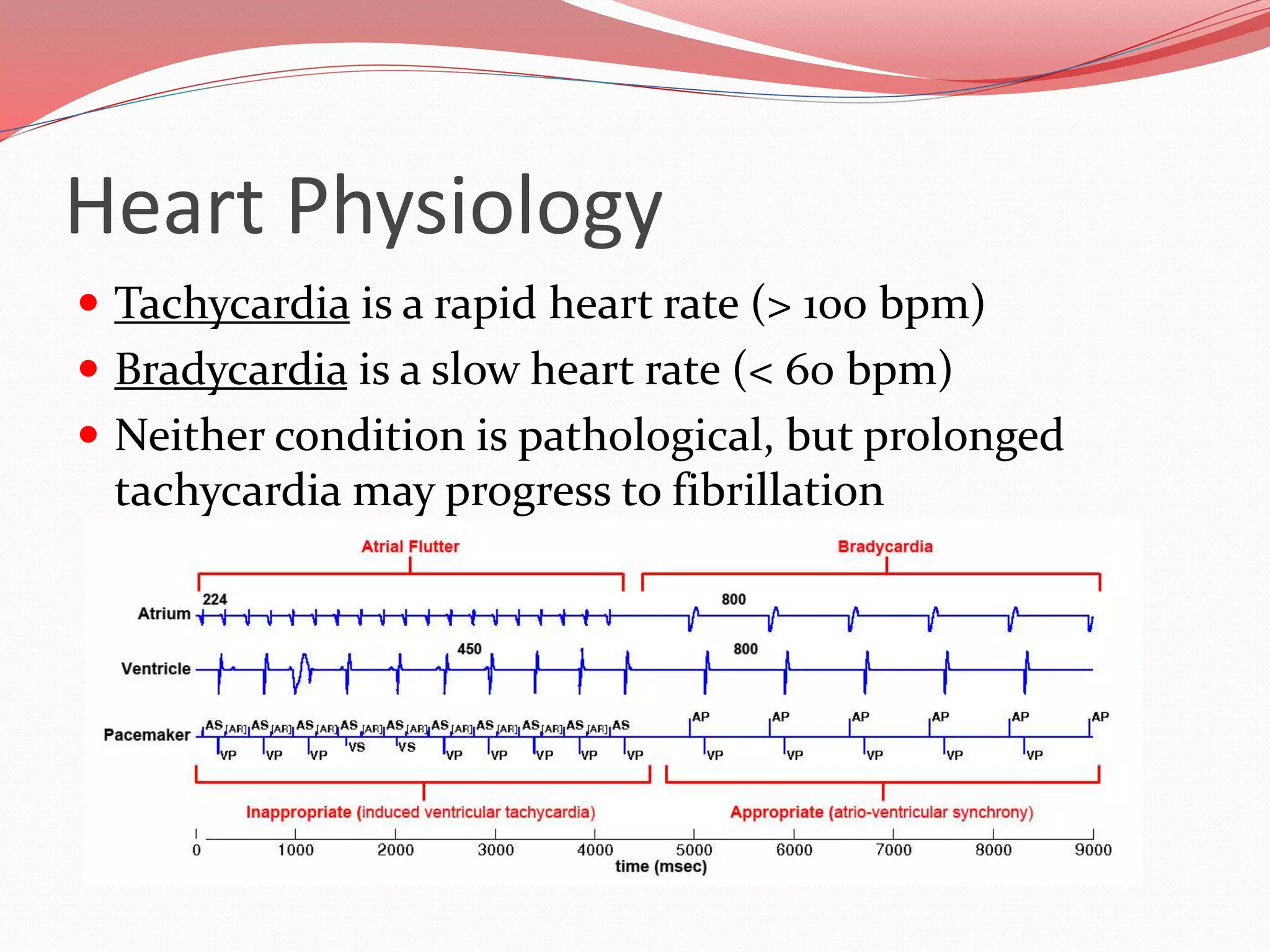
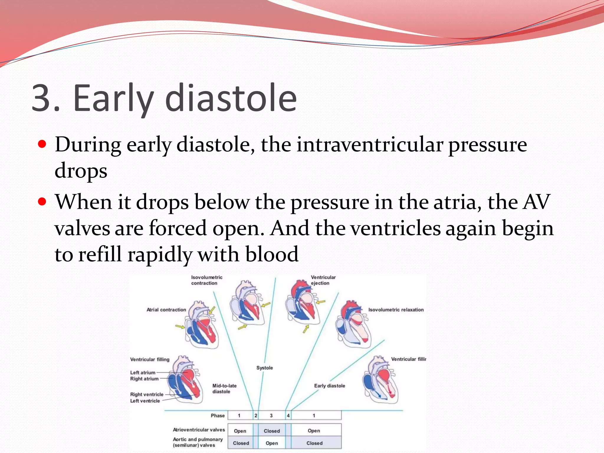
The document discusses the cardiovascular system and heart. It describes how the cardiovascular system transports blood throughout the body using the heart as a pump. It then provides details on the structure and layers of the heart, how it functions as a double pump with two circuits (pulmonary and systemic), and the roles of the heart valves and chambers. It also discusses heart physiology including the conduction system that regulates heartbeat, conditions like tachycardia and fibrillation, and the purpose of pacemakers.
This presentation is an overview of the description of the 4 stages of the cardiac cycle (atrial diastole, atrial systole, ventricular systole, ventricular diastole) as well as explaining the mechanism of the cardiac cycle.
This is a presentation explaining the process of writing reflective essays. It includes structuring the essay using a reflective model and suggestions for introductions and conclusions.
The human heart heart length, width, and thickness are 12 cm, 8.5 cm, and 6 cm, respectively. In addition, the mean weight of the heart is 280-340 g in males and 230-280 g in females.
Location and orientation with the thorax
Structure of the heart
Structure of the Heart Wall
Chambers of the Heart
Valves of the Heart
Pathway of blood through the heart
Cardiac Muscle Tissue
Conducting System and Innervation
Four Steps of Cardiac Conduction
Blood Supply to the Heart
Cardiovascular System, Heart, Blood Vessel, ECG, Hypertension, Arrhythmia Audumbar Mali
Cardiovascular System,
Human Anatomy and Physiology-I,
The Blood Vessels,
The Heart,
The Electrocardiogram,
The Vascular Pathways,
As per PCI syllabus,
Atherosclerosis,
Coronary bypass operation,
Heart Transplants and Artificial Hearts
B. Pharm SEM -I; Unit V- Cardiovascular system. Heart – anatomy of heart, blood circulation, elements of conduction system of heart and heart beat, its
regulation by autonomic nervous system, cardiac output, cardiac cycle. Regulation of
blood pressure, pulse, electrocardiogram
Slide 1: Title Slide
Extrachromosomal Inheritance
Slide 2: Introduction to Extrachromosomal Inheritance
Definition: Extrachromosomal inheritance refers to the transmission of genetic material that is not found within the nucleus.
Key Components: Involves genes located in mitochondria, chloroplasts, and plasmids.
Slide 3: Mitochondrial Inheritance
Mitochondria: Organelles responsible for energy production.
Mitochondrial DNA (mtDNA): Circular DNA molecule found in mitochondria.
Inheritance Pattern: Maternally inherited, meaning it is passed from mothers to all their offspring.
Diseases: Examples include Leber’s hereditary optic neuropathy (LHON) and mitochondrial myopathy.
Slide 4: Chloroplast Inheritance
Chloroplasts: Organelles responsible for photosynthesis in plants.
Chloroplast DNA (cpDNA): Circular DNA molecule found in chloroplasts.
Inheritance Pattern: Often maternally inherited in most plants, but can vary in some species.
Examples: Variegation in plants, where leaf color patterns are determined by chloroplast DNA.
Slide 5: Plasmid Inheritance
Plasmids: Small, circular DNA molecules found in bacteria and some eukaryotes.
Features: Can carry antibiotic resistance genes and can be transferred between cells through processes like conjugation.
Significance: Important in biotechnology for gene cloning and genetic engineering.
Slide 6: Mechanisms of Extrachromosomal Inheritance
Non-Mendelian Patterns: Do not follow Mendel’s laws of inheritance.
Cytoplasmic Segregation: During cell division, organelles like mitochondria and chloroplasts are randomly distributed to daughter cells.
Heteroplasmy: Presence of more than one type of organellar genome within a cell, leading to variation in expression.
Slide 7: Examples of Extrachromosomal Inheritance
Four O’clock Plant (Mirabilis jalapa): Shows variegated leaves due to different cpDNA in leaf cells.
Petite Mutants in Yeast: Result from mutations in mitochondrial DNA affecting respiration.
Slide 8: Importance of Extrachromosomal Inheritance
Evolution: Provides insight into the evolution of eukaryotic cells.
Medicine: Understanding mitochondrial inheritance helps in diagnosing and treating mitochondrial diseases.
Agriculture: Chloroplast inheritance can be used in plant breeding and genetic modification.
Slide 9: Recent Research and Advances
Gene Editing: Techniques like CRISPR-Cas9 are being used to edit mitochondrial and chloroplast DNA.
Therapies: Development of mitochondrial replacement therapy (MRT) for preventing mitochondrial diseases.
Slide 10: Conclusion
Summary: Extrachromosomal inheritance involves the transmission of genetic material outside the nucleus and plays a crucial role in genetics, medicine, and biotechnology.
Future Directions: Continued research and technological advancements hold promise for new treatments and applications.
Slide 11: Questions and Discussion
Invite Audience: Open the floor for any questions or further discussion on the topic.
(May 29th, 2024) Advancements in Intravital Microscopy- Insights for Preclini...Scintica Instrumentation
Intravital microscopy (IVM) is a powerful tool utilized to study cellular behavior over time and space in vivo. Much of our understanding of cell biology has been accomplished using various in vitro and ex vivo methods; however, these studies do not necessarily reflect the natural dynamics of biological processes. Unlike traditional cell culture or fixed tissue imaging, IVM allows for the ultra-fast high-resolution imaging of cellular processes over time and space and were studied in its natural environment. Real-time visualization of biological processes in the context of an intact organism helps maintain physiological relevance and provide insights into the progression of disease, response to treatments or developmental processes.
In this webinar we give an overview of advanced applications of the IVM system in preclinical research. IVIM technology is a provider of all-in-one intravital microscopy systems and solutions optimized for in vivo imaging of live animal models at sub-micron resolution. The system’s unique features and user-friendly software enables researchers to probe fast dynamic biological processes such as immune cell tracking, cell-cell interaction as well as vascularization and tumor metastasis with exceptional detail. This webinar will also give an overview of IVM being utilized in drug development, offering a view into the intricate interaction between drugs/nanoparticles and tissues in vivo and allows for the evaluation of therapeutic intervention in a variety of tissues and organs. This interdisciplinary collaboration continues to drive the advancements of novel therapeutic strategies.
Introduction:
RNA interference (RNAi) or Post-Transcriptional Gene Silencing (PTGS) is an important biological process for modulating eukaryotic gene expression.
It is highly conserved process of posttranscriptional gene silencing by which double stranded RNA (dsRNA) causes sequence-specific degradation of mRNA sequences.
dsRNA-induced gene silencing (RNAi) is reported in a wide range of eukaryotes ranging from worms, insects, mammals and plants.
This process mediates resistance to both endogenous parasitic and exogenous pathogenic nucleic acids, and regulates the expression of protein-coding genes.
What are small ncRNAs?
micro RNA (miRNA)
short interfering RNA (siRNA)
Properties of small non-coding RNA:
Involved in silencing mRNA transcripts.
Called “small” because they are usually only about 21-24 nucleotides long.
Synthesized by first cutting up longer precursor sequences (like the 61nt one that Lee discovered).
Silence an mRNA by base pairing with some sequence on the mRNA.
Discovery of siRNA?
The first small RNA:
In 1993 Rosalind Lee (Victor Ambros lab) was studying a non- coding gene in C. elegans, lin-4, that was involved in silencing of another gene, lin-14, at the appropriate time in the
development of the worm C. elegans.
Two small transcripts of lin-4 (22nt and 61nt) were found to be complementary to a sequence in the 3' UTR of lin-14.
Because lin-4 encoded no protein, she deduced that it must be these transcripts that are causing the silencing by RNA-RNA interactions.
Types of RNAi ( non coding RNA)
MiRNA
Length (23-25 nt)
Trans acting
Binds with target MRNA in mismatch
Translation inhibition
Si RNA
Length 21 nt.
Cis acting
Bind with target Mrna in perfect complementary sequence
Piwi-RNA
Length ; 25 to 36 nt.
Expressed in Germ Cells
Regulates trnasposomes activity
MECHANISM OF RNAI:
First the double-stranded RNA teams up with a protein complex named Dicer, which cuts the long RNA into short pieces.
Then another protein complex called RISC (RNA-induced silencing complex) discards one of the two RNA strands.
The RISC-docked, single-stranded RNA then pairs with the homologous mRNA and destroys it.
THE RISC COMPLEX:
RISC is large(>500kD) RNA multi- protein Binding complex which triggers MRNA degradation in response to MRNA
Unwinding of double stranded Si RNA by ATP independent Helicase
Active component of RISC is Ago proteins( ENDONUCLEASE) which cleave target MRNA.
DICER: endonuclease (RNase Family III)
Argonaute: Central Component of the RNA-Induced Silencing Complex (RISC)
One strand of the dsRNA produced by Dicer is retained in the RISC complex in association with Argonaute
ARGONAUTE PROTEIN :
1.PAZ(PIWI/Argonaute/ Zwille)- Recognition of target MRNA
2.PIWI (p-element induced wimpy Testis)- breaks Phosphodiester bond of mRNA.)RNAse H activity.
MiRNA:
The Double-stranded RNAs are naturally produced in eukaryotic cells during development, and they have a key role in regulating gene expression .
Astronomy Update- Curiosity’s exploration of Mars _ Local Briefs _ leadertele...
Cardiovascular System
1.
2. The Cardiovascular System
Cardiovascular system:
organ system that
distributes blood to all
parts of the body
Major function –
transportation, using
blood as the transport
vehicle
3. The Cardiovascular System
This system carries oxygen, nutrients, cell wastes,
hormones and other substances vital for body
homeostasis to and form cells
The force to move blood around the body is provided
by the pumping heart and blood pressure
4.
5. The Heart
The human heart is
approximately the size of
a fist, and weighs less
than a pound
It is enclosed within the
inferior mediastinum,
the medial cavity of the
thorax, and flanked on
each side by the lungs
6. The Heart
The pointed apex is
directed toward the left
hip and rests at about the
fifth intercostal space
The broad aspect, or
base, points toward the
right shoulder and lies
beneath the second rib
7. The Heart
The heart is enclosed by a double-walled sac called the
pericardium
The superficial loosely fitted part is called the fibrous
pericardium
Protects and anchors the heart
9. The Heart
Deep to the fibrous
pericardium is the
slippery, two-layer
serous pericardium
The parietal layer lines
the interior of the
fibrous pericardium
10. The Heart
The parietal layer attaches to the large arteries leaving
the heart and then makes a U-turn and continues
inferiorly over the heart surface as the visceral layer, or
epicardium
11. The Heart
A slippery lubricating fluid is produced by the serous
pericardial membranes which allows the heart to beat
easily in a relative frictionless environment
12. The Heart
Inflammation of the pericardium, pericarditis, often
results in a decrease in the serous fluid
The cause the pericardial layers to stick, forming
painful adhesions that interfere with heart movements
13. The Heart
The heart walls are composed of three layers:
1. outer epicardium
2. myocardium
3. endocardium
14. The Heart
The myocardium
consists of thick bundles
of the cardiac muscle
twisted into ringlike
arrangements
This is the layer of the
heart that actually
contracts
Reinforced by dense,
fibrous connective tissue
(“heart skeleton”)
15. The Heart
The endocardium is a thin, glistening sheet of
endothelium that lines the heart chambers
Continuous with the linings of the blood vessels
leaving and entering the heart
16. The Heart
The heart has four hollow chambers:
2 atria – receiving chambers
2 ventricles – filling chambers
17. The Heart
Blood flows into the
atria under low
pressure from the
veins, and continues
into the ventricles
18. The Heart
The ventricles are thick-
walled discharging
chambers
They are the pumps of the
heart
When they contract, blood
is propelled out of the
heart and into circulation
19. The Heart
The right ventricle forms most of the heart’s anterior
surface
The left ventricle forms the apex
20. The Heart
The septum that divides
the heart longitudinally
is the interventricular
septum or the interatrial
septum based on the
chambers it separates
21. The Heart
The heart functions as a double pump
The right side works as the pulmonary circuit pump
Receives relatively oxygen-poor blood from the veins of
the body through the large superior and inferior vena
cavae
23. The Heart
The blood then pumps
out through the
pulmonary trunk which
splits into the left and
right pulmonary arteries
The pulmonary arteries
carry blood to the lungs,
where oxygen is picked
up and carbon dioxide is
unloaded
24. The Heart
Oxygen-rich blood drains from the lungs and is
returned to the left side of the heart through the four
pulmonary veins
This circuit is call pulmonary circulation
Its only function is to carry blood to the lungs for gas
exchange and then return it to the heart
26. The Heart
Blood returned to the left side of the heart is pumped
out of the heart into the aorta
The systemic arteries branch from the aorta to supply
the body tissues with blood
27. The Heart
Oxygen-poor blood
circulates from the
tissues back to the right
atrium via the systemic
veins, which empty their
blood into either the
superior or inferior vena
cava
28. The Heart
This second circuit, from the left side of the heart
through the body tissues and back to the right side of
the heart is called systemic circulation
It supplies oxygen and nutrient-rich blood to all body
organs
29. The Heart
Because the left ventricle is the systemic pump that
pumps blood over a much longer pathway through the
body, its walls are thicker than those of the right
ventricle
It is a more powerful pump
30. The Heart
The heart also has four
valves:
2 that separate the
atria from the
ventricles
2 that separate the
ventricles from their
arteries
All of these valves
prevent back flow
31. The Heart
The atrioventricular (AV)
valves are between the atria
and ventricles
On the left is the bicuspid or
mitral valve
On the right is the tricuspid
valve
They are all anchored by the
chordae tendineae
32. The Heart
When the heart is relaxed and blood is passively filling
its chambers, the AV-valve flaps hang limply into the
ventricles
As the ventricles contract, they press on the blood in
their chamber, and the intraventricular pressure rises
33. The Heart
The semilunar valves
guard the bases of the
large arteries leaving the
ventricular chambers
On the right is the
pulmonary valve
On the left is the aortic
valve
34. The Heart
When the ventricles are
contracting these valves
are forced open and
flattened against the
arterial walls
When the ventricles are
relaxed the blood flows
back towards the heart
This prevents arterial
blood from reentering
the heart
35. The Heart
The coronary arteries branch from the base of the
aorta and encircle the heart in the coronary sulcus (AV
groove) at the junction of the atria and ventricles
36. The Heart
The coronary
arteries and their
major branches are
compressed when
the ventricles are
contracting and fill
when the heart is
relaxed
37. The Heart
The myocardium is drained by several cardiac veins,
which empty into the coronary sinus
The coronary sinus, in turn, empties into the right
atrium
38. The Heart
When the heart beats rapidly the myocardium can
received an inadequate amount of blood
This can result in crushing chest pain called angina
pectoris
39. The Heart
Pain due to angina pectoris is
a warning sign
If angina is prolonged,
oxygen-deprived heart cells
may die forming an infarct
The resulting myocardial
infarction is a “heart attack”
40.
41. Heart Physiology
The heart pumps the
body’s 6 quart supply of
blood through the blood
vessels over 1000 times per
day
In reality, the heart pumps
about 6000 quarts of
blood in a single day
42. Heart Physiology
Cardiac muscles cells can
and do contract
spontaneously and
independently, even if all
nervous connections are
severed
These contractions occur in
a regular and continuous
way
43. Heart Physiology
Although cardiac muscle
can beat independently,
the muscle cells on
different areas of the heart
have different rhythms
Atrial cells – 60 bpm
Ventricular cells – 20-40
bpm
44. Heart Physiology
Two systems act to regulate heart activity:
1. Autonomic nervous system – brakes and accelerator
Acts to decrease or increase heart rate
2. Intrinsic conduction system (nodal system)
Composed of specialized tissue that is a cross between
muscle and nervous tissue
Causes heart muscle depolarization from the atria to
the ventricles
Enforces contraction rate ~ 75bpm
46. Heart Physiology
Components of the Intrinsic Conduction System
include:
The sinoatrial (SA) node is a crescent shaped node in
the right atrium
The atrioventricular (AV) node is at the junction of the
atria and ventricles
The atrioventricular (AV) bundle (bundle of His)
Branch bundles in the interventricular septum
Purkinje fibers which spread with the muscle of the
ventricle walls
48. Heart Physiology
The SA node has the
highest rate of
depolarization in the
whole system
It starts each heartbeat
and sets the pace for the
whole heart and is
therefore called the
pacemaker
50. Heart Physiology
The impulse travels from the SA node through the
atria to the AV node, causing the atria to contract
51. Heart Physiology
At the AV node, the
impulse is delayed to give
the atria time to finish
contracting
It then passes rapidly
through the AV bundle, the
bundle branches, and the
Purkinje fibers, causing a
“wringing” contraction of
the ventricles that begins
at the apex and moves
toward the atria
52. Heart Physiology
This contraction effectively ejects blood superiorly into
the large arteries leaving the heart
53. Heart Physiology
Tachycardia is a rapid heart rate (> 100 bpm)
Bradycardia is a slow heart rate (< 60 bpm)
Neither condition is pathological, but prolonged
tachycardia may progress to fibrillation
54. Fibrillation is a rapid,
uncoordinated
shuddering of the heart
muscle
Fibrillation makes the
heart totally useless as a
pump and is a major
cause of death from
heart attacks in adults
Heart Physiology
55. Heart Physiology
A pacemaker is a small
device, about the size of a
half dollar piece, placed
under the skin near the
heart to help control the
heartbeat.
A pacemaker is implanted
as part of what's often
referred to as "cardiac
resynchronization
therapy."
56. Heart Physiology
People may need a pacemaker for a
variety of reasons — mostly due to
one of a group of conditions called
arrhythmias, in which the heart's
rhythm is abnormal
They can be implanted temporarily
to treat a slow heartbeat after a
heart attack, surgery or overdose of
medication
Pacemakers can also be implanted
permanently to correct bradycardia
or to help treat heart failure
58. Cardiac Cycle and Heart Sounds
In a healthy heart, the atria contract simultaneously
When they start to relax, contraction of the ventricles
begins
Systole and diastole mean heart contraction and
relaxation respectively
59. Cardiac Cycle and Heart Sounds
Because most of the pumping work is done by the
ventricles, these terms always refer to the contraction
and relaxation of the ventricles unless otherwise stated
60. Cardiac Cycle and Heart Sounds
The term cardiac
cycle refers to the
events of one
complete heartbeat,
during which both
atria and ventricles
contract and then
relax
61. Cardiac Cycle and Heart Sounds
The average heart beats 75 times per minute
The average length of a cardiac cycle is 0.8 seconds
The cardiac cycle occurs in three major steps:
1. mid-to-late diastole
2. ventricular systole
3. early diastole
62. 1. Mid-to-late diastole
The heart is in complete relaxation
Pressure in the heart is low
Blood is flowing passively into and through the atria
and into the ventricles from pulmonary and systemic
circulations
63. 1. Mid-to-late diastole
The semilunar valves are closed
The AV valves are open
Then the atria contract and force the blood into the
ventricles
65. 2. Ventricular systole
The pressure within the
ventricles increases rapidly,
closing the AV valves
When the intraventricular
pressure is higher than the
pressure in the large arteries
leaving the heart, the semilunar
valves are forced open, and blood
rushes out of the ventricles
The atria are relaxed, and again
are filling with blood
67. 3. Early diastole
At the end of systole, the ventricles relax, the
semilunar valves snap shut, and for a moment the
ventricles are completely closed chambers
68. 3. Early diastole
During early diastole, the intraventricular pressure
drops
When it drops below the pressure in the atria, the AV
valves are forced open. And the ventricles again begin
to refill rapidly with blood
70. Heart Sounds
When using a stethoscope, the heart beat usually has
two distinct sounds – “lup” and “dup”
These are caused by the closing of the two sets of
valves
“lup” – AV valves
“dup” – semilunar valves
71.
72. Cardiac Output
Cardiac Output (CO) is
the amount of blood
pumped out by each side
of the heart in 1 minute
It is the product of heart
rate (HR) and stroke
volume (SV)
73. Cardiac Output
In general, stroke volume increases as the force of
ventricular contraction increases
Let’s look at normal resting heart rate and volume:
CO = HR x SV
CO = (75 bpm) x (70 ml per beat)
CO = 5250 ml/min
74. Cardiac Output
A healthy heart pumps
out about 60% of blood
in the ventricles (~70 ml)
per heart beat
The critical factor is how
much the cardiac muscle
cells stretch just before
contracting
75. Cardiac Output
The important factor
stretching the heart
muscle is venous
return, the amount of
blood entering the
heart and distending
the ventricles
The more the heart
muscles stretch, the
stronger the
contraction
76. Cardiac Output
If one side of the heart suddenly begins to pump more
blood than the other, the increased venous return to
the opposite ventricle will force it to pump out an
equal amount, thus preventing backup of blood in the
circulation
77. Cardiac Output
The enhanced squeezing action
of active skeletal muscles from
exercise speeds up venous return
Severe blood loss or rapid heart
rate, decreases stroke volume,
creating less venous return
78. Factors Modifying Basic Heart Rate
Heart contraction does not depend on the nervous
system, but it can be changed temporarily by the ANS
It is also modified by chemicals, hormones and ions
79. Neural (ANS) Control
During times of physical or emotional stress, the
nerves of sympathetic division stimulate the SA and
AV nodes and the cardiac muscles
The heart beats more rapidly
80. Neural (ANS) Control
When the demand declines, the heart adjusts, the
parasympathetic nerves slow and steady the heart rate
Gives the heart time to recover and rest
81. Neural (ANS) Control
In patients with Congestive Heart Failure (CHF), or
other heart disease the heart pumps weakly
Some medications can be used to enhance contractile
force and stroke volume of the heart, improving
cardiac output
83. Neural (ANS) Control
Various hormones and ions have a dramatic effect on
heart activity
Epinephrine – mimics sympathetic nerves, increases
heart rate
Thyroxine – increase heart rate
Electrolyte imbalance – prolonged contractions,
arrhythmias, decrease output
84. Physical Factors
Resting heart rate is fastest in the fetus and then
gradually decreases
Faster heart rate in females than males
High body temperature also increase heart rate, Low
body temperature decreases heart rate