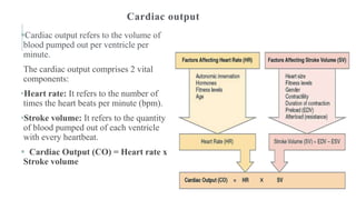
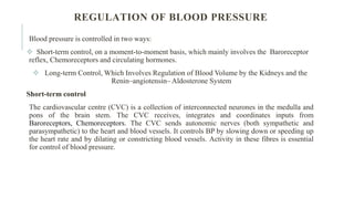
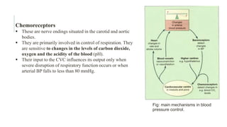
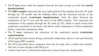
CARDIOVASCULAR
SYSTEM
By- Samruddhi S. Khonde
Asst. Prof
P. R. Patil Institute of Pharmacy
https://orcid.org/0000-0002-7955-6399
ANATOMY OF THE HEART
The heart, a muscular pump made up of cardiac muscle fibers, could be considered a
muscle rather than an organ. It has four chambers, or cavities, and beats an average of
60–100 beats per minute (bpm) or about 100,000 times in one day. It is about 12 cm (5
in.) long, 9 cm (3.5 in.) wide at its broadest point, and 6 cm (2.5 in.) thick, with an
average mass of 250 g (8 oz) in adult females and 300 g (10 oz) in adult males.
Each time the cardiac muscle contracts, blood is ejected from the heart and pushed
throughout the body within the blood vessels.
The heart is located in the mediastinum in the center of the chest cavity; however, it is
not exactly centered; more of the heart is on the left side of the mediastinum than the
right.
At about the size of a fist and shaped like an upside-down pear, the heart lies directly
behind the sternum. The tip of the heart at the lower edge is called the apex.
HEART LAYERS
The wall of the heart is quite thick and is composed of three
layers
1. The endocardium is the inner layer of the heart lining the
heart chambers. It is a very smooth, thin layer that serves to
reduce friction as the blood passes through the heart
chambers.
2. The myocardium is the thick, muscular middle layer of
the heart. Contraction of this muscle layer develops the
pressure required to pump blood through the blood vessels.
3. The epicardium is the outer layer of the heart. The heart
is enclosed within a double-layered pleural sac, called the
pericardium. The epicardium is the visceral pericardium, or
inner layer of the sac. The outer layer of the sac is the
parietal pericardium. Fluid between the two layers of the sac
reduces friction as the heart beats.
HEART CHAMBERS
The heart is divided into four chambers or cavities.
There are two atria, or upper chambers, and two ventricles, or lower
chambers.
These chambers are divided into right and left sides by walls called the
interatrial septum and the interventricular septum. The atria are the
receiving chambers of the heart.
Blood returning to the heart via veins first collects in the atria. The
ventricles are the pumping chambers. They have a much thicker
myocardium and their contraction ejects blood out of the heart and into
the great arteries.
Heart Valves
Four valves act as restraining gates to control the direction of blood flow.
They are situated at the entrances and exits to the ventricles. Properly
functioning valves allow blood to flow only in a forward direction by
blocking it from returning to the previous chamber
VALVES
The four valves are:
1. Tricuspid valve: an atrioventricular valve (AV), meaning that it controls the
opening between the right atrium and the right ventricle. Once the blood enters the
right ventricle, it cannot go back up into the atrium again. The prefix tri-, meaning
three, indicates that this valve has three leaflets or cusps.
2. Pulmonary valve: a semilunar valve, with the prefix semi- meaning half and the
term lunar meaning moon, indicate that this valve looks like a half moon. Located
between the right ventricle and the pulmonary artery, this valve prevents blood that has
been ejected into the pulmonary artery from returning to the right ventricle as it
relaxes.
3. Mitral valve: also called the bicuspid valve, indicating that it has two cusps. Blood
flows through this atrioventricular valve to the left ventricle and cannot go back up into
the left atrium.
4. Aortic valve: a semilunar valve located between the left ventricle and the aorta.
Blood leaves the left ventricle through this valve and cannot return to the left ventricle.
BLOOD CIRCULATION
Blood circulation is a vital physiological process that ensures the continuous transport of
oxygen, nutrients, hormones, and waste products throughout the body. It is a complex
system involving the heart, blood vessels, and blood.
The heart pumps blood into two closed circuits with each beat—
- systemic circulation (The left side of the heart pumps) and
- pulmonary Circulation (pulmon- = lung) (The right side of the heart pumps)
The two circuits are arranged in series: the output of one becomes the input of the other.
 organs involved in blood circulation
systemic circulation- Heart, Blood Vessels, Capillaries Veins
pulmonary Circulation : Lungs
•Blood Circulation
•The right side pumps for pulmonary circulation, receiving all dark-red deoxygenated blood returning from
systemic circulation.
•Blood ejected from the right ventricle flows into the pulmonary trunk, branches into pulmonary arteries.
•In the lungs, carbon dioxide is exchanged for oxygen. Oxygenated blood returns to the left atrium of the heart
through the pulmonary veins.
•The left side of the heart pumps for systemic circulation.
•On receiving oxygen-rich blood from the lungs, the left ventricle ejects blood into the aorta, which divides into
smaller systemic arteries that carry it to all organs except for the air sacs of the lungs.
•Systemic tissues give rise to arterioles, which lead to extensive beds of systemic capillaries. Blood exchanges
nutrients and gases across these capillary walls, unloading oxygen and picking up carbon dioxide.
•In most cases, blood flows through only on capillaries and then enters a systemic venule. Venules carry
deoxygenated blood away from tissues and merge to form larger systemic veins.
•Ultimately, blood flows back to the right atrium.
BLOOD PRESSURE
• Blood pressure is the force or pressure that the blood exerts on the walls of the
blood vessels. Systemic arterial blood pressure maintains the essential flow of
blood into and out of the organs of the body.
• If it becomes too high, blood vessels can be damaged, causing clots or bleeding
from sites of blood vessel rupture. If it falls too low, then blood flow through
tissue beds may be inadequate. This is particularly dangerous for such essential
organs as the heart, brain or kidneys.
• The systemic arterial blood pressure, usually called simply arterial blood
pressure, is the result of the discharge of blood from the left ventricle into the
already full aorta.
• Blood pressure varies according to the time of day, the posture, gender and age
of the individual. Blood pressure falls at rest and during sleep. It increases with
age and is usually higher in women than in men.
Systolic and diastolic pressure
When the left ventricle contracts and pushes blood into the aorta, the pressure produced within the arterial
system is called the systolic blood pressure. In adults it is about 120 mmHg or 16 kPa.
When complete cardiac diastole occurs and the heart is resting following the ejection of blood, the
pressure within the arteries is much lower and is called diastolic blood pressure. In an adult this is about
80 mmHg or 11 kPa.
The difference between systolic and diastolic blood pressures is the pulse pressure.
Arterial blood pressure is measured with a sphygmomanometer and is usually expressed with the systolic
pressure written above the diastolic pressure:
BP = 120/ 80 mmHg or BP = 16/11mmHg
Factors determining blood pressure
Blood pressure is determined by cardiac output and peripheral resistance. Change in either of these
parameters tends to alter systemic blood pressure, although the body’s compensatory mechanisms usually
adjust for any significant change. Blood pressure = cardiac output × peripheral resistance
Cardiac output
Cardiac output refers to the volume of
blood pumped out per ventricle per
minute.
The cardiac output comprises 2 vital
components:
•Heart rate: It refers to the number of
times the heart beats per minute (bpm).
•Stroke volume: It refers to the quantity
of blood pumped out of each ventricle
with every heartbeat.
 Cardiac Output (CO) = Heart rate x
Stroke volume
REGULATION OF BLOOD PRESSURE
Blood pressure is controlled in two ways:
 Short-term control, on a moment-to-moment basis, which mainly involves the Baroreceptor
reflex, Chemoreceptors and circulating hormones.
 Long-term Control, Which Involves Regulation of Blood Volume by the Kidneys and the
Renin–angiotensin– Aldosterone System
Short-term control
The cardiovascular centre (CVC) is a collection of interconnected neurones in the medulla and
pons of the brain stem. The CVC receives, integrates and coordinates inputs from
Baroreceptors, Chemoreceptors. The CVC sends autonomic nerves (both sympathetic and
parasympathetic) to the heart and blood vessels. It controls BP by slowing down or speeding up
the heart rate and by dilating or constricting blood vessels. Activity in these fibres is essential
for control of blood pressure.
Baroreceptors –
 These are nerve endings sensitive to pressure changes (stretch) within the vessel, situated in
the arch of the aorta and in the carotid sinuses.
 A rise in blood pressure in these arteries stimulates the baroreceptors, increasing their input
to the cardiovascular centre .
 The cardiovascular centre (CVC) responds by increasing parasympathetic nerve activity to
the heart; this slows the heart down.
 At the same time, sympathetic stimulation to the blood vessels is inhibited, causing
vasodilatation. The net result is a fall in systemic blood pressure.
 Conversely, if pressure within the aortic arch and carotid sinuses falls, the rate of
baroreceptor discharge also falls.
 The CVC responds by increasing sympathetic drive to the heart to speed it up. Sympathetic
activity in blood vessels is also increased, leading to vasoconstriction.
 Both these measures counteract the falling blood pressure. Baroreceptor control of blood
pressure is also called the baroreceptor reflex.
Chemoreceptors
 These are nerve endings situated in the carotid and aortic
bodies.
 They are primarily involved in control of respiration. They
are sensitive to changes in the levels of carbon dioxide,
oxygen and the acidity of the blood (pH).
 Their input to the CVC influences its output only when
severe disruption of respiratory function occurs or when
arterial BP falls to less than 80 mmHg.
Fig: main mechanisms in blood
pressure control.
The impulse of cardiac contraction are transmitted through the conduction system of heart.
The system of heart is made up of
 Sinoatrial (Sa) Node
 Atrioventricular ( AV) Node
 Atrioventricular (AV) Bundle (Purkinje Fibres)
The conduction of impulses occurs in the following sequence
The origin of impulse of cardiac contraction is at the Sinoatrial node. it is present at the opening of
superior vena cava into the right atrium. SA node is called as Pacemaker of the heart.
The impulses then pass through the atrial muscle to Atrioventricular node. It lies in the upper part of
atrioventricular septum.
 The impulse passed to the Bundle of His. It is a specialised bundle of nerve and muscle tissue. it is the
only muscular connection between the atria and ventricle.
The bundle of his passes through interventricular septum later divides into the branches called
Purkinje fibre. The right and left branches of fibre supplied the two ventricles.
ELEMENTS OF CONDUCTION SYSTEM OF HEART AND
HEART BEAT
Fig: Flowchart of Conduction system of heart
Nerve supply to the heart:
The heart is influenced by autonomic (sympathetic and parasympathetic) nerves originating in the
cardiovascular centre in the medulla oblongata.
The vagus nerves (parasympathetic) supply mainly the SA and AV nodes and atrial muscle.
Parasympathetic stimulation reduces the rate at which impulses are produced, decreasing the rate and
force of the heartbeat.
The sympathetic nerves supply the SA and AV nodes and the myocardium of atria and ventricles.
Sympathetic stimulation increases the rate and force of the heartbeat.
THE CARDIAC CYCLE
The event occurring in the heart from the beginning of one heartbeat to the beginning of other
heartbeat is called as cardiac cycle.
The term systole refers to the phase of contraction while diastole refers to the phase of
relaxation.
The normal number of cardiac cycles per minute ranges from 60 to 80. each cycle lasts about 0.8
sec
Cardiac cycle divided into the 3 Stages.
Stages of the Cardiac Cycle
• Atrial systole — contraction of the atria
• Ventricular systole — contraction of the ventricles
• Complete cardiac diastole — relaxation of the atria and ventricles.
Complete cardiac
diastole
Fig: Stages of the Cardiac Cycle
Period of each cycle = 0.8 second
The superior vena cava and the inferior vena cava transport deoxygenated blood into the right atrium at the
same time as the four pulmonary veins bring oxygenated blood into the left atrium.
The SA node triggers a wave of contraction that spreads over the myocardium of both atria, emptying the atria
and completing ventricular filling (atrial systole 0.1s).
When same the electrical impulse reaches the AV node, it slowed down, delaying atrioventricular transmission.
This allows the atria to finish emptying into the ventricles before the ventricles begin to contract.
After this brief delay, the AV node triggers its own electrical impulse, which quickly spreads to the ventricular
muscle via the AV bundle,(the bundle of Purkinje fibres and branches). This results in a wave of contraction
from the apex of the heart and across the walls of both ventricles pumping the blood into the pulmonary artery
and the aorta (ventricular systole 0.3s).
After contraction of the ventricles there is complete cardiac diastole, a period of 0.4 seconds, when atria and
ventricles are relaxed
During this time the myocardium recovers ready for the next heartbeat, and the atria refill ready for the next
cycle. The valves of the heart and of blood vessels open and close according to the pressure within the chambers
of the heart.
When the ventricular pressure rises above that in the pulmonary artery and in the aorta, the pulmonary and
aortic valves open and blood flows into these vessels. When the ventricles relax and the pressure within them
falls, the reverse process occurs. First the pulmonary and aortic valves close, then the atrioventricular valves
open and the cycle begins again. This sequence of opening and closing valves ensures that the blood flows in
only one direction.
The Cardiac Cycle
Electrocardiogram
The body tissues and fluids conduct electricity well, so the electrical activity in the heart can be recorded on
the skin surface using electrodes positioned on the limbs and/or the chest. This recording, called an
Electrocardiogram (ECG).
 It shows the spread of the electrical signal generated by the SA node as it travels through the atria, the AV
node and the ventricles.
 The normal ECG tracing shows five waves which, by convention, have been named P. Q, R, S and T.
 P is of atrial origin, hence called the atrial complex, while QRS-T being of ventricular origin, and are
collectively known as the ventricular complex.
Fig: Electrocardiogram (ECG).
 The P wave arises when the impulse from the SA node sweeps over the atria (atrial
depolarisation).
 The QRS complex represents the very rapid spread of the impulse from the AV node
through the AV bundle and the Purkinje fibres and the electrical activity of the
ventricular muscle (ventricular depolarisation). Note the delay between the
completion of the P wave and the onset of the QRS complex. This represents the
conduction of the impulse through the AV node (p. 92), which is much slower than
conduction elsewhere in the heart, and allows atrial contraction to finish completely
before ventricular contraction starts.
 The T wave represents the relaxation of the ventricular muscle (ventricular
repolarisation).
 Atrial repolarisation occurs during ventricular contraction, and so is not seen because
of the larger QRS complex.
 The ECG described above originates from the SA node and is called sinus rhythm.
The rate of sinus rhythm is 60-100 b.p.m.
 A faster heart rate is called tachycardia and a slower heart rate, bradycardia.

Cardiovascular system.pptx

  • 1.
    CARDIOVASCULAR SYSTEM By- Samruddhi S.Khonde Asst. Prof P. R. Patil Institute of Pharmacy https://orcid.org/0000-0002-7955-6399
  • 2.
    ANATOMY OF THEHEART The heart, a muscular pump made up of cardiac muscle fibers, could be considered a muscle rather than an organ. It has four chambers, or cavities, and beats an average of 60–100 beats per minute (bpm) or about 100,000 times in one day. It is about 12 cm (5 in.) long, 9 cm (3.5 in.) wide at its broadest point, and 6 cm (2.5 in.) thick, with an average mass of 250 g (8 oz) in adult females and 300 g (10 oz) in adult males. Each time the cardiac muscle contracts, blood is ejected from the heart and pushed throughout the body within the blood vessels. The heart is located in the mediastinum in the center of the chest cavity; however, it is not exactly centered; more of the heart is on the left side of the mediastinum than the right. At about the size of a fist and shaped like an upside-down pear, the heart lies directly behind the sternum. The tip of the heart at the lower edge is called the apex.
  • 4.
    HEART LAYERS The wallof the heart is quite thick and is composed of three layers 1. The endocardium is the inner layer of the heart lining the heart chambers. It is a very smooth, thin layer that serves to reduce friction as the blood passes through the heart chambers. 2. The myocardium is the thick, muscular middle layer of the heart. Contraction of this muscle layer develops the pressure required to pump blood through the blood vessels. 3. The epicardium is the outer layer of the heart. The heart is enclosed within a double-layered pleural sac, called the pericardium. The epicardium is the visceral pericardium, or inner layer of the sac. The outer layer of the sac is the parietal pericardium. Fluid between the two layers of the sac reduces friction as the heart beats.
  • 5.
    HEART CHAMBERS The heartis divided into four chambers or cavities. There are two atria, or upper chambers, and two ventricles, or lower chambers. These chambers are divided into right and left sides by walls called the interatrial septum and the interventricular septum. The atria are the receiving chambers of the heart. Blood returning to the heart via veins first collects in the atria. The ventricles are the pumping chambers. They have a much thicker myocardium and their contraction ejects blood out of the heart and into the great arteries. Heart Valves Four valves act as restraining gates to control the direction of blood flow. They are situated at the entrances and exits to the ventricles. Properly functioning valves allow blood to flow only in a forward direction by blocking it from returning to the previous chamber
  • 7.
    VALVES The four valvesare: 1. Tricuspid valve: an atrioventricular valve (AV), meaning that it controls the opening between the right atrium and the right ventricle. Once the blood enters the right ventricle, it cannot go back up into the atrium again. The prefix tri-, meaning three, indicates that this valve has three leaflets or cusps. 2. Pulmonary valve: a semilunar valve, with the prefix semi- meaning half and the term lunar meaning moon, indicate that this valve looks like a half moon. Located between the right ventricle and the pulmonary artery, this valve prevents blood that has been ejected into the pulmonary artery from returning to the right ventricle as it relaxes. 3. Mitral valve: also called the bicuspid valve, indicating that it has two cusps. Blood flows through this atrioventricular valve to the left ventricle and cannot go back up into the left atrium. 4. Aortic valve: a semilunar valve located between the left ventricle and the aorta. Blood leaves the left ventricle through this valve and cannot return to the left ventricle.
  • 9.
    BLOOD CIRCULATION Blood circulationis a vital physiological process that ensures the continuous transport of oxygen, nutrients, hormones, and waste products throughout the body. It is a complex system involving the heart, blood vessels, and blood. The heart pumps blood into two closed circuits with each beat— - systemic circulation (The left side of the heart pumps) and - pulmonary Circulation (pulmon- = lung) (The right side of the heart pumps) The two circuits are arranged in series: the output of one becomes the input of the other.  organs involved in blood circulation systemic circulation- Heart, Blood Vessels, Capillaries Veins pulmonary Circulation : Lungs
  • 11.
    •Blood Circulation •The rightside pumps for pulmonary circulation, receiving all dark-red deoxygenated blood returning from systemic circulation. •Blood ejected from the right ventricle flows into the pulmonary trunk, branches into pulmonary arteries. •In the lungs, carbon dioxide is exchanged for oxygen. Oxygenated blood returns to the left atrium of the heart through the pulmonary veins. •The left side of the heart pumps for systemic circulation. •On receiving oxygen-rich blood from the lungs, the left ventricle ejects blood into the aorta, which divides into smaller systemic arteries that carry it to all organs except for the air sacs of the lungs. •Systemic tissues give rise to arterioles, which lead to extensive beds of systemic capillaries. Blood exchanges nutrients and gases across these capillary walls, unloading oxygen and picking up carbon dioxide. •In most cases, blood flows through only on capillaries and then enters a systemic venule. Venules carry deoxygenated blood away from tissues and merge to form larger systemic veins. •Ultimately, blood flows back to the right atrium.
  • 12.
    BLOOD PRESSURE • Bloodpressure is the force or pressure that the blood exerts on the walls of the blood vessels. Systemic arterial blood pressure maintains the essential flow of blood into and out of the organs of the body. • If it becomes too high, blood vessels can be damaged, causing clots or bleeding from sites of blood vessel rupture. If it falls too low, then blood flow through tissue beds may be inadequate. This is particularly dangerous for such essential organs as the heart, brain or kidneys. • The systemic arterial blood pressure, usually called simply arterial blood pressure, is the result of the discharge of blood from the left ventricle into the already full aorta. • Blood pressure varies according to the time of day, the posture, gender and age of the individual. Blood pressure falls at rest and during sleep. It increases with age and is usually higher in women than in men.
  • 13.
    Systolic and diastolicpressure When the left ventricle contracts and pushes blood into the aorta, the pressure produced within the arterial system is called the systolic blood pressure. In adults it is about 120 mmHg or 16 kPa. When complete cardiac diastole occurs and the heart is resting following the ejection of blood, the pressure within the arteries is much lower and is called diastolic blood pressure. In an adult this is about 80 mmHg or 11 kPa. The difference between systolic and diastolic blood pressures is the pulse pressure. Arterial blood pressure is measured with a sphygmomanometer and is usually expressed with the systolic pressure written above the diastolic pressure: BP = 120/ 80 mmHg or BP = 16/11mmHg Factors determining blood pressure Blood pressure is determined by cardiac output and peripheral resistance. Change in either of these parameters tends to alter systemic blood pressure, although the body’s compensatory mechanisms usually adjust for any significant change. Blood pressure = cardiac output × peripheral resistance
  • 14.
    Cardiac output Cardiac outputrefers to the volume of blood pumped out per ventricle per minute. The cardiac output comprises 2 vital components: •Heart rate: It refers to the number of times the heart beats per minute (bpm). •Stroke volume: It refers to the quantity of blood pumped out of each ventricle with every heartbeat.  Cardiac Output (CO) = Heart rate x Stroke volume
  • 15.
    REGULATION OF BLOODPRESSURE Blood pressure is controlled in two ways:  Short-term control, on a moment-to-moment basis, which mainly involves the Baroreceptor reflex, Chemoreceptors and circulating hormones.  Long-term Control, Which Involves Regulation of Blood Volume by the Kidneys and the Renin–angiotensin– Aldosterone System Short-term control The cardiovascular centre (CVC) is a collection of interconnected neurones in the medulla and pons of the brain stem. The CVC receives, integrates and coordinates inputs from Baroreceptors, Chemoreceptors. The CVC sends autonomic nerves (both sympathetic and parasympathetic) to the heart and blood vessels. It controls BP by slowing down or speeding up the heart rate and by dilating or constricting blood vessels. Activity in these fibres is essential for control of blood pressure.
  • 16.
    Baroreceptors –  Theseare nerve endings sensitive to pressure changes (stretch) within the vessel, situated in the arch of the aorta and in the carotid sinuses.  A rise in blood pressure in these arteries stimulates the baroreceptors, increasing their input to the cardiovascular centre .  The cardiovascular centre (CVC) responds by increasing parasympathetic nerve activity to the heart; this slows the heart down.  At the same time, sympathetic stimulation to the blood vessels is inhibited, causing vasodilatation. The net result is a fall in systemic blood pressure.  Conversely, if pressure within the aortic arch and carotid sinuses falls, the rate of baroreceptor discharge also falls.  The CVC responds by increasing sympathetic drive to the heart to speed it up. Sympathetic activity in blood vessels is also increased, leading to vasoconstriction.  Both these measures counteract the falling blood pressure. Baroreceptor control of blood pressure is also called the baroreceptor reflex.
  • 17.
    Chemoreceptors  These arenerve endings situated in the carotid and aortic bodies.  They are primarily involved in control of respiration. They are sensitive to changes in the levels of carbon dioxide, oxygen and the acidity of the blood (pH).  Their input to the CVC influences its output only when severe disruption of respiratory function occurs or when arterial BP falls to less than 80 mmHg. Fig: main mechanisms in blood pressure control.
  • 18.
    The impulse ofcardiac contraction are transmitted through the conduction system of heart. The system of heart is made up of  Sinoatrial (Sa) Node  Atrioventricular ( AV) Node  Atrioventricular (AV) Bundle (Purkinje Fibres) The conduction of impulses occurs in the following sequence The origin of impulse of cardiac contraction is at the Sinoatrial node. it is present at the opening of superior vena cava into the right atrium. SA node is called as Pacemaker of the heart. The impulses then pass through the atrial muscle to Atrioventricular node. It lies in the upper part of atrioventricular septum.  The impulse passed to the Bundle of His. It is a specialised bundle of nerve and muscle tissue. it is the only muscular connection between the atria and ventricle. The bundle of his passes through interventricular septum later divides into the branches called Purkinje fibre. The right and left branches of fibre supplied the two ventricles. ELEMENTS OF CONDUCTION SYSTEM OF HEART AND HEART BEAT
  • 19.
    Fig: Flowchart ofConduction system of heart
  • 20.
    Nerve supply tothe heart: The heart is influenced by autonomic (sympathetic and parasympathetic) nerves originating in the cardiovascular centre in the medulla oblongata. The vagus nerves (parasympathetic) supply mainly the SA and AV nodes and atrial muscle. Parasympathetic stimulation reduces the rate at which impulses are produced, decreasing the rate and force of the heartbeat. The sympathetic nerves supply the SA and AV nodes and the myocardium of atria and ventricles. Sympathetic stimulation increases the rate and force of the heartbeat.
  • 21.
    THE CARDIAC CYCLE Theevent occurring in the heart from the beginning of one heartbeat to the beginning of other heartbeat is called as cardiac cycle. The term systole refers to the phase of contraction while diastole refers to the phase of relaxation. The normal number of cardiac cycles per minute ranges from 60 to 80. each cycle lasts about 0.8 sec Cardiac cycle divided into the 3 Stages. Stages of the Cardiac Cycle • Atrial systole — contraction of the atria • Ventricular systole — contraction of the ventricles • Complete cardiac diastole — relaxation of the atria and ventricles.
  • 22.
    Complete cardiac diastole Fig: Stagesof the Cardiac Cycle Period of each cycle = 0.8 second
  • 23.
    The superior venacava and the inferior vena cava transport deoxygenated blood into the right atrium at the same time as the four pulmonary veins bring oxygenated blood into the left atrium. The SA node triggers a wave of contraction that spreads over the myocardium of both atria, emptying the atria and completing ventricular filling (atrial systole 0.1s). When same the electrical impulse reaches the AV node, it slowed down, delaying atrioventricular transmission. This allows the atria to finish emptying into the ventricles before the ventricles begin to contract. After this brief delay, the AV node triggers its own electrical impulse, which quickly spreads to the ventricular muscle via the AV bundle,(the bundle of Purkinje fibres and branches). This results in a wave of contraction from the apex of the heart and across the walls of both ventricles pumping the blood into the pulmonary artery and the aorta (ventricular systole 0.3s). After contraction of the ventricles there is complete cardiac diastole, a period of 0.4 seconds, when atria and ventricles are relaxed During this time the myocardium recovers ready for the next heartbeat, and the atria refill ready for the next cycle. The valves of the heart and of blood vessels open and close according to the pressure within the chambers of the heart. When the ventricular pressure rises above that in the pulmonary artery and in the aorta, the pulmonary and aortic valves open and blood flows into these vessels. When the ventricles relax and the pressure within them falls, the reverse process occurs. First the pulmonary and aortic valves close, then the atrioventricular valves open and the cycle begins again. This sequence of opening and closing valves ensures that the blood flows in only one direction. The Cardiac Cycle
  • 25.
    Electrocardiogram The body tissuesand fluids conduct electricity well, so the electrical activity in the heart can be recorded on the skin surface using electrodes positioned on the limbs and/or the chest. This recording, called an Electrocardiogram (ECG).  It shows the spread of the electrical signal generated by the SA node as it travels through the atria, the AV node and the ventricles.  The normal ECG tracing shows five waves which, by convention, have been named P. Q, R, S and T.  P is of atrial origin, hence called the atrial complex, while QRS-T being of ventricular origin, and are collectively known as the ventricular complex. Fig: Electrocardiogram (ECG).
  • 26.
     The Pwave arises when the impulse from the SA node sweeps over the atria (atrial depolarisation).  The QRS complex represents the very rapid spread of the impulse from the AV node through the AV bundle and the Purkinje fibres and the electrical activity of the ventricular muscle (ventricular depolarisation). Note the delay between the completion of the P wave and the onset of the QRS complex. This represents the conduction of the impulse through the AV node (p. 92), which is much slower than conduction elsewhere in the heart, and allows atrial contraction to finish completely before ventricular contraction starts.  The T wave represents the relaxation of the ventricular muscle (ventricular repolarisation).  Atrial repolarisation occurs during ventricular contraction, and so is not seen because of the larger QRS complex.  The ECG described above originates from the SA node and is called sinus rhythm. The rate of sinus rhythm is 60-100 b.p.m.  A faster heart rate is called tachycardia and a slower heart rate, bradycardia.