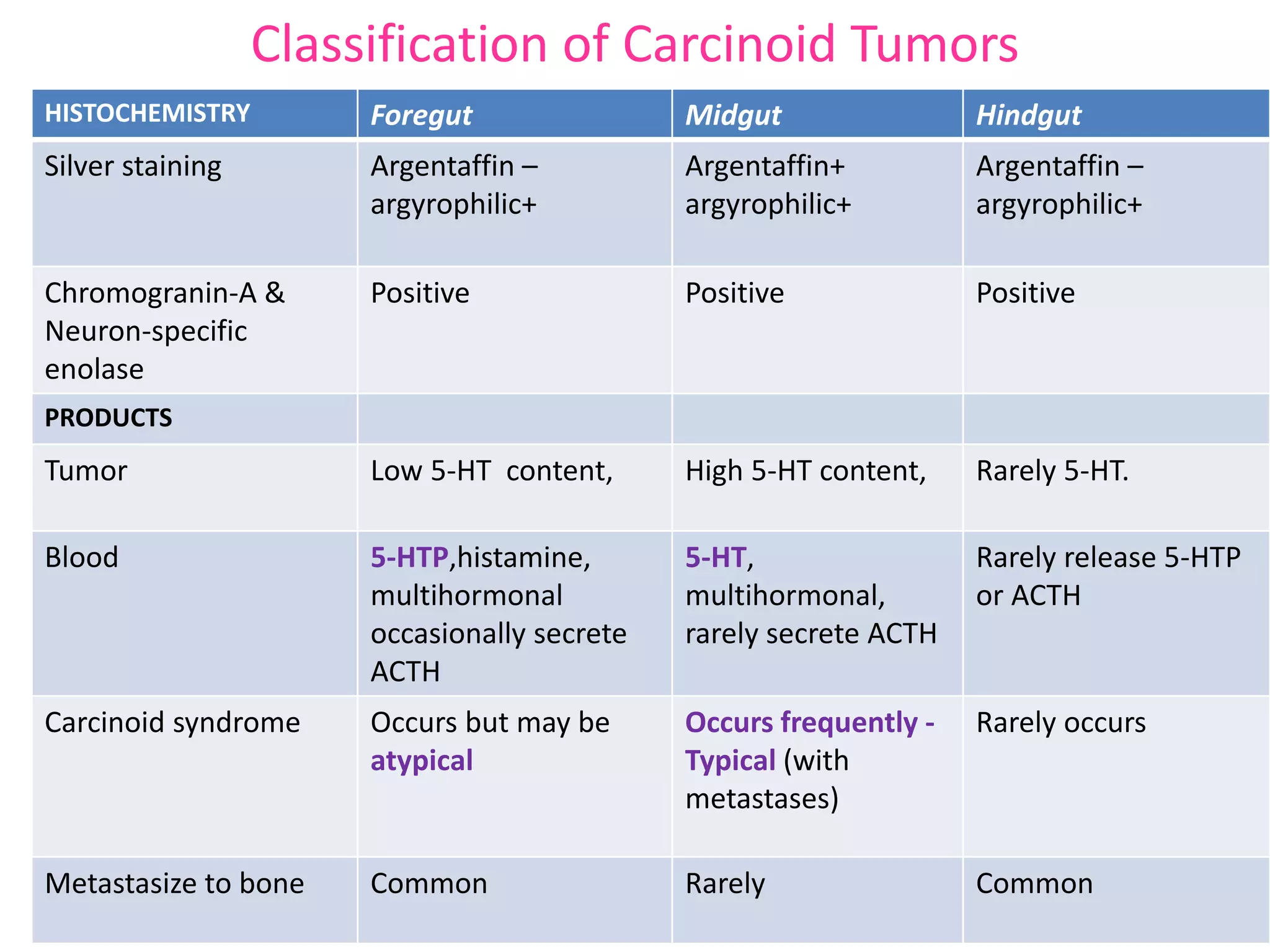
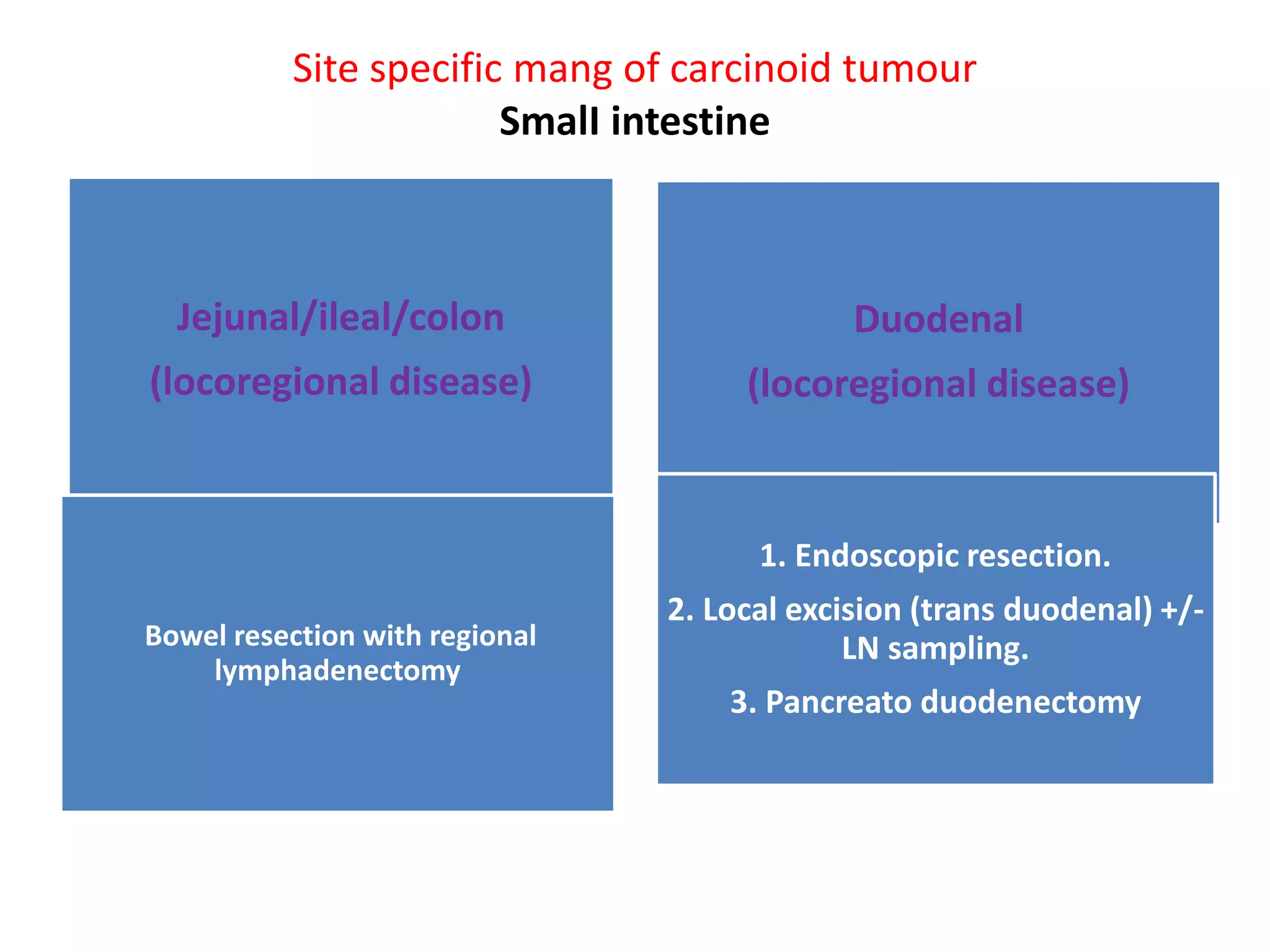
Carcinoid tumors are slow-growing neuroendocrine tumors that commonly arise in the gastrointestinal tract and lungs. The document discusses carcinoid tumors in depth, including their definition, sites of origin, histology, staging, clinical features, diagnostic testing, and management approaches. Treatment involves surgical resection when possible, with additional therapies for advanced or metastatic disease aimed at controlling hormone secretion and tumor growth.
2. D E F I N I T I O N
These are slow growing tumors of
neuroendocrine system that
belongs to amine precursor uptake
and decarboxylation (APUD)
system.
Oberndorfer in 1907
Karzinoid : carcinoma like
are thought of arise from enterochromaffin cells , also
known as kulchitsky cells.
3. Carcinoid tumours
• Neuroendocrine tumors - comprise a broad family of tumors,
the most common of which are ;
- carcinoid tumors (most commonly arising in the lungs and
bronchi (so-called bronchopulmonary), small intestine,
appendix, rectum, and thymus) and
- pancreatic neuroendocrine tumors.
- Other less common neuroendocrine tumors include those
arising in the parathyroid, thyroid, adrenal, and pituitary
glands).
4. • The SEER database estimated that the
incidence of neuroendocrine tumors in the
United States was 5.25 cases per 100,000
people in the year 2004.
• True Incidence and Prevalence of NETs Most
Likely Underestimated.
• Approx 2/3rd of carcinoid tumours arise in GIT
and 1/3rd in lungs or thymus.
• Carcinoids are the Second Most Prevalent
Type of GI Malignancy.
6. • Carcinoid tumors are composed of monotonous sheets of
small round cells with uniform nuclei and cytoplasm, and
mitotic figures are rare.
• Pathologists cannot differentiate benign from malignant
carcinoid tumors based on histologic analysis.
• Malignancy can only be determined if there is invasion or
distant metastases
7. Site of origin
NETs are generally classified as
foregut,
midgut, or
hindgut
depending on their embryonic origin.
Foregut
• Thymus
• Esophagus
• Lung
• Stomach
•Duodenum
Pancreas
Midgut
• Appendix
• small bowel
• Cecum
• Ascending
colon
Hindgut
• Distal large
bowel
• Rectum
8. Classification of Carcinoid Tumors
HISTOCHEMISTRY Foregut Midgut Hindgut
Silver staining Argentaffin –
argyrophilic+
Argentaffin+
argyrophilic+
Argentaffin –
argyrophilic+
Chromogranin-A &
Neuron-specific
enolase
Positive Positive Positive
PRODUCTS
Tumor Low 5-HT content, High 5-HT content, Rarely 5-HT.
Blood 5-HTP,histamine,
multihormonal
occasionally secrete
ACTH
5-HT,
multihormonal,
rarely secrete ACTH
Rarely release 5-HTP
or ACTH
Carcinoid syndrome Occurs but may be
atypical
Occurs frequently -
Typical (with
metastases)
Rarely occurs
Metastasize to bone Common Rarely Common
9.
10. • Histologic Classification
• Neuroendocrine tumors are classified
histologically based on tumor differentiation (well
or poorly differentiated) and tumor grade (grades
1–3).
• Most neuroendocrine tumors fall into 3 broad
histologic categories:
- well-differentiated, low-grade (G1);
- well-differentiated, intermediate-grade (G2); and
- poorly differentiated, high-grade (G3).
11. Grading Of NET (european NET society/WHO)
Low grade Intermediate
grade
High grade
Typical carcinoid
Mitotic count (10HPF):<2
Ki-67 index: </= 2
Atypical carcinoid
Ki67 is most often
positive.
Mitotic count (10HPF):
2-20
Ki-67 index: 3-20
Small cell carcinoma
Large cell
neuroendocrine
carcinoma
Mitotic count (10HPF):
>20
Ki-67 index:>20
12. • Grade is generally defined by mitotic count and/or Ki-67 index, whichever
is higher.
• In some cases, however, tumors may not fall clearly into one category.
• For example, a morphologically well- differentiated neuroendocrine tumor
with a low mitotic index may have a Ki-67 proliferation index that falls into
the high-grade category.
• While technically classified as a high-grade tumor, clinical judgment should
be used in making treatment decisions for such cases
• The classification of lung and thymus carcinoids - does not include Ki-67
and includes the assessment of necrosis.
13. TNM staging 7th edn, 2010
STOMACH,
DUODENUM,
JEJUNUM,ILEUM
AMPULLA COLON, RECUM APPENDIX
T1 Lamina
propria/submucosa &
</= 1Cm
</= 1Cm T1a:<1cm
T1b:1- 2 cms
T1a:</=1cm
T1b:>1- 2 cms
T2 Muscularis propria or
>1cm
>1cm Muscularis propria
or >2cm
>2 to </= 4cms
T3 subserosa Invades pancreas
or RP or non
peritonalised
tissue
Subserosa or non
peritonalised
pericolic &
perirectal tissues
> 4cms or
extension to
ileum
T4 serosal Invade other
organs
Peritonium & other
organs
Other strs,
abdo wall &
14. No/N1; Mo/M1
• Staging
Stage 0 -Tis No Mo
Stage I -T1 No Mo
Stage IIa -T2 No Mo
Stage IIb -T3 No Mo
Stage IIIa -T4 No Mo
Stage IIIb -anyT N1 Mo
Stage IV -anyT anyN M1
17. CLINICAL FEATURES
Systemic symptoms are consequences of the peptide
released by the tumor
Carcinoid syndrome
Cushing’s Syndrome
Acromegaly
Peptic ulcer disease
Heart problem
18. • Specific markers for carcinoid tumors
• Biochemical markers: often elevated in NETs and can be a surrogate marker
of symptoms of hormone excess or tumor growth.
• Chromogranin A levels are elevated in approximately 80% of patients with
GI NETs;
- sensitivity is 75% and specificity 85%.
- Falsely positive elevated -PPI or H2 receptor antagonist, hepatic or renal
failure.
• Urine 5-hydroxyindoleacetic acid (5-HIAA), the primary metabolite of
serotonin, is elevated in carcinoid syndrome. ---
- Sensitivity of 35% and specificity of 100%.
- Abnormal levels (>5 mg/24 hours) of 5-HIAA are diagnostic of carcinoid
syndrome.
- to avoid false positive results -avoid some diet & medications.
• ACTH
19. Systematic Approach to Diagnosing NETs
GI Endoscopy
Bronchoscopy
CT scan/ MRI
PET scan
20. Endoscopy:
Gastric and rectal carcinoids are often
asymptomatic and are thus most commonly
imaged on screening or diagnostic endoscopy.
Endoscopic ultrasound has aided in the
characterization of these tumors.
21. CT scan
•Primary tumor localization is rare with CT because of its submucosal location and often
small size
•CT is effective at identifying the mesenteric stranding/fibrosis that often accompanies
carcinoid tumors, as well as mesenteric nodal involvement
•The desmoplastic response can also cause changes in the mesenteric vasculature that can
be identified with CT-angiography
•CT is most effective for identifying hepatic metastasis, with sensitivity greater than 85%
•Metastatic carcinoid tumors are highly vascular and hence display bright enhancement
during arterial-phase imaging
•The role of magnetic resonance imaging (MRI) in the diagnosis of a primary carcinoid
tumor is limited, and it adds little or no information above that provided by CT
22. SRS has been used to image carcinoid tumors.
-Most NETs express somatostatin receptors that can bind and
internalize the currently available octapeptide somatostatin
analogues octreotide and lanreotide.
-This feature can be exploited both in terms of somatostatin
receptor-based treatments and scintrigraphy (SRS).
-Based on 111In-labeled diethylenetriamine pentaacetic acid
(DPTA) Octreoscan. 80% to 90% sensitivity and specificity in
identifying carcinoid neoplasms
The technology is also limited in patients who do not exhibit
carcinoid syndrome, for whom the sensitivity is only 60% for
identification of the primary tumor
23. PET has had mixed results in imaging carcinoid tumors
FDG-PET appears to be sensitive and specific in identifying carcinoid tumors that exhibit high
proliferative activity or are poorly differentiated.
(PET) scanning with Ga-68 DOTATATE is a newer somatostatin-based scintrigraphy that is thought
to be more sensitive than 111In scans.
131I-metaiodobenzylguanidine (MIBG) scans – limited studies have shown
results roughly equivalent to those of CT, with a sensitivity of 55% to 85% and a specificity of
95%
especially helpful in patients who are being maintained on long-term octreotide therapy or
whose tumors secrete catecholamines.
CT-enteroclysis
a newer imaging modality designed to image tumors of the small bowel. It has a sensitivity of
86% and a specificity of 100% for small bowel NETs..
Echocardiography
is also included in the diagnostic workup to either confirm or exclude
carcinoid heart disease.
24. Magnetic resonance image showing a single focus of metastatic disease (arrow) in the right
lobe of the liver in a patient with a carcinoid tumor and elevated 5-hydroxyindoleacetic
acid. B, A 111In-labeled pentetreotide scan of the same patient shows multiple hepatic
lesions and two involved para-aortic lymph nodes.
25. General Management Principles
• Management of NETs falls under two main
categories:
1) Therapies directed at tumor control and
2) Therapies directed at controlling symptoms of
hormone excess.
26. General Management Principles
• Surgical resection remains the mainstay of treatment for resectable
NETs of the GI tract and lungs.
• In addition to prolonged survival, surgery can also palliate
symptoms of obstruction, diarrhea, flushing,and/or pain with
eating.
• In the setting of unresectable disease, antitumor therapy is only
indicated in the setting of symptoms, tumor bulk, and/or disease
progression.
• In newly diagnosed asymptomatic patients with unresectable or
metastatic disease, it is often reasonable to monitor closely without
any active treatment.
27. MANAGEMENT BY PRIMARY TUMOR
SITE
• Esophageal Neuroendocrine Tumors
- very uncommon (<1% of GI NETs)
- distal esophagus just proximal to the esophageal-gastric junction
- Endoscopic ultrasonography and SRS are indicated for staging because
lymph node metastases occur in 50% of patients.
- Endoscopic mucosal resection (EMR) is done if there is no evidence
of lymph node metastases and the tumor is amenable to complete
excision.
- Open resection is done for patients with lymph node metastases. The
operative procedure is most commonly an Ivor Lewis
esophagogastrectomy.
28. GASTRIC CARCINOIDS
• 9% of GI NETs , arise from the enterochromaffin-like (ECL) cells of
the stomach that occur in the gastric fundus and body
3/1000 gastric neoplasms
3 Types-
Type I - Atrophic gastritis
Type II – ZES
Type III – No hypergastrinemias
Hypergastinemia
29. Gastric carcinoid tumors- Three subtypes
• Two subtypes are associated with hypergastrinemic states, either
Chronic atrophic gastritis (type I; 80%) or
Zollinger-Ellison syndrome (type II; 6%), almost always as part of MEN-1,--
there is allelic loss at the MEN-1 locus on chromosome band 11q13, and thus
fundic gastric carcinoid tumors are now included in the spectrum of MEN-1
tumors.
These tumors generally
pursue a benign course,
with 9% to 30% LN metastases, distant mets 10-20%
usually multicentric and small,
with infiltration restricted to the mucosa and submucosa,
size- few mm to 1.5 cm
30. Gastric carcinoid tumors- cont..,
The third type of gastric carcinoid
Type III-(15-20%) -sporadic ECL tumors occurs without
hypergastrinemia,
- often large (mean size 3 cm) & solitary,
- pursue a more aggressive course,
- Most lesions invade the full thickness wall of the stomach.
- located in the body or fundus.
- Regional lymph node and liver metastases in 70% of patients
- 50% have atypical histologic features, and some patients
develop carcinoid syndrome.
31. Site specific mang of Gastric carcinoid
Loco regional disease
Hypergastrinemic
Tumour </= 2cms/
solitary or multiple
1. Observe or
2. Endo resection or
3. octreotide for ZE
pts
Patients……………
Tumour > 2cms/
solitary or multiple
1. Endo resection, if
possible or
2. Surgical
resection
Pts with normal
gastrin
Radical gastric
resection+Ln
removal
32. • <4% of all GI NETs
• more common in the proximal duodenum
• usually small <5 mm
• (30% to 70%) spread to regional LNs such that adjacent lymph
node sampling is recommended
• not commonly spread to distant sites
• In contrast to jejunoileal carcinoid tumors, duodenal carcinoid
tumors are often discovered by endoscopy.
Duodenal carcinoid tumours
33. Site specific mang of carcinoid tumour
SmalI intestine
Jejunal/ileal/colon
(locoregional disease)
Duodenal
(locoregional disease)
Bowel resection with regional
lymphadenectomy
1. Endoscopic resection.
2. Local excision (trans duodenal) +/-
LN sampling.
3. Pancreato duodenectomy
34. Small intestinal carcinoid tumors
• Small bowel NETs are most common , 42% of all GI NETs, and
they are most prevalent in ileum within 20 cm of the ileal-
cecal valve .
• Ileal NETs are commonly very small (2 to 4 mm) and multiple
within the wall of the ileum, with adjacent large lymph node
metastases that cause cicatrization and venous conjestion
that can result in small bowel obstruction .
• Approximately 50% of patients will have liver metastases or
peritoneal carcinomatosis at the time of diagnosis.
• Cholecystectomy is also indicated because most of these
patients with either lymph node or liver metastases will
require the longterm use of somatostatin analogues that can
cause gallstones when administered long-term.
35. Appendiceal carcinoid Tumors
• In the past carcinoid tumors were most frequently reported in
the appendix (approximately 40%);
however, more recently small intestine and the bronchus &
lung are the most common sites
• A carcinoid tumor is discovered in approx one out of every 200 to
300 appendectomies.
- Most occur in the tip of the appendix.
- The majority (i.e., 90%) are less than 1 cm in diameter
without metastases
36. Site specific mang of carcinoid tumour
Appendix
</=2cms & confined to
appendix, not present at
the base- SIMPLE
APPENDICECTOMY
>2cms or incomplete
resection, invasion
through the wall,
presence at the base, or
lymph node metastases-
REEXPLORATION/RIGHT
HEMI-COLECTOMY
Rectal
</= 2 cms-
resection(trans-anal or
endoscopic excision, if
possible)
>2cms-LAR OR APR
37. • Colonrectal carcinoid Tumors
• 4% of GI NETs and 1% to 5% of colorectal tumors.
• Mc site Rt colon
• majority of colon NETs are well differentiated
but some are large, less differentiated, and exophytic and
have a higher incidence of lymph node and liver
metastases.
• The 5-year survival for colon NETs is 37%,
• Rectal carcinoid Tumors
• 27% of GI NETs and 1% to 2% of all rectal tumors
38. Carcinoid Syndrome
Malignant carcinoid syndrome is a clinical entity marked by flushing,
diarrhea, abdominal cramping, wheezing, heart valve dysfunction, and
pellagra
Flushing
(63%-74%)
Cardiac disease
(14%-41%)
Diarrhea
(68%-73%)
Dermatitis
(5%)
Abdominal pain
(10%-34%)
Bronchoconstriction
(3%-18%)
39. • It is found in 10% to 18% of all patients with carcinoid
tumors, but the incidence increases to 40% to 50% in
patients with advanced disease
• Carcinoid tumors produce a large number of substances,
including serotonin, tachykinins, and histamine.
• It is the actions of serotonin that lead to many of the
manifestations of carcinoid syndrome.
• Develops in the presence of hepatic metastasis, which
precludes hepatic inactivation of the active metabolites of
serotonin.
40. CARCINOID SYNDROME
91% CASES – Only AFTER DM TO LIVER
EXCEPTION
☻ Lung carcinoids
☻ Ovarian carcinoids
☻ Pancreatic carcinoids
41. The development of carcinoid syndrome is related to the embryologic origin of the
original tumor
•TYPICAL CARCINOID SYNDROME
- Midgut carcinoids are the most common source of carcinoid syndrome because these
tumors produce high levels of serotonin
- More than 90% of all patients suffering from carcinoid syndrome have a midgut
primary
•ATYPICAL CARCINOID SYNDROME
-Foregut carcinoids lack the aromatic amino acid decarboxylase required to convert 5-
hydroxytryptamine (5-HT) to serotonin and therefore cannot produce the classic
carcinoid syndrome.
- In patients with gastric carcinoid tumors. secondary to large release of histamine
from the tumor rather than serotonin.
- Hindgut carcinoids also lack the ability to convert tryptophan to serotonin, and thus
even if metastatic lesions are present, carcinoid syndrome will not develop.
42.
43. TREATMENT OF CARCINOID
SYNDROME
Avoid stress and precipitating factors
Diuretics – heart failure
Bronchodilators – wheezing
Antidiarrhoeal agents, niacin supplementation
Somatostatin analogues (octreotide) – 100 mg s/c 8 hourly – 3000 mg/ day
Before surgery – 150-250 mg s/c 6-8 hrly 24-48 hrs prior to anaesthesia
Inhibitors of serotonin synthesis are emerging as a new class of
agents to treat carcinoid syndrome. Telotristat etiprate
44. Metastatic Disease
Cytoreductice Rxs for metastatic carcinoids
• Surgery/ local ablative therapy
• Hepatic artery chemoembolisation
• Systemic CT
45. Surgical Therapy
Approach to the treatment of metastatic disease is based on symptoms.
-Resection of the primary site in the setting of unresectable metastases is generally not indicated
if the primary site remains asymptomatic and is relatively stable.
-Patients with limited hepatic metastases or other sites of disease can undergo complete
resection of the primary tumor and metastases with curative intent.
-Noncurative debulking surgery can also be considered in select cases, especially if the patient
is symptomatic either from tumor bulk or hormone production
-Such resection aids in avoiding complications from growth of the primary tumor in terms of
bleeding, obstruction, and abdominal pain, especially with midgut carcinoids because of their
propensity to cause intense fibrosing reactions
46. -By using a resection or ablative approach to hepatic metastases, the
patient can be palliated and even enjoy a long-term survival
benefit.
-Surgical therapy for metastatic carcinoid tumors is aimed at removing
more than 90% of the tumor burden via either anatomic or wedge
resections, with acceptable morbidity and mortality rates of 15%
and 1.2%, respectively
-By using this cytoreductive approach, symptoms from metastatic
carcinoid are reduced in 95% of patients, with a median symptom-
free duration of 45 months.
- Five-year survival rates improve from 40% to 50% in patients who do
not undergo surgery to 60% to 82% in patients after hepatic
resection of their metastatic disease
47. • Patients whose tumor is unresectable or in whom reduction of greater
than 90% of the tumor burden is not feasible
Liver directed therapies include (CAT-2B)
-radiofrequency ablation (RFA),
-cryotherapy,
-hepatic arterial occlusion,
-embolization, and
-chemoembolization.
• Orthotopic liver transplantation
- its true role in the treatment of patients with metastatic carcinoid remains
unclear
-Patients who appear to have the greatest survival rates are those with well-
differentiated tumors that exhibit low proliferative activity
-In this select group, orthotopic liver transplantation can yield a 5-year
survival rate of up to 69%
48. ANTITUMOR Medical Therapy
focused mainly on relief of the symptoms of carcinoid syndrome
Somatostatin Analogues
-The most commonly used therapy.
-The somatostatin receptors are blocked with a somatostatin analogue, thereby
relieving the flushing, diarrhea, and other symptoms of carcinoid syndrome.
-The most commonly prescribed agent has been octreotide
-However, the short half-life of octreotide necessitates continuous infusion or twice-daily
subcutaneous injections.
-The long-acting release (LAR) formulation of octreotide is commonly used
-Standard doses of octreotide LAR are 20 to 30 mg intramuscularly every 4 weeks.
-Therapeutic levels are not achieved for 10 to 14 days after LAR injection.
Short-acting octreotide (usually 150–250 mcg subcutaneously 3 times daily)
-Lanreotide is administered every 10 to 14 days(60 to 120 mg).
Side effects of the treatment include sinus bradycardia, arrhythmias, gallbladder stones,
steatorrhea, hypothyroidism, and hypoglycemia
49. • PROMID-The best clinical evidence came from this
trial,
- Phase 3 study RCT
- 85 patients
- Metastatic midgut carcinoids to
- Octreotide LAR 30 mg versus placebo.
- Significant improvement of median time to progression
from 6 months in the placebo arm to 14.3 months in
the treatment arm.
• CLARINET – Lanreotide Vs plecebo
- Improvement in PFS.
50. Interferon-Alpha:category 3
-Because of its potential side effects interferon is
usually not initiated until failure of
somatostatin analog treatment.
Mammalian Target of Rapamycin
Inhibitors(Everolimus)
-approved by the US FDA for the treatment of
metastatic pancreatic NETs based on
improvement in PFS from 4.6 months to 11
months in a randomized study of metastatic
pancreatic NETs (RADIANT-3).
51. • Angiogenesis Inhibitors
-NETs are highly vascular tumors that frequently overexpress
the VEGF receptor.
• Bevacizumab (a monoclonal antibody to circulating VEGF)-
investigated in phase II clinical trials, showing disease
stability in 95% of pts when combined with octrotide.
• Sunitinib, (an oral TKI, inhibits VEGF receptors 1, 2, and 3 as
well as PDGF, cKIT)-
- Demonstrated improved PFS compared to placebo in a
randomized phase 3 study of advanced pancreatic NETs,
which led to its approval by the US FDA for this indication in
2010
52. Systemic CT
• Poorly differentiated NETs, regardless of primary site, are typically
treated like small-cell lung cancer with a platinum/etoposide
based regimen.
• no best systemic CT is yet recognised and the role of CT
continues to be debated.
60. GIST
• Gastrointestinal stromal tumors (GIST) are
mesenchymal neoplasms of the gastrointestinal
tract-interstitial cell of Cajal
• GISTs are rare cancers, which were defined as a
distinct disease in the 1990s, having been
classified within smooth muscle neoplasms for
decades from their first description in the 1960s.
• Coincidentally, in 2000, they became targetable
by new tyrosine-kinase inhibitors (TKI).
61. • GISTs can arise anywhere along the GI tract,
Stomach (60%) and
Small intestine (30%) are the most common primary
sites.
Duodenum (4% to 5%) and
Rectum (4%) are the less common primary sites, and
Esophagus (<1%) and colon & appendix (1% to 2%)
rare site
• Some GISTs have been labeled as extragastrointestinal, apparently
arising from the mesentery, omentum,and retroperitoneum;
however, it remains unknown whether these are lesions detached
from their gastrointestinal origin and/or are metastases from an
unknown primary tumor.
62. Clinical features:
• Variety of symptoms, which may include early satiety,
abdominal discomfort due to pain or swelling,
intraperitoneal hemorrhage, GI bleeding, or fatigue related
to anemia.
• Some patients may present with an acute abdomen (as a
result of tumor rupture, GI obstruction, or appendicitis-like
pain),which requires immediate medical attention.
• Liver metastases and/or dissemination within the
abdominal cavity are the most common clinical
manifestations of malignancy
63. • outward growth of many GISTs within the
gastrointestinal wall is one of the reasons why
several are diagnosed relatively late,
• One-fourth of GISTs are discovered
incidentally during diagnostic assessments
(whether an endoscopic procedure,
ultrasound, or computed tomography [CT]
scan) done for other reasons.
64. PATHOLOGY
• Morphologically, GISTs can be made up of spindle
cells (in more than two-thirds of cases),
epithelioid cells, or both.
• there are no pathologic clues to make a
distinction between malignant GISTs and benign.
• All GISTs are currently considered malignant
neoplasms, although with a highly variable risk of
distant relapse, which is negligible in a significant
proportion of them.
65. • Immunohistochemical Pattern
• Most GISTs (95%) express KIT (CD117).
• Approximately 80% of GISTs have a mutation in the gene encoding
the KIT receptor tyrosine kinase;
• Another 5% to 10% of GISTs have a mutation in the gene encoding
the related PDGFRA receptor tyrosine kinase.
• Other commonly expressed markers include CD34 antigen (70%),
smooth muscle actin, (25%), and desmin (less than 5%).
• Immunohistochemical status does not reflect the mutational status
with regard to KIT and PDGFRA, per se, so that it has no concrete
predictive value for sensitivity to TKIs.
• KIT positivity alone may not be sufficient to confirm the diagnosis
and, conversely, the absence of KIT and/or PDFGRA mutations does
not exclude the diagnosis of GIST
66. • Approximately 10% to 15% of GISTs are WT for
KIT and PDGFRA.
(1) SDH-deficient GISTs;
(2) neurofibromatosis (NF)-1–related GISTs; or
(3) others, including those with the BRAF V600E
mutation.
• Immunohistochemically, these GIST are negative
to SDHB staining.
67. • DOG1 is calcium dependent, receptor-
activated chloride channel protein.
• it is expressed in GISTs independent of
mutation type.
• DOG1 expression was not different between
the KIT/PDGFRA mutant or wild-type GIST
68.
69. PRINCIPLES OF BIOPSY FOR GIST
• GISTs are soft and fragile tumors. EUS-FNA biopsy of primary site is
preferred over percutaneous biopsy (due to the risk for hemorrhage and
intra-abdominal tumor dissemination).
• Biopsy is necessary to confirm the diagnosis of primary GIST prior to the
initiation of preoperative therapy.
• Percutaneous image-guided biopsy may be appropriate for confirmation of
metastatic disease
• Testing for mutations in KIT and PDGFRA is strongly recommended.
• Testing for germline mutations in the SDH genes should be considered for
patients with wild-type GIST (lacking KIT or PDGFRA mutations).
70. PRINCIPLES OF SURGERY FOR GIST
Primary (Resectable) GIST
• Surgery is the primary treatment for all patients with
GISTs (2 cm or greater) that are resectable without
significant risk of morbidity.
• Aim is to resect the tumor with histologically negative
margins.
• wedge or segmental resection of the involved gastric or
intestinal tract, with margins that can be less wide than
for an adenocarcinoma.
• complex multi-visceral resection should be avoided. If
the surgeon feels that a multi-visceral resection may be
required, then multidisciplinary consultation is
indicated regarding a course of preoperative imatinib.
71. • Lymphadenectomy is usually not required given the low
incidence of nodal metastases.
• As GIST tends to be very friable, every effort should be
made not to violate the pseudocapsule of the tumor.
• A macroscopically complete resection with negative or
positive microscopic margins (R0 or R1 resection,
respectively) is associated with a better prognosis than a
macroscopically incomplete excision (R2 excision)
(exception are GIST of the rectum-where microscopically
positive margins are clearly associated with a higher risk of
local failure.)
72. Laparoscopic approach may be considered in
favorable anatomic locations (greater
curvature or anterior wall of the stomach,
jejunum, and ileum) by surgeons with
appropriate laparoscopic experience.
• Resection specimens should be removed from
the abdomen in a plastic bag to prevent
spillage or seeding of port sites.
73. Targeted Therapy
• Imatinib, a selective inhibitor of the KIT
protein tyrosine kinase, has produced durable
clinical benefit and objective responses in
most patients with GIST.
• In February 2002, the FDA approved of
imatinib for the treatment of patients with
KIT-positive
74. Sunitinib
• TKI inhibiting KIT and PDGFRA but also displaying
antiangiogenic activity by the inhibition of
vascular endothelial growth factor receptor
(VEGFR)1, 2, and 3
• Standard second-line therapy.(Cat-1)
• January 2006, sunitinib received FDA approval.
• It was shown to be effective at increasing
progression-free survival by 5 months in a
randomized trial versus placebo in patients failing
(or intolerant) to imatinib
75. Regorafenib,
• another TKI with activity on KIT and PDGFRA as
well as VEGFR1, 2, and 3, and thus with
antiangiogenic properties,
• standard third-line therapy for advanced GIST
patients. (Cat-1)
• In fact, it was shown to be effective as a third-line
therapy in patients failing both imatinib and
sunitinib, by providing a median advantage of 4
months of progression-free survival over placebo
in a randomized clinical trial
76.
77. • Two separate phase III studies
1) EORTC 62005 study and
2) S0033/CALGB 150105 study
have assessed the efficacy of imatinib at two
initial dose levels (400 mg daily vs. 800 mg daily)
in patients with metastatic or unresectable GIST.
• Both studies showed equivalent response rates
and OS for both dose levels.
• Higher dose of imatinib was associated with more
side effects than the lower dose in both studies.
78. • Primary resistance is defined as the evidence of clinical progression developing
during the first 6 months of imatinib therapy and it is most commonly seen in
patients with;
- KIT exon 9 mutations,
- PDGFRA exon 18 D842V mutations, or
- tumors that lack identifiable activating mutation in
KIT or PDGFRA (SDH-deficient / WT GIST.
• Secondary resistance is seen in patients who have been on imatinib for more than
6 months with an initial response or disease stabilization followed by progression,
Mechanism- occurrence of new mutations to the same primarily mutated
oncogene, or, less frequently,
- oncogene amplifications or alterations of alternative pathways.
• Dose escalation to 800 mg/day or switching to sunitinib is a reasonable option for
patients progressing on imatinib 400 mg/day
79. • Termination of imatinib in patients with GIST
that is refractory to imatinib has been shown
to result in a flare phenomenon, which in turn
indicates that even in patients with
progressive disease on imatinib therapy, there
are some tumor cells for which imatinib may
still be effective.
80. Preoperative imatinib
The RTOG 0132/ACRIN 6665
• First prospective study that evaluated the efficacy
of preoperative imatinib (600 mg/day) in patients
with potentially resectable primary disease (30
patients) or potentially resectable recurrent or
metastatic disease (22 patients).
• The estimated 2-year OS rate was 93% and 91%
for patients with primary GIST and those with
recurrent or metastatic GIST, respectively.
• The estimated 2-year PFS rate was 83% and 77%,
respectively.
81. • Indications:
- treatment for patients with GIST that is resectable with negative
margins but with a significant risk of morbidity.
- big abdominal masses that may be felt by the surgeon as implying a
significant risk of tumor rupture during surgery
• Close monitoring is essential, because some patients may rapidly become
unresectable.
• The guidelines recommend an initial dose of 400 mg daily. Patients with
documented KIT exon 9 mutations may benefit from dose escalation up to
800 mg daily (given as 400 mg twice daily), as tolerated.
82. • Baseline CT with or without MRI is recommended prior to the start of preoperative
imatinib.
• imatinib should be continued until maximal response (defined as no further
improvement between 2 successive CT scans, which can take as long as 6–12
months).
• Diagnostic CT is indicated every 8 to12 weeks. If there is no progression,
continuation of the same dose of imatinib is recommended and resection should
be considered, if possible.
• Surgery is recommended
-If bleeding and/or symptoms are present.
-If there is progression
• dosing can be stopped right before surgery and resumed as soon as the patient is
able to tolerate oral medications following surgery regardless of surgical margins-
life long.
83. Response Assessment
• The radiologic response, as assessed through CT scans and MRI, is
marked by
- tumor shrinkage and/or
- changes in tumor tissue.
• Response Evaluation Criteria in Solid Tumors (RECIST) criteria for
tumor response assessment are based on the measurement on one
diameter of selected target lesions.
• Choi criteria were worked out in GISTs to accommodate these
patterns of response, by factoring tumor hypodensity on CT scans in
addition to a decrease in size.
• The EORTC has developed metabolic response criteria for tumors
evaluated with PET.
84. • A positive PET scan may turn negative in a few
days.
• Of course, this does not correspond to the
disappearance of the tumor lesion, but rather be
the consequence of the metabolic switch off that
the tumor undergoes when an effective TKI
targets its cells.
• The reverse is true as well, so that any stop of
therapy rapidly entails a switch on of functional
imaging.
85. Post-operative imatinib
• Surgery does not routinely cure GIST.
Complete resection is possible in
approximately 85% of patients with primary
tumors.
• At least 50% of these patients will develop
recurrence or metastasis following complete
resection and the 5-year survival rate is about
50%. Median time is about 2 years.
86. ACOSOG Z9001
• randomized study phase III, double-blind,
• Patients with primary localized GISTs (3 cm or greater in size)
postoperative imatinib 400 mg (359 patients) or placebo (354
patients) for one year after complete resection.
• Results:
• The RFS rate at one year was significantly higher in the imatinib arm
compared to placebo (98% and 83%, respectively, P < .001).
• OS was not different in both arms (99.2% vs. 99.7%, respectively; P
= .47).
87. SSGXVIII/AIO:
• randomized phase III study
• suggest that postoperative imatinib
administered for 36 months improves RFS and
OS compared to 12 months for patients with a
high estimated risk of recurrence after
surgery.(mitotic index of >5 mitoses/50 HPF,
size >5 cm, non-gastric location, and tumor
rupture).
88. • In patients who have received preoperative
imatinib, regardless of surgical margins until
progression.
• Postoperative imatinib should be initiated
following resection, if the patient had not
received prior imatinib therapy.
89. Unresectable or Metastatic GIST
Imatinib (CAT-1)is the primary therapy for
advanced, unresectable or metastatic GIST.
Surgery may be indicated for:
• Limited disease progression refractory to imatinib.
• Locally advanced or previously unresectable
tumors after a favorable response to preoperative
imatinib.(if complete resection can be obtained in
primary metastatic disease)
90. • Several retrospective studies have demonstrated
survival benefit of cytoreductive surgery
following preoperative imatinib in patients with
advanced or metastatic GIST responding to
preoperative imatinib.
• Imatinib should be continued if resection is not
feasible for metastatic GIST until progression.
• The dose of imatinib should not be increased if
patients remain stable without objective
progression of the disease.
91. • Recurrence following complete resection
should be managed as described for
unresectable or metastatic disease, because
recurrent disease represents locoregional
metastatic or infiltrative spread of the
malignancy and carries essentially the same
prognosis as distant metastases overall.
92. • Current risk classification systems are based on
the combination of mitotic count, tumor size,
and site of origin.
• the combination of these three factors allows one
to forecast a risk of relapse by using tools such as
-The Armed Forces Institute of Pathology (AFIP)
risk classification,
-The Memorial Sloan Kettering Cancer
Center(MSKCC) nomogram, or
-The contour maps.
93. Prognostic Factors
• Tumor size and the mitotic rate are the most widely used pathologic
features for the risk stratification of GIST.
• The metastatic rate was 86% for tumors >10 cm with a mitotic index
of >5 mitoses/50 HPFs, whereas tumors of the same size with a
mitotic index of <5 mitoses/50 HPFs have a relatively low metastatic
rate of 11%.
• With small intestinal GIST, tumors >10 cm with a mitotic index of ≤5
mitoses/50 HPF had a metastatic rate of 50%, which is a contrast to
that reported for gastric GIST
• In addition to the tumor size and mitotic rate, tumor site has also
been included in the guidelines developed by Miettinen and
colleagues for the risk stratification of primary GIST.
94. • According to these guidelines,
• Gastric GISTs have an overall indolent behavior
and those that are ≤2 cm (irrespective of the
mitotic index) are essentially benign,
• Small intestinal GISTs tend to be more aggressive
than gastric GISTs.
• Rectal GISTs are also very aggressive and tumors
<2 cm with a mitotic index of >5 mitoses/50 HPFs
have a higher risk of recurrence and malignant
potential.
95. • KIT & PDGFR mutations can be found in high-
grade tumors as well as in small incidental
GIST and tumors that have a benign course.
• Therefore, KIT mutational status is not used in
the routine prognostic assessment of a
primary GIST.