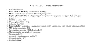
The most common type of cancer arising in the kidney: Renal cell carcinoma(RCC)(also known as Hypernephroma or Grawitz tumor).
Renal cell carcinoma accounts for over 3% of all adult malignancies and has several histological subtypes.
Approximately 85% of kidney tumors are renal cell carcinoma, and approximately 70% of these have a Clear cell histology. Its diagnostic work-up, staging and management.
Non-clear cell renal cell carcinoma (RCC) encompasses diverse subtypes, each requiring tailored therapeutic approaches. Papillary RCC may benefit from immunotherapy or vascular endothelial growth factor receptor (VEGFR) inhibitors. Chromophobe RCC often sees mTOR inhibitors or VEGFR inhibitors as initial treatments. For collecting duct and renal medullary carcinomas, cytotoxic chemotherapy is recommended.
Translocation RCC may respond well to lenvatinib plus pembrolizumab, while unclassified RCC patients might consider immunotherapy-based regimens. Sarcomatoid features in non-clear cell RCC lean towards immunotherapy.
Clinical trials are encouraged due to limited high-quality data, emphasizing the need for personalized strategies based on histologic subtypes. Overall, these recommendations aim to optimize outcomes in the diverse landscape of non-clear cell RCC.
The evolution of radiation treatment planning and delivery, with innovative techniques (3DCRT, IMRT, IGRT, IGBT), particle therapy allowing for better definition of target and sensitive structure volumes and more precise quantification of dose, has introduced more complexity into the evaluation of radiation effects on OARs.
Acute radiation syndrome (ARS) or acute radiation sickness is an acute illness caused by irradiation of the entire body (or most of the body) by a high dose penetrating radiation in a very short period of time (usually a matter of minutes). The major cause of this syndrome is depletion of immature parenchymal stem cells in specific tissues.
Classically, acute radiation syndrome is subdivided into three sub-syndromes:
the hematopoietic syndrome,
the gastrointestinal syndrome,
the cerebrovascular syndrome
others include :
pulmonary syndrome, cutaneous radiation injury
radiation-induced multi organ dysfunction (failure) syndrome.
PHYSICS AND CHEMISTRY OF RADIATION ABSORPTION 1.pptxDr Monica P
Radiobiology is the study of the action of Ionizing radiations on the living things.
The absorption of energy from the radiation in biologic material leads to either of the following two processes: EXCITATION, IONIZATION
Tumor lysis occurs when cancer cells release their contents into the blood stream, either spontaneously or following antineoplastic therapy leading to an influx of electrolytes and nucleic acids into the circulation.
The sudden development of hyperkalemia, hyperuricemia and hyperphosphatemia can have life-threatening end-organ effects on the myocardium, kidneys and CNS.
Hypocalcemia is a consequence of hyperphosphatemia in TLS.
Symptoms are variable from the metabolic derangements of TLS.
Pituitary tumors: Most common type of pituitary tumor is pituitary adenoma. Most pituitary adenomas develop in adenohypophysis.
Pituitary tumors account for 12-19% of all primary brain tumors, making them 3rd most common primary brain tumors in adults.
These tumors are broadly classified based on whether they secrete excessive amounts of pituitary hormones or not.
2/3rd of the pituitary adenomas are secreting type.
Explore natural remedies for syphilis treatment in Singapore. Discover alternative therapies, herbal remedies, and lifestyle changes that may complement conventional treatments. Learn about holistic approaches to managing syphilis symptoms and supporting overall health.
MANAGEMENT OF ATRIOVENTRICULAR CONDUCTION BLOCK.pdfJim Jacob Roy
Cardiac conduction defects can occur due to various causes.
Atrioventricular conduction blocks ( AV blocks ) are classified into 3 types.
This document describes the acute management of AV block.
These simplified slides by Dr. Sidra Arshad present an overview of the non-respiratory functions of the respiratory tract.
Learning objectives:
1. Enlist the non-respiratory functions of the respiratory tract
2. Briefly explain how these functions are carried out
3. Discuss the significance of dead space
4. Differentiate between minute ventilation and alveolar ventilation
5. Describe the cough and sneeze reflexes
Study Resources:
1. Chapter 39, Guyton and Hall Textbook of Medical Physiology, 14th edition
2. Chapter 34, Ganong’s Review of Medical Physiology, 26th edition
3. Chapter 17, Human Physiology by Lauralee Sherwood, 9th edition
4. Non-respiratory functions of the lungs https://academic.oup.com/bjaed/article/13/3/98/278874
Prix Galien International 2024 Forum ProgramLevi Shapiro
June 20, 2024, Prix Galien International and Jerusalem Ethics Forum in ROME. Detailed agenda including panels:
- ADVANCES IN CARDIOLOGY: A NEW PARADIGM IS COMING
- WOMEN’S HEALTH: FERTILITY PRESERVATION
- WHAT’S NEW IN THE TREATMENT OF INFECTIOUS,
ONCOLOGICAL AND INFLAMMATORY SKIN DISEASES?
- ARTIFICIAL INTELLIGENCE AND ETHICS
- GENE THERAPY
- BEYOND BORDERS: GLOBAL INITIATIVES FOR DEMOCRATIZING LIFE SCIENCE TECHNOLOGIES AND PROMOTING ACCESS TO HEALTHCARE
- ETHICAL CHALLENGES IN LIFE SCIENCES
- Prix Galien International Awards Ceremony
New Directions in Targeted Therapeutic Approaches for Older Adults With Mantl...i3 Health
i3 Health is pleased to make the speaker slides from this activity available for use as a non-accredited self-study or teaching resource.
This slide deck presented by Dr. Kami Maddocks, Professor-Clinical in the Division of Hematology and
Associate Division Director for Ambulatory Operations
The Ohio State University Comprehensive Cancer Center, will provide insight into new directions in targeted therapeutic approaches for older adults with mantle cell lymphoma.
STATEMENT OF NEED
Mantle cell lymphoma (MCL) is a rare, aggressive B-cell non-Hodgkin lymphoma (NHL) accounting for 5% to 7% of all lymphomas. Its prognosis ranges from indolent disease that does not require treatment for years to very aggressive disease, which is associated with poor survival (Silkenstedt et al, 2021). Typically, MCL is diagnosed at advanced stage and in older patients who cannot tolerate intensive therapy (NCCN, 2022). Although recent advances have slightly increased remission rates, recurrence and relapse remain very common, leading to a median overall survival between 3 and 6 years (LLS, 2021). Though there are several effective options, progress is still needed towards establishing an accepted frontline approach for MCL (Castellino et al, 2022). Treatment selection and management of MCL are complicated by the heterogeneity of prognosis, advanced age and comorbidities of patients, and lack of an established standard approach for treatment, making it vital that clinicians be familiar with the latest research and advances in this area. In this activity chaired by Michael Wang, MD, Professor in the Department of Lymphoma & Myeloma at MD Anderson Cancer Center, expert faculty will discuss prognostic factors informing treatment, the promising results of recent trials in new therapeutic approaches, and the implications of treatment resistance in therapeutic selection for MCL.
Target Audience
Hematology/oncology fellows, attending faculty, and other health care professionals involved in the treatment of patients with mantle cell lymphoma (MCL).
Learning Objectives
1.) Identify clinical and biological prognostic factors that can guide treatment decision making for older adults with MCL
2.) Evaluate emerging data on targeted therapeutic approaches for treatment-naive and relapsed/refractory MCL and their applicability to older adults
3.) Assess mechanisms of resistance to targeted therapies for MCL and their implications for treatment selection
Pulmonary Thromboembolism - etilogy, types, medical- Surgical and nursing man...VarunMahajani
Disruption of blood supply to lung alveoli due to blockage of one or more pulmonary blood vessels is called as Pulmonary thromboembolism. In this presentation we will discuss its causes, types and its management in depth.
HOT NEW PRODUCT! BIG SALES FAST SHIPPING NOW FROM CHINA!! EU KU DB BK substit...GL Anaacs
Contact us if you are interested:
Email / Skype : kefaya1771@gmail.com
Threema: PXHY5PDH
New BATCH Ku !!! MUCH IN DEMAND FAST SALE EVERY BATCH HAPPY GOOD EFFECT BIG BATCH !
Contact me on Threema or skype to start big business!!
Hot-sale products:
NEW HOT EUTYLONE WHITE CRYSTAL!!
5cl-adba precursor (semi finished )
5cl-adba raw materials
ADBB precursor (semi finished )
ADBB raw materials
APVP powder
5fadb/4f-adb
Jwh018 / Jwh210
Eutylone crystal
Protonitazene (hydrochloride) CAS: 119276-01-6
Flubrotizolam CAS: 57801-95-3
Metonitazene CAS: 14680-51-4
Payment terms: Western Union,MoneyGram,Bitcoin or USDT.
Deliver Time: Usually 7-15days
Shipping method: FedEx, TNT, DHL,UPS etc.Our deliveries are 100% safe, fast, reliable and discreet.
Samples will be sent for your evaluation!If you are interested in, please contact me, let's talk details.
We specializes in exporting high quality Research chemical, medical intermediate, Pharmaceutical chemicals and so on. Products are exported to USA, Canada, France, Korea, Japan,Russia, Southeast Asia and other countries.
Lung Cancer: Artificial Intelligence, Synergetics, Complex System Analysis, S...Oleg Kshivets
RESULTS: Overall life span (LS) was 2252.1±1742.5 days and cumulative 5-year survival (5YS) reached 73.2%, 10 years – 64.8%, 20 years – 42.5%. 513 LCP lived more than 5 years (LS=3124.6±1525.6 days), 148 LCP – more than 10 years (LS=5054.4±1504.1 days).199 LCP died because of LC (LS=562.7±374.5 days). 5YS of LCP after bi/lobectomies was significantly superior in comparison with LCP after pneumonectomies (78.1% vs.63.7%, P=0.00001 by log-rank test). AT significantly improved 5YS (66.3% vs. 34.8%) (P=0.00000 by log-rank test) only for LCP with N1-2. Cox modeling displayed that 5YS of LCP significantly depended on: phase transition (PT) early-invasive LC in terms of synergetics, PT N0—N12, cell ratio factors (ratio between cancer cells- CC and blood cells subpopulations), G1-3, histology, glucose, AT, blood cell circuit, prothrombin index, heparin tolerance, recalcification time (P=0.000-0.038). Neural networks, genetic algorithm selection and bootstrap simulation revealed relationships between 5YS and PT early-invasive LC (rank=1), PT N0—N12 (rank=2), thrombocytes/CC (3), erythrocytes/CC (4), eosinophils/CC (5), healthy cells/CC (6), lymphocytes/CC (7), segmented neutrophils/CC (8), stick neutrophils/CC (9), monocytes/CC (10); leucocytes/CC (11). Correct prediction of 5YS was 100% by neural networks computing (area under ROC curve=1.0; error=0.0).
CONCLUSIONS: 5YS of LCP after radical procedures significantly depended on: 1) PT early-invasive cancer; 2) PT N0--N12; 3) cell ratio factors; 4) blood cell circuit; 5) biochemical factors; 6) hemostasis system; 7) AT; 8) LC characteristics; 9) LC cell dynamics; 10) surgery type: lobectomy/pneumonectomy; 11) anthropometric data. Optimal diagnosis and treatment strategies for LC are: 1) screening and early detection of LC; 2) availability of experienced thoracic surgeons because of complexity of radical procedures; 3) aggressive en block surgery and adequate lymph node dissection for completeness; 4) precise prediction; 5) adjuvant chemoimmunoradiotherapy for LCP with unfavorable prognosis.
2. • The five tissues from outermost to
innermost are:
1. Gerota fascia
2. Perirenal fat
3. Adrenal gland (which is embedded in the
perirenal fat up to the kidney)
4. Renal capsule
5. Renal cortex
3. • RENAL CELL CARCINOMA
• The most common type of cancer arising in the kidney: Renal cell carcinoma(RCC)(also
known as Hypernephroma or Grawitz tumor).
• Renal cell carcinoma accounts for over 3% of all adult malignancies and has several
histological subtypes.
• RCC is a tumor of older age group and is most commonly seen between the ages of 50 to
70 years and has a male to female ratio of 2:1.
• Approximately 85% of kidney tumors are renal cell carcinoma, and approximately 70%
of these have a Clear cell histology.
• Medullary renal carcinoma is a rare and aggressive RCC variant which is exclusively seen
in Sickle-cell trait positive patients.
4. • About 4% of renal cell cancers are the result of rare hereditary condition. The hereditary
conditions are:
1. Von Hippel-Lindau (VHL) Syndrome – gene mutated is vHL on Ch 3p25
2. Hereditary Leiomyomatosis and Aggressive Papillary Carcinoma Syndrome
3. Hereditary Papillary Renal Carcinoma (HPRC)
4. Birt-Hogg-Dube Syndrome(BHD)
5. Tuberous Sclerosis Syndrome(TSS)
• When hereditary, it usually presents early and may be bilateral and multicentric
• Autosomal dominant polycystic kidney disease does not appear to increase the incidence of
RCC.
5. • Risk factors for Renal cell carcinoma are:
• Strongest risk factor for RCC – Smoking
• Major risk factor especially in women – Obesity
• Hypertension, Diabetes, Hepatitis C
• Chronic renal failure
• Occupational exposure to certain chemicals such as Trichloroethylene(an industrial solvent
used as a metal degreaser)
• Factors associated with reduced risk of kidney cancer are:
• Moderate amounts of alcohol consumption (upto about 2 drinks per day)
• Fruits and vegetables rich diet
• Long term fatty fish consumption
6. • Typical traid symptoms seen are: Hematuria, palpable flank mass and flank pain. This is seen in
only 5% to 10% of patients.
• Typical presentation of RCC - Suspicious mass involving the kidney that has been visualized
using a radiographic study (often a CT scan).
• Paraneoplastic syndromes associated with RCC:
• 1. Hypercalcemia
• 2. Elevated LFTs
• 3. Hypertension
• These paraneoplastic syndromes arise in 20% of pts.
7. • D/D OF RENAL MASS
Benign tumors of the kidney: Malignant tumors of the kidney
1. Angiomyolipomas
2. Fibromas
3. Lipomas
4. Lymphangiomas
5. Oncocytomas
6. Hemangiomas
1. : Renal cell carcinoma(RCC)- most
common type
2. Medullary renal carcinoma
3. Familial renal cell carcinomas
8. DIAGNOSTIC WORK-UP
• General :
• History and physical examination
• If ≤ 46𝑦𝑟𝑠 then refer to a hereditary cancer clinic for further evaluation
• Biopsy: It is avoided if resection is considered.
• Indications for needle biopsy are:
• If clinically indicated or if a non-renal cell cancer is suspected(eg. Lymphoma)
• for small lesions to confirm diagnosis or guide surveillance
• Cryosurgery is planned
• If radiofrequency ablation surgeries if planned
9. • Radiographic studies done are:
• abdominal +/- pelvis CT (preferred as it shows calcification and better visualization of other
body parts)
• MRI is done (than CT) when evaluating inferior venacava and right atrium for tumor
involvement
• CT urography is considered as it allows imaging of both the renal parenchyma and collecting
system.
• To rule out metastasis: Chest X-ray of CT chest;
• Bone scan if patient has bone pain or elevated ALP
• MRI brain with or without contrast depending on renal function if clinical symptoms are
suggestive of brain metastases.
• Lymph nodes larger than 1cm in short axis diameter or nodes that appear to have distorted
architecture on imaging – s/o nodal metastasis.
10. • Laboratory studies:
• Urine analysis
• CBC
• Comprehensive metabolic panel( LDH, serum corrected calcium, LFT, RFT)
• PETCT in RCC is not a standard tool to diagnose kidney cancer or follow for evidence of
relapse after nephrectomy.
11. TNM STAGING OF RCC
T Category T Criteria
TX Primary tumor cannot be assessed
T0 No evidence of primary tumor
T1 Tumor ≤ 7 cm in greatest dimension, limited to
the kidney
T1a Tumor ≤ 4 cm in greatest dimension, limited to
the kidney
T1b Tumor > 4 cm but ≤ 7 cm in greatest dimension
limited to the kidney
T2 Tumor > 7 cm in greatest dimension, limited to
the kidney
T2a Tumor > 7 cm but ≤ 10 cm in greatest dimension,
limited to the kidney
T2b Tumor > 10 cm, limited to the kidney
T Category T Criteria
T3 Tumor extends into major veins or perinephric
tissues, but not into the ipsilateral adrenal gland
and not beyond Gerota’s fascia
T3a Tumor extends into the renal vein or its segmental
branches, or invades the pelvicalyceal system, or
invades perirenal and/or renal sinus fat but not
beyond Gerota’s fascia
T3b Tumor extends into the vena cava below the
diaphragm
T3c Tumor extends into the vena cava above the
diaphragm or invades the wall of the vena cava
T4 Tumor invades beyond Gerota’s fascia (including
contiguous extension into the ipsilateral adrenal
gland)
12. N Category N Criteria
NX Regional lymph nodes cannot be assessed
N0 No regional lymph node metastasis
N1 Metastasis in regional lymph node(s)
M Category M Criteria
M0 No distant metastasis
M1 Distant metastasis
13. HISTOLOGIC GRADE (FURHRMAN GRADING SYSTEM)
GRADE (G) G DEFINITION
GX Grade cannot be assessed
G1 Nucleoli absent or inconspicuous and basophilic at 400x
magnification
G2 Nucleoli conspicuous and eosinophilic at 400x magnification,
visible but not prominent at 100x magnification
G3 Nucleoli conspicuous and eosinophilic at 100x magnification
G4 Marked nuclear pleomorphism and/or multinucleate giant
cells and/or rhabdoid and/or sarcomatoid differentiation
14. • PATHOLOGIC CLASSIFICATION OF RCC
• WHO classification:
1. Clear cell RCC (CCRCC) - most common (80-90%)
2. Multilocular cystic renal neoplasm of low malignant potential
3. Papillary RCC (10-15%) : 2 subtypes Type 1 (low grade, better prognosis) and Type 2 (high grade, poor
prognosis)
4. HLRCC-associated RCC
5. Chromophobe RCC (4-5%)
6. Collecting duct RCC
7. Renal medullary carcinoma : very aggressive tumor, mostly seen in young black patients with sickle cell trait
8. MiT family translocation RCC
9. Succinate dehydrogenase (SDH) deficient RCC
10. Mucinous tubular and spindle cell carcinoma
11. Tubulocystic RCC
12. Acquired cystic disease-associated RCC
13. Clear cell papillary RCC
14. Unclassified RCC
15.
16. TUMOR RELATED PATIENT RELATED LABORATORY RELATED
• Stage
• tumor size
• tumor grade
• histologic type
• tumor necrosis
• sarcomatoid formation
• ≥ 2 sites of organ metastases
• Asymptomatic versus local
symptoms versus systemic
symptoms
• weight loss
• paraneoplastic syndromes
• An interval of less than a year
from original diagnosis to start
of systemic therapy
• Thrombocytosis
• Elevated ESR or CRP
• Nuclear grade, sarcomatoid component, tumor size, stage, presence of tumor necrosis increase the
likelihood of lymph node involvement
PROGNOSTIC FACTORS
17. MANAGEMENT OF KIDNEY CANCER
• Therapeutic foundation for management of kidney cancer: Surgery
• Role of radiotherapy: in palliative management of RCC
• Metastatic RCC treatment has evolved over time and now with availability of TKI and
immunotherapy, the older cytokines are rarely used- IL2 and INF alpha.
18. STAGE I – TREATMENT
Primary treatment in Stage I (T1a and T1b): Surgery:
1. Preferred is Partial nephrectomy
2. Ablative techniques
3. Active surveillance
4. Radical nephrectomy
Post primary treatment, patient will be on surveillance.
During surveillance - radiological imaging of abdomen is done
within 6months post surgery / 1-6months following ablative
therapy; then annually for >3yrs
Chest x-ray or CT is done annually to assess pulmonary
metastases, for atleast 5yrs
Renal mass biopsy is considered at initiation of active
surveillance or at follow-up
19. STAGE II - TREATMENT
Primary treatment is :
1. Partial nephrectomy or
2. Radical nephrectomy
Post primary treatment, patient will be on surveillance or
clinical trial
Follow-up will be as in stage I
20. STAGE III – TREATMENT
Primary treatment:
1. Radical nephrectomy or
2. Partial nephrectomy if clinically indicated
Post primary treatment, if its Clear cell histology, then
1. clinical trial is preferred, or
2. the patient will be on surveillance or
3. Adjuvant Sunitinib is considered in Stage III disease, clear cell
histology and high risk for relapse which is given for 1year.
(Evidence – S-TRAC trial)
If its non- clear cell histology, then the patient will be on
surveillance
Follow-up guidelines are same as in stage I
21. STAGE IV – TREATMENT
If the lesion is surgically resectable, then Tissue sampling is
considered.
And then the primary treatment will be either Cytoreductive
nephrectomy or Systemic therapy.
Systemic therapy is preferred in Clear cell histology with
poor risk features.
If the lesion is surgically unresectable then Tissue sampling
is done.
Followed by that, treatment will be either
Clinical trial or
Metastasectomy or SBRT or Ablative techniques for
oligometastatic disease
And best supportive care – includes palliative RT,
bisphosphonates, or RANK ligand inhibitors for bony
metastases.
22. RADICAL NEPHRECTOMY:
It includes a perifascial resection of the kidney, perirenal fat, regional lymph nodes and ipsilateral
adrenal gland.
It is the preferred treatment if the tumor extends into the inferior venacava.
PARTIAL NEPHRECTOMY :
It is a nephron- sparing surgery
It is preferred in cases like – RCC in a solitary kidney, RCC in one kidney with inadequate
contralateral renal function, and bilateral synchronous RCC.
The goals of nephron-sparing surgery should be optimal locoregional tumor control while
minimizing ischemia time to ideally less than 30 minutes.
23. LYMPH NODE DISSECTION:
In EORTC phase III trial, which compared radical nephrectomy with a complete lymph node
dissection to radical nephrectomy alone showed that there is no significant differences in OS,
time to progression of disease, or PFS between the two study groups.
NCCN kidney cancer panel recommends regional lymph node dissection for patients with
palpable or enlarged lymph nodes detected on preoperative imaging tests.
24. ADRENALECTOMY :
Ipsilateral adrenal gland resection should be considered for patients with large upper pole
tumors or abnormal-appearing adrenal glands on CT.
Adrenalectomy is not indicated when imaging shows a normal adrenal gland or if the tumor
is not high risk, based on size and location..
ACTIVE SURVEILLANCE AND ABLATIVE TECHNIQUES:
Active surveillance is defined as the initial monitoring of tumors using abdominal imaging
techniques with delayed intervention when indicated.
25. • Patients with local symptoms like hematuria, pain, hypertension or ther paraneoplastic
syndromes may benefit from palliative nephrectomy. Spontaneous regression of metastatic
renal cell cancer after nephrectomy has been reported.
• Incidence of spontaneous regression of metastatic foci induced by nephrectomy was 0.8%
according to extensive literature review.
• Cytoreductive surgery is performed to prolong or increase the response of metastatic
disease in response to systemic therapy.
• Radiofrequency Ablation (RFA) is a potential treatment option in an elderly patients with
favourable lesions (<4cm and in the periphery of the kidney), or in patients with solitary
kidney.
26. • Relative contraindications for Cryoablation and RFA are:
• Distant metastases
• Tumors >5cm
• Tumors in the hilum or central collecting system
• Life expectancy <1year
27. SYSTEMIC THERAPY FOR STAGE IV DISEASE
TWO TYPES OF RISK MODELS ARE: (Risk stratification)
1. Memorial Sloan Kettering Cancer center (MSKCC) Prognostic model
2. International Metastatic RCC Database Consortium (IMDC) Criteria
28. MSKCC Prognostic model is for Treatment with new therapies against
advanced RCC
Here, the prognostic factors are:
1. Interval from diagnosis to treatment of <1year
2. KPS (performance status) <80%
3. Serum LDH > 1.5times the upper limit of normal
4. Corrected Serum Calcium > the upper limit of normal
5. Serum Hemoglobin < lower limit of normal
Based on these factors, the prognostic risk groups are:
Low risk group – No prognostic factors
Intermediate risk group – 1 or 2 prognostic factors
Poor risk group – 3 or more prognostic factors
29. IMDC Criteria is for Overall survival in patients with metastatic renal cell
carcinoma treated with vascular endothelial growth factor- targerted agents
The prognostic factors are:
1. <1year from time of diagnosis to systemic therapy
2. KPS <80%
3. Serum Hemoglobin < lower limt of normal (N : 12 g/dl)
4. Calcium > upper limit of normal (N : 8.5 – 10.2 mg/dl)
5. Neutrophil > upper limt of normal (N – 2.0 – 7.0 x 10⁹ /L)
6. Platelets > upper limit of normal (N- 1.5 – 4 lakhs)
Prognostic risk groups are:
Favorable risk group : No prognostic factors
Intermediate risk group : 1 or 2 prognostic factors
Poor risk groups : 3 to 6 prognostic factors
30. HISTORY ABOUT SYSTEMIC THERAPY IN KIDNEY CANCER
TREATMENT:
CYTOKINE ERA (BEFORE 2005)
BLOOD VESSEL TARGETING TREATMENT / SIGNALLING PATHWAY (2005 TO
2015) : Targeted therapy with Sorafenib, Sunitinib, Ervolimus, etc
CHECKPOINT INHIBITORS ERA (TARGETED IMMUNOTHERAPY ERA) (2015 TO
2018): Nivolumab (approved based on Checkpoint 05 trial), combinations of
apilumimab and nivolumab
Combinations approved in 2019 ( Axitibin+ Temrolizumab , Axitibin + Nivolumab)
31. • FIRST LINE SYSTEMIC
THERAPY FOR CCRCC
• SUBSEQUENT
THERAPY FOR CCRCC
• FIRST LINE THERAPY
FOR NON-CCRCC
• PAZOPANIB
• SUNITINIB
• BEVACIZUMAB+INTER
FERON
• TEMSIROLIMUS
• AXITINIB
• HIGH DOSE IL-2
• SORAFENIB
• CABOZANTINIB
• NIVOLUMAB
• AXITINIB
• EVEROLIMUS
• PAZOPANIB
• SORAFENIB
• SUNITNIB
• BEVACIZUMAB
• HIGH DOSE IL-2
• TEMSIROLIMUS
• SUNITINIB
• AXITINIB
• BEVACIZUMAB
• CABOZANTINIB
• ERLOTINIB
• EVEROLIMUS
• LENVATINIB+EVEROLI
MUS
• NIVOLUMAB
• PAZOPANIB
• SORAFENIB
• TEMSIROLIMUS
32. THE FIRST-LINE THERAPY FOR CLEAR CELL HSTOLOGY IS CHOSEN
BASED ON THE RISK GROUP:
1. FAVORABLE RISK
2. POOR/ INTERMEDIATE RISK
PREFERRED REGIMENS IN FAVORABLE RISK GROUPS ARE:
Axitinib + Pembrolizumab
Cabozantinib + Nivolumab
Lenvatinib + Pembrolizumab
Sunitinib
35. PAZOPANIB: multikinase inhibitor targeting c-KIT, FGFR, PDGFR and VEGFR.
SUNITINIB: multikinase inhibitor targeting c-KIT, FGFR, FLT-3, CSF-1R, PDGFR, VEGFR and RET.
BEVACIZUMAB: recombinant monoclonal antibody that binds VEGF-A.
TEMSIROLIMUS & EVEROLIMUS: mTOR inhibitors
AXITINIB: small molecule tyrosine kinase inhibitor targeting VEGF, c-KIT and PDGFR.
SORAFENIB: small inhibitor of several tyrosine protein kinases such as VEGFR, PDGFR and Raf
family kinases.
CABOZANTINIB: small molecule inhibitor of the tyrosine kinases c-Met and VEGFR2.
NIVOLUMAB: an antibody that selectively blocks the interaction between PD-1 and its ligands.
ERLOTINIB: a receptor tyrosine kinase inhibitor of EGFR.
LENVATINIB: a multikinase inhibitor targeting VEGFR, FGFR, PDGFR, c-KIT and RET.
36.
37.
38.
39. ROLE OF RADIATION THERAPY IN RCC TREATMENT
• According to National Cancer Database in US, there is decreasing trend in the utilization of
RT from 1998 to 2010.
• However, there is an increased interest in dose escalation using single fractions and other
hypofractionated regimens for the primary treatment of kidney lesions and palliation of cranial
and extracranial metastases.
40. ROLE OF RT IN NEOADJUVANT TREATMENT
• NA-RT is not recommended in patients with resectable RCC.
• Evidence: 2 European studies done to test the efficacy of neoadjuvant/preoperative RT
in RCC. (RT given is 30Gy in 15fractions and 40Gy in 20fractions)
41. ROLE OF RT IN ADJUVANT TREATMENT
• Adjuvant RT is not recommended in RCC after complete resection.
• Evidence: 2 prospective randomized studies testing the value of adjuvant RT did not demonstrate an
advantage to patients receiving RT after surgery.
• In the first study, mortality was due to fatal hepatotoxicity in 4patients who received RT to right
sided nephrectomy bed.
• In the second study, there were significant complications involving the stomach, duodenum and liver
in 44% patients. Mortality was seen in 19% patients due to radiation induced complications.
42. SBRT FOR PRIMARY TREATMENT IN RCC
• Many small phase I and II trails are ongoing for SBRT in primary treatment of renal cancers.
• In these trials, all tumors were treated with a single 25Gy fraction to the 70% isodose.
• Local tumor control rate 9months post-SBRT was 98%.
• There was a measurable size reduction in 38 lesions including complete remission in 19.
43. WBRT FOR BRAIN METASTASES
• Retrospective study of 60 patients : 3arms
• WBRT with 30Gy/ 10# and
• WBRT with 40Gy/20# or 45Gy/15#
• Local control (LC) at 6mnths was 21 and 57%
• LC at 12months was 7% and 35%
• OS at 6months was 29% and 52%; at 12months was 13% and 47%.
• Conclusion: escalating the WBRT dose beyond 30Gy/10# could improve the outcomes in
RCC patients with brain metastases.
44. SRS FOR BRAIN METASTASES
• A retrospective study of 280 consecutive patients with metastatic brain tumors (of which 80
were RCC) treated with gamma knife radiosurgery (GKS) observed that to control
symptomatic peritumoral edema, a higher marginal dose of 25 Gy or more was necessary
• Conclusion:
• Lesions greater than or equal to 3cm undergo resection
• Lesions >2cm with symptomatic peritumoral edema undergo resection (because a 25Gy
was not considered safe for tumors >2cm) and those without gamma knife radiosurgery.
• Lesions less than or equal to 2cm receive GKS.
45. CONVENTIONAL RT FOR EXTRACRANIAL METASTASES
• Palliative RT is effective at relieving symptoms from metastatic RCC.
• Patient with a solitary bone metastasis may have a long survival time and a sufficient
radiation dose should be recommended to allow durable pain relief.
• If a surgery is used to remove a metastatic lesion, then postoperative RT is indicated to
prevent its recurrence.
46. Efficacy of SBRT for RCC
Extracranial stereotactic radiotherapy for primary and metastatic renal cell carcinoma
Patient population and intervention:
•58 patients with 162 lesions treated with SBRT (lung metastases most common)
•50 patients with metastatic disease and 8 patients with primary or inoperable recurrent disease
•Most common fractionation schedules: 8 Gy x4, 10 Gy x4, & 15 Gy x3
•Co-planar or non-coplanar conformal static fields with CT verification
Results:
•30% of lesions with complete regression, 60% with partial regression or no change
•3 local recurrences, local control of ~ 90% with median follow-up of 13 –37 months
•Majority of patients developed new metastatic lesions and many were re-treated to new sites of metastatic disease, with
suggested survival benefit
Adverse events:
•23 of 58 patients with adverse effects
–50% Grade I-II, and most common cough, nausea, and pain
–5x patients requiring steroid treatment for radiation pneumonitis
–1x Grade 5 gastric hemorrhage after treatment for pancreatic metastasis
47. Radiation for Primary RCC
Pooled analysis of SABR for primary RCC: A report
from the International Radiosurgery Oncology
Consortium for Kidney (IROCK)
Patient population and intervention:
•223 patients from 9 institutions
•70% male, and mean age of 72
•Mean maximal tumor dimension of 43.6 mm +/-
27.7 mm
•118 patients received single-fraction SBRT (median
BED 87.5 Gy) and 105 patients received multi-
fraction SBRT (median BED 80 Gy). Dose range of
14-26 Gy, median of 25 Gy.
–Patients receiving single-fraction SBRT were
younger, had better performance status, and had
smaller tumors
48. Results:
•Local control at 2 & 4 years = 97.8%
•2-year: CSS = 95.7%, OS = 82.1%, PFS = 77.4%
•4-year: CSS = 91.9%, OS = 70.7%, PFS = 65.4%
•3 patients with local recurrence, 16 with distant recurrence (1 of with both local and distant)
•Mean change in EGFR -5.5 +/-13.3 mL
•Larger maximum tumor size and multi-fraction SBRT associated with inferior CSS and PFS in both regimens
•Larger maximum tumor size associated with worse OS
Adverse events:
•36% with Grade 1 or 2 toxicity only (nausea more common in single-fraction 17% versus 6.8%)
•1 patient with Grade 3 nausea and Grade 2 bowel toxicity
•1 patient with Grade 4 bowel toxicity
•1 patient with Grade 4 gastritis, followed by Grade 4 bowel toxicity
49. Inferior Vena Cava Tumor Thrombus
•Inferior vena cava tumor thrombus (IVC-TT)
–Level 3 and 4 tumor thrombus may involve more extensive
surgical resection with increased morbidity
–Patients with comorbidities may not be candidates for surgical
resection of advanced lesions
SBRT used successfully to treat patients with IVC-TT
–Of 2 patients reported in literature:
•One patient demonstrated ongoing response at 24 months after
treatment to level 4 recurrent IVC-TT lesion
•One patient with metastatic disease and level 4 IVC-TT had
better than expected clinical course with 18-month survival after
treatment
50. Mayo Clinic RCC Tumor Thrombus Classification
o Level Definition
o 0 Limited to renal vein or its tributaries
o 1 Extends into IVC < 2 cm above renal vein orifice
o 2 Extends into IVC > 2 cm above renal vein orifice, but below hepatic
veins
o 3 Extends above hepatic veins but below diaphragm
o 4 Extends above diaphragm
51. Ongoing Phase II Clinical Trials
NCT02141919: SABR for Patients with Primary Renal Cancer
•Estimated enrollment: 16 patients
•Inclusion: Biopsy proven renal cancer ≤ 5 cm with growth ≥ 2 mm in a 1-year period
•Exclusion: No prior abdominal radiation, RFA, cryoablation or evidence of metastatic disease for ≥ 3 ears prior to
registration
•Technique: 12 Gy in 3 fractions, 10 Gy in 4 fractions, 8 Gy in 5 fractions
•Primary outcome: 2-year tumor growth and viability
•Secondary outcomes: Growth rate, renal function, disease progression, adverse events
NCT01890590: A Phase II Study of Cyberknife Radiosurgery for RCC
•Estimated enrollment: 46 patients
•Inclusion: Biopsy proven T1N0M0 RCC ≤ 8 cm, serum creatinine < 3 mg/dL, no coagulopathy, no transaminitis
•Exclusion: Prior abdominal EBRT, prior invasive malignancy within 2 years, inability to target tumor or achieve dose
constrains
•Technique: ≥ 1 gold fiducials required, 3-4 fractions delivered with Cyberknife platform
•Primary outcome: Local control
•Secondary outcome: Adverse events, quality of life
52. Ongoing Phase II Clinical Trials
NCT02613819: Focal Ablative Stereotactic Radiosurgery for Cancers of the Kidney (FASTRACK II)
•Estimated enrollment: 70 patients
•Inclusion: Biopsy proven renal cancer in high-risk, medically inoperable patients or those who decline surgery Exclusion:
Tumors > 8 cm, high-dose radiation to overlapping region, < 30 mLs/min GFR, recent cytotoxic chemotherapy, no
concurrent chemo or targeted agents
•Technique: ≤ 4 cm size 26 Gy in 1 fraction, > 4 cm 42 Gy in 3 fractions
•Primary outcome: 1-year local progression
•Secondary outcomes: Tolerability, survival, distant failure rate, renal function change
NCT03747133: SABR for Renal Tumors
•Estimated enrollment: 30 patients
•Inclusion: Solid kidney mass (primary RCC or metastasis) ≤ 6 cm, inoperable, high-risk or declined surgery
•Exclusion: ≥ 5 active metastases, prior abdominal XRT leading to excessive cumulative kidney dose, concurrent systemic
therapy, ESRD, familial syndrome with renal cancer predisposition
•Technique: 27.5 –40 Gy in 5 fractions
•Primary outcome: Renal impairment
•Secondary outcome: Local control, acute and late toxicity, CKD progression, QOL
53. Ongoing Phase II Clinical Trials
NCT03108703: Assessment of QoL Outcomes with SBRT for RCC (AQuOS-RCC)
•Estimated enrollment: 30 patients
•Inclusion: Biopsy proven renal cancer, radiologic growth on surveillance in medically inoperable patients or those
who decline surgery, ≥ 2.5 cm or recurrence after ablative therapy
•Exclusion: Prior abdominal radiation
•Technique: 35 –40 Gy in 5 fractions
•Primary outcome: QoL up to 5-years
•Secondary outcomes: Oncologic outcomes, treatment-related toxicity, cost-effectiveness
54.
55. SUPPORTIVE CARE IN METASTATIC RCC
Supportive care remains a mainstay of therapy for all patients with metastatic RCC.
In a brain metastases, surgery can be done. Alternative to surgery is Stereotactic radiotherapy
if brain metastases is in limited volume.
If the patient has multiple brain metastases then whole brain irradiation is recommended.
56. In a selected patients with malignant spinal cord compression, or impending or actual fractures
in weight-bearing bones, with the rest of disease burden is limited or patients remain
symptomatic, then surgery may be appropriate.
In a painful bone metastases, Radiation therapy with Bisphosphonates is considered for
palliation.
The bone-modifying agents such as Bisphosphonates( Zolindronic acid) and RANK-L inhibitor
(Denosumab) are recommended in selected patients with bony metastases and creatinine
clearance greater than or equal to 30mL/min. Daily supplementation of calcium and vitamin D
are strongly recommended in these patients.