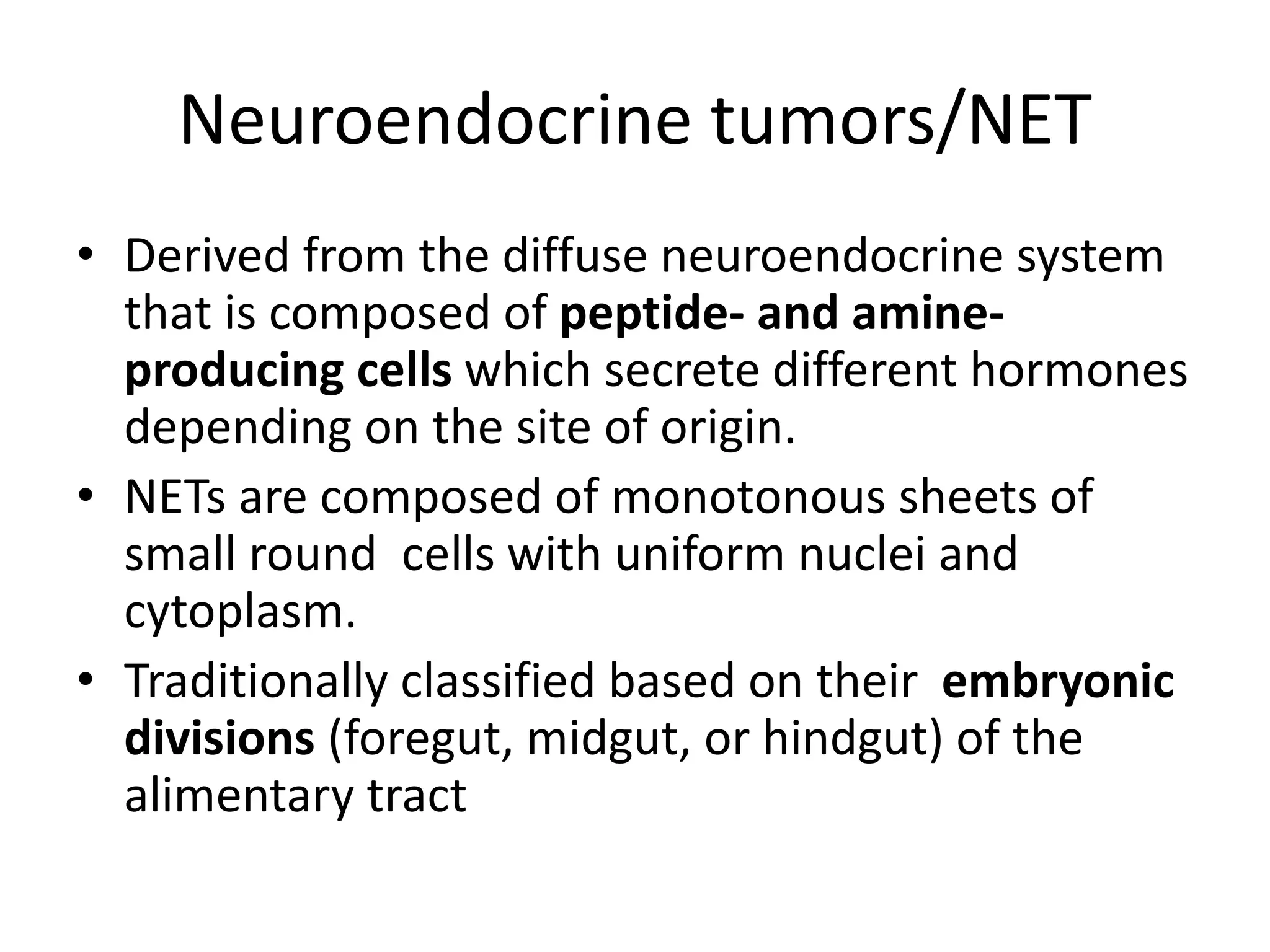
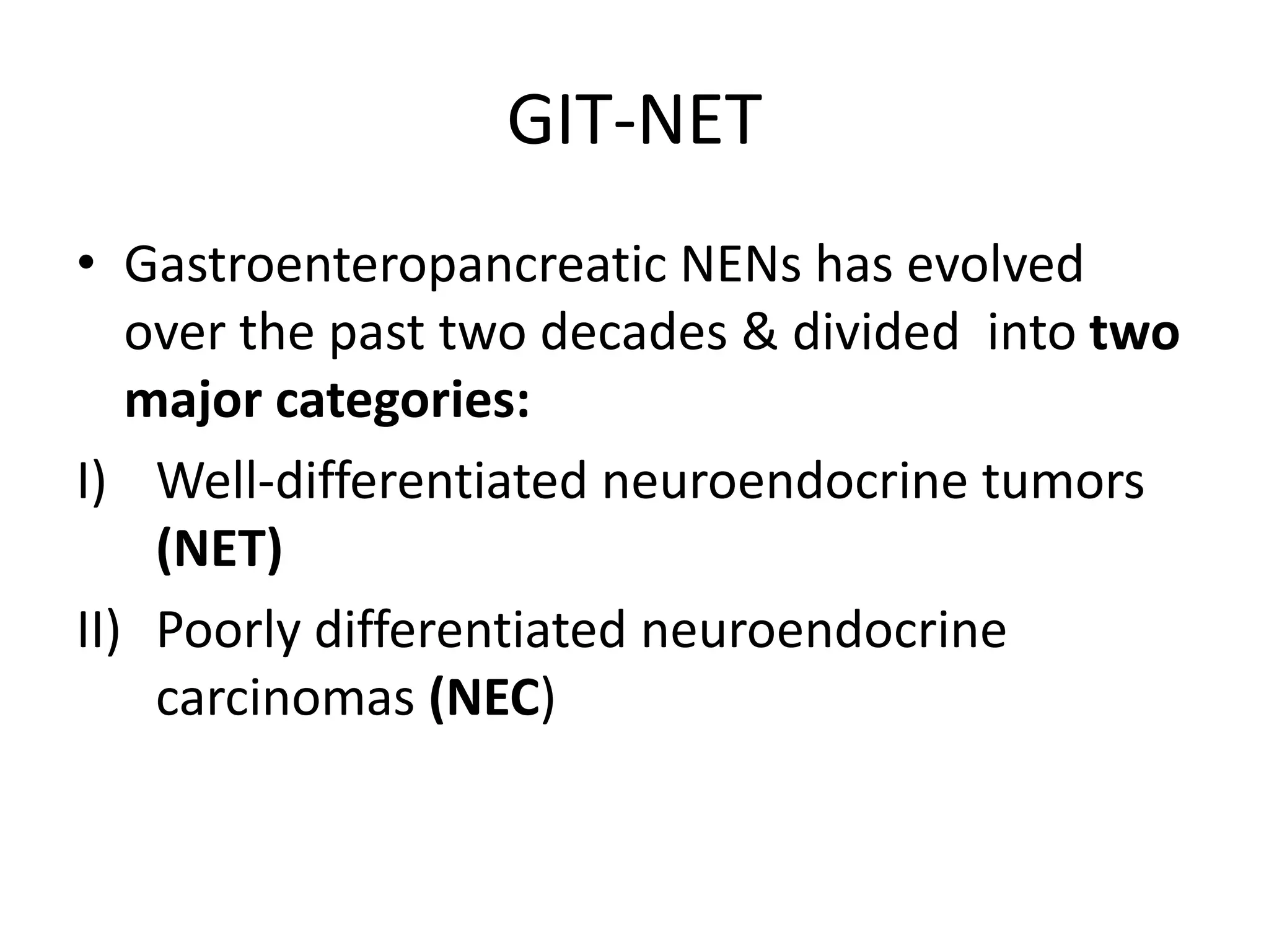
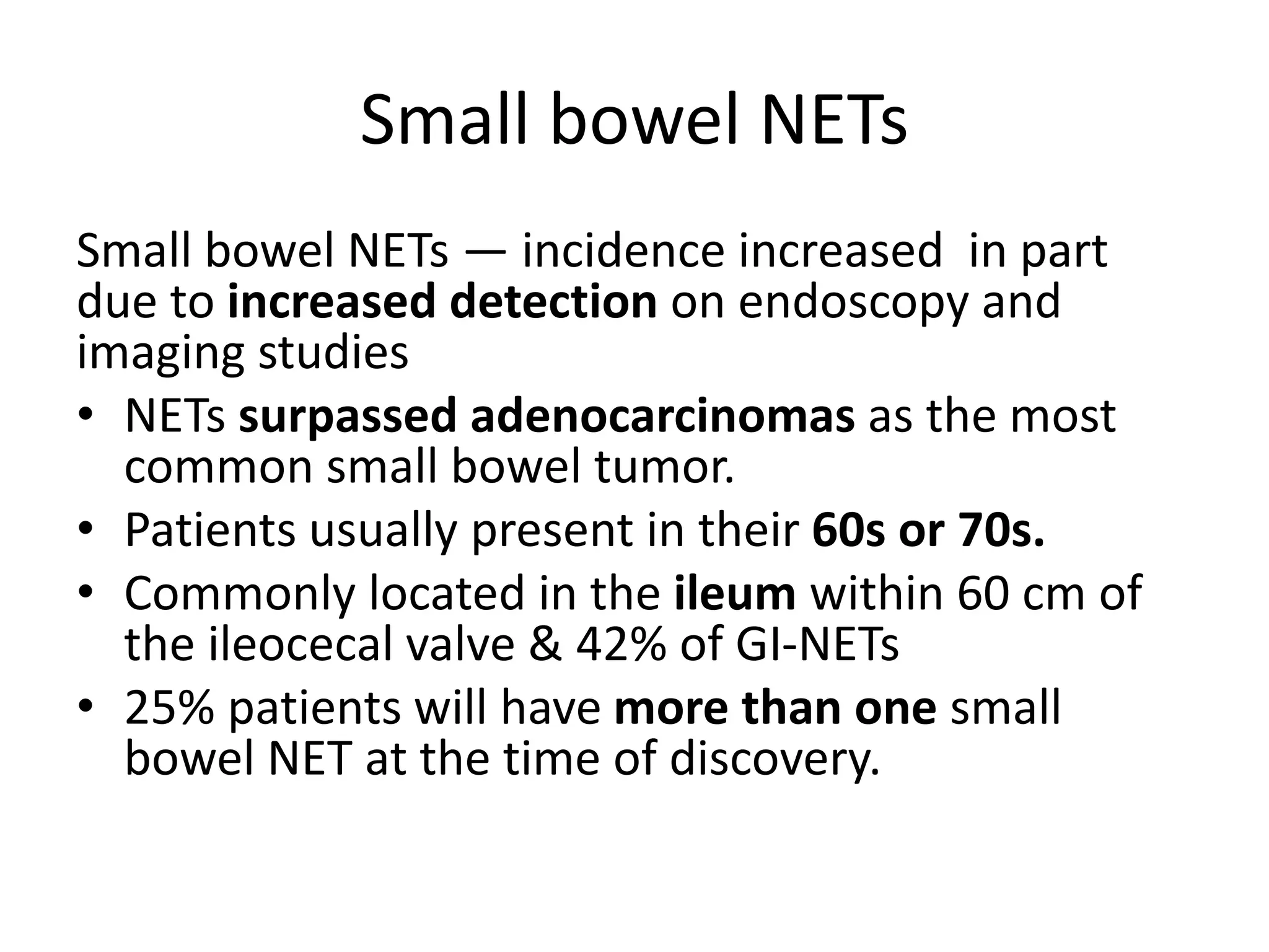
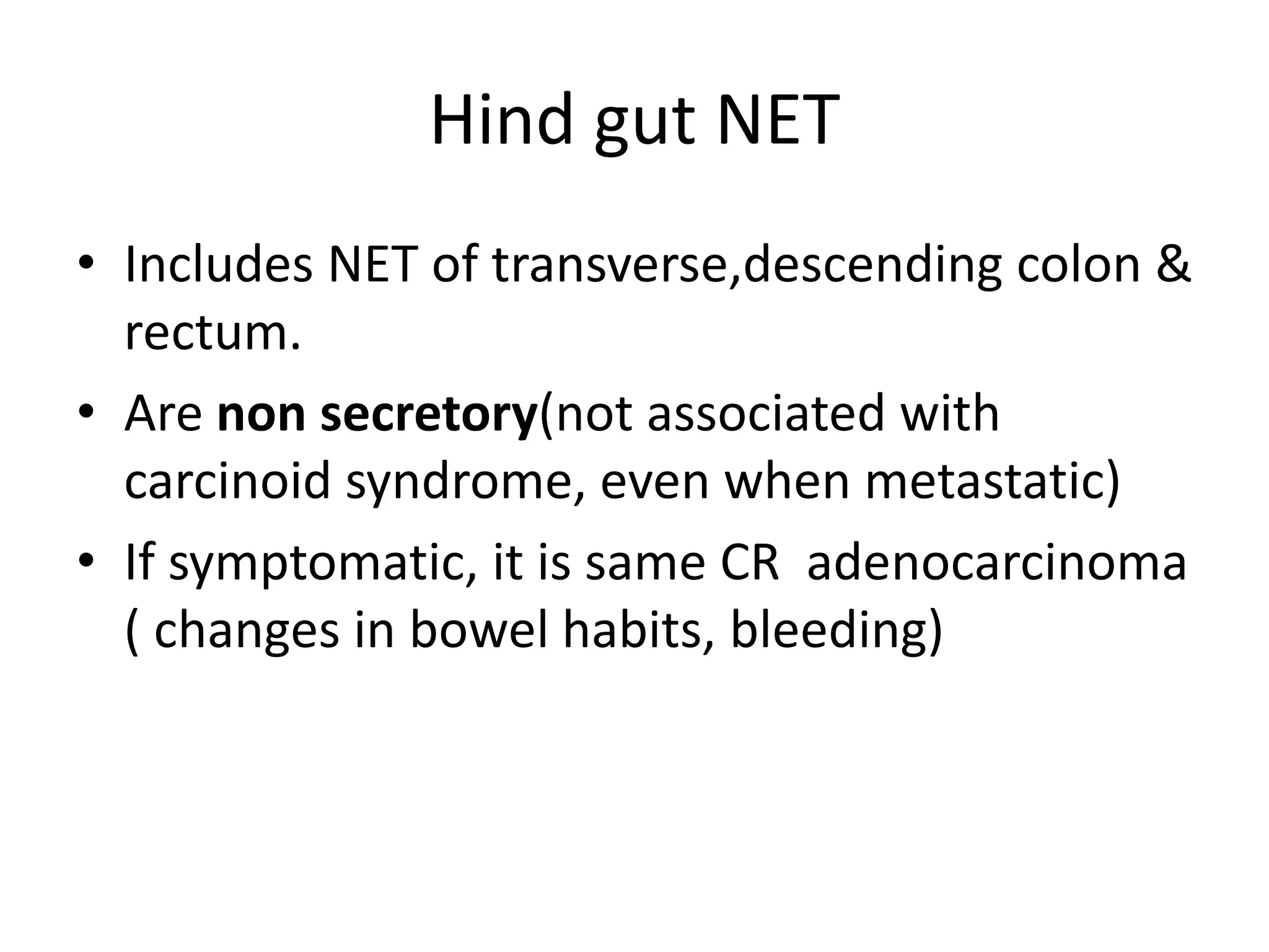
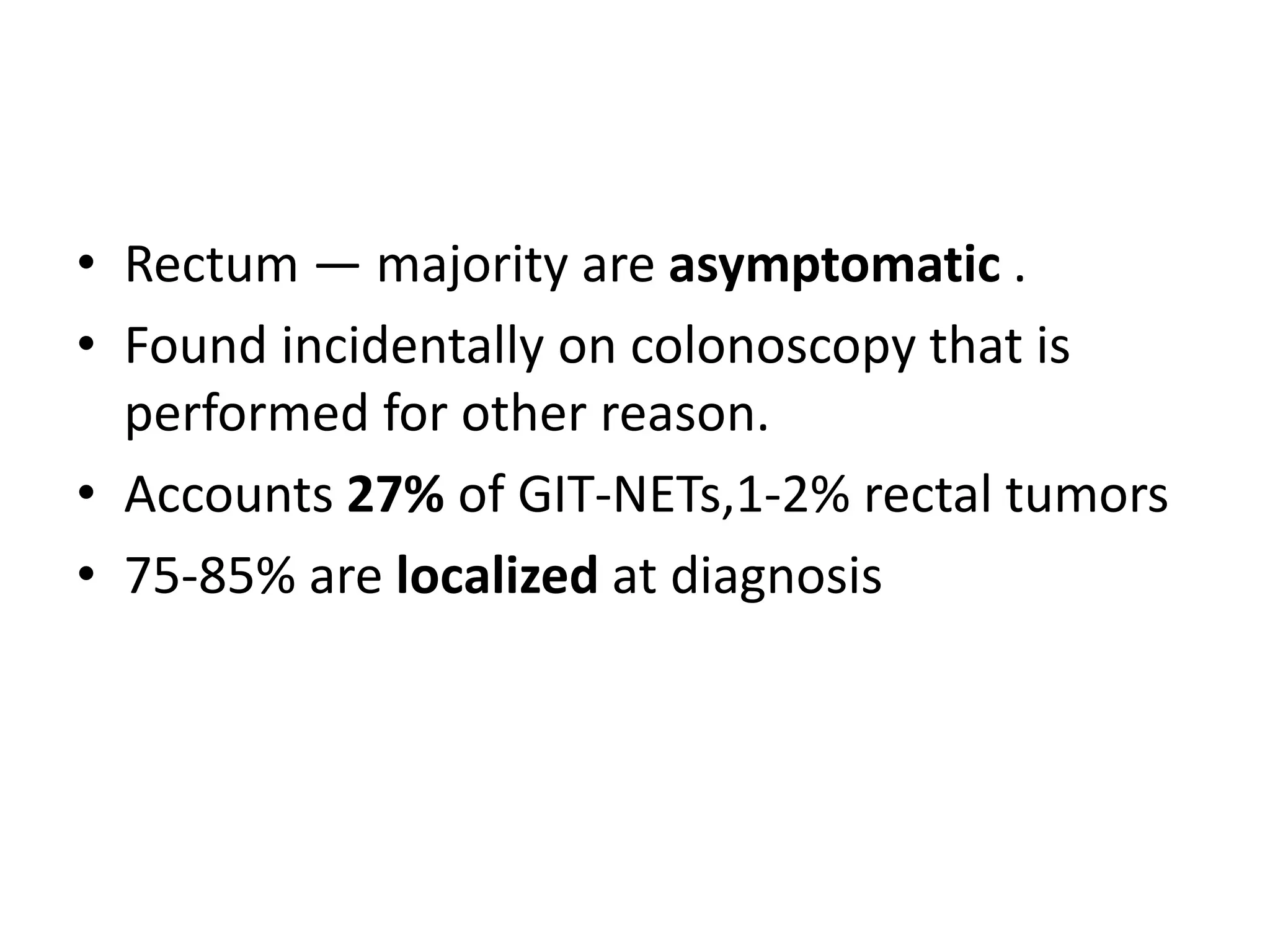
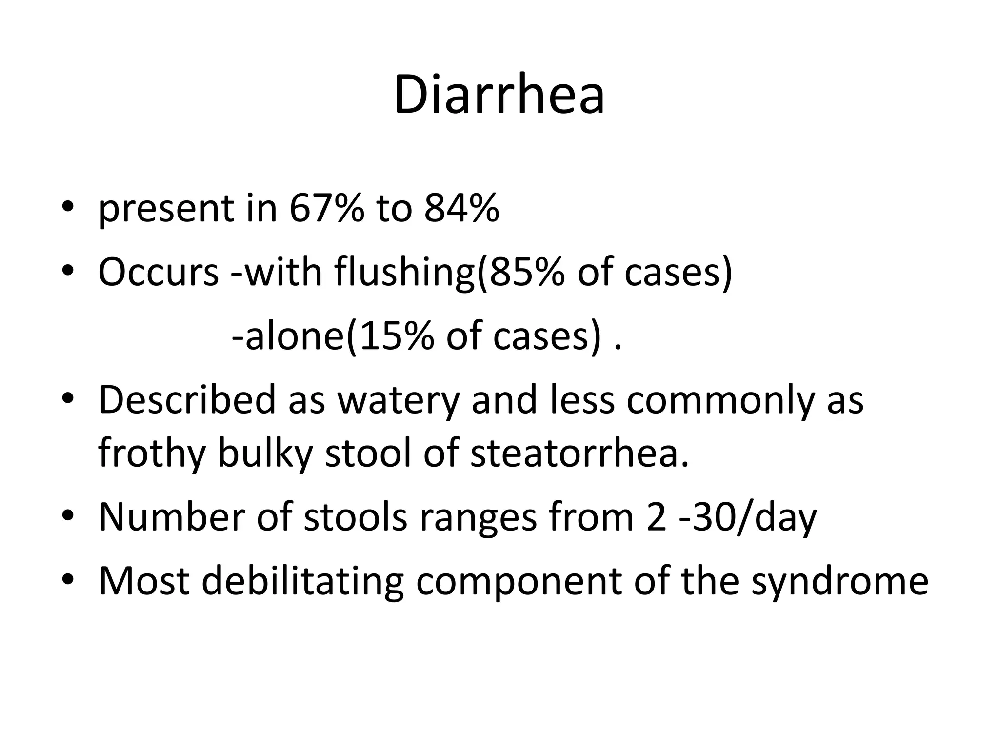
GIT-Neuroendocrine Tumors/NET
Dr Gebrekirstos Hagos
Clinical Oncology R-I
AAU-SoM
October 30,2018
Outlines of presentation:
• Introduction
• NET-Distribution
-classification -Grade
-Epidemiology - Pathophysiology
-Molecular biology - Clinical features
Introduction
• Neuroendocrine cells are cells that receive
neuronal input (neurotransmitter) release
messaging molecules (hormone) to the blood.
• These hormones control many body functions.
• So they brings neural and endocrine
integration,a process know as neuroendocrine
integration.
Introduction…
• Pituitary gland, parathyroid glands and inner
layer of adrenal gland (adrenal medulla) are
almost made up of neuroendocrine cells.
• Other NEC are found scattered through out,
mainly GIT, Respiratory tract and pancreases.
• These NEC found scattered throughout these
organs are referred to as the diffuse
neuroendocrine system
Neuroendocrine cells/NEC
• This diffuse neuroendocrine cells don’t form
an actual organ like the adrenal or thyroid
glands(Instead scattered)
• GIT has more neuroendocrine cells than any
other part of the body so is commonest origin
of NET(64% of NET cases).
Neuroendocrine tumors/NET
• Derived from the diffuse neuroendocrine system
that is composed of peptide- and amine-
producing cells which secrete different hormones
depending on the site of origin.
• NETs are composed of monotonous sheets of
small round cells with uniform nuclei and
cytoplasm.
• Traditionally classified based on their embryonic
divisions (foregut, midgut, or hindgut) of the
alimentary tract
GIT-NET
• Gastroenteropancreatic NENs has evolved
over the past two decades & divided into two
major categories:
I) Well-differentiated neuroendocrine tumors
(NET)
II) Poorly differentiated neuroendocrine
carcinomas (NEC)
GIT-NET…Well differentiated
• Show a solid, trabecular, with fairly uniform
nuclei, coarsely stippled chromatin, and finely
granular cytoplasm.
• Traditionally referred to as carcinoid tumors in
GIT and pancreatic NETs (islet cell tumors) in
pancreases.
GIT-NET…Poorly differentiated
• Are high-grade carcinomas that resemble
small cell or large cell NEC of the lung.
• Often associated with a rapid clinical course,
so bad prognosis compared to well-
differentiated NET.
• Well-differentiated NETs, are further
subdivided into low grade and intermediate
grade based on proliferative rate.
Grading GIT-NET
• WHO classification of GIT-NET is based on
proliferative rate ,divided in to:
- Low-grade(G1) well-
-Intermediate-grade(G2) differentiated
-High-grade tumor(G3) poorly differentiated
Proliferative rate is measured using either
mitotic counts or Ki-67 labeling index.
GIT-NET
• progression from a lower grade (G1 or G2)
well-differentiated NET to a G3 NET can occur.
• Evidence of progression can be:
-increase in the proliferative rate
-change in tumor morphology(↑ nuclear
atypia, or development of significant necrosis)
Molecular pathogenesis
• Little is known about the induction & growth of
carcinoid tumors.
• Gastric NET ↑ hypergastrinemia conditions (pernicious
anemia, atrophic gastritis, ZES).
• other growth factors in some carcinoid tumors are :
- transforming growth factor-alpha
-insulin like growth factor- 1
-VEGF
-acidic and basic fibroblast growth factor
-epidermal growth factor
Epidemiology
• Generally GIT-NET are uncommon, but GIT
common primary site NETs(6.2/100,000)
• Incidence is increasing due to multifactorial
-increased awareness (if you don’t suspect
NET you will not Dx it!)
-improved endoscopic methods detection
Small bowel NETs (midgut carcinoids) are more
common than both foregut and hindgut
Esophageal NET
• Rare (<1% of GI NET)
• Common in men >60 yrs
• Seen in distal esophagus, proximal to GEJ
• On EUS they are sub mucosal mass, dimple
the mucosa.
Stomach-NET
• Subdivided into three categories which have different
biologic behavior and prognosis
• Type 1 -70-80 % all gastric NETs
-associated with chronic atrophic gastritis
-more common in women
-derived from enterochromaffin-like (ECL) cells
-non functioning
- Endoscopically →<1 cm
→multiple
→ polypoid lesions
→ small central ulceration
• Type 2 — associated with Zollinger-Ellison
syndrome, in the setting of MEN-1
-6-8% gastric NETs
-Men=women
-develop in a hyperplasia-dysplasia-neoplasia
sequence
-arise from ECL cells
-stimulated by elevated serum gastrin
levels
Gastric NET
• Type 3
-sporadic, no association with hypergastrinemia.
-15-20% of gastric NETs
-solitary, and grow more rapidly.
-Present with metastasis at Dx
-M:F,3:1
Small bowel NETs
Small bowel NETs — incidence increased in part
due to increased detection on endoscopy and
imaging studies
• NETs surpassed adenocarcinomas as the most
common small bowel tumor.
• Patients usually present in their 60s or 70s.
• Commonly located in the ileum within 60 cm of
the ileocecal valve & 42% of GI-NETs
• 25% patients will have more than one small
bowel NET at the time of discovery.
Small bowel NET…
• Many are asymptomatic at presentation and
are found incidentally.
• If symptomatic ,abdominal pain(51%) followed
bowel obstruction(31%),abdominal
mass(17)and GI bleeding(11%).
• Metastases to LN/liver are common, even if
the primary tumor is <2 cm in size.
• Commonly presented with carcinoid
syndrome.
Small bowel NET…
• Pts with metastatic NET with unknown
primary, they can harbor occult primary in
the ileum.
• So exploration and palpation of the bowel
can allow detection of the sub mucosal
primary which feel like little pea in the bowel
wall.
• Appendix — NETs are the most common
neoplasms in the appendix.
• NET is discovered incidentally (1 in 300
appendectomies, commonly at the tip).
• Common at age of 40s or 50s
• more common in women
Hind gut NET
• Includes NET of transverse,descending colon &
rectum.
• Are non secretory(not associated with
carcinoid syndrome, even when metastatic)
• If symptomatic, it is same CR adenocarcinoma
( changes in bowel habits, bleeding)
• Colon — in pts at their 70s during evaluation
for abdominal pain, anorexia, or weight loss.
• Incidence of functioning tumors is very low.
• More on Rt side of colon.
• Rectum — majority are asymptomatic .
• Found incidentally on colonoscopy that is
performed for other reason.
• Accounts 27% of GIT-NETs,1-2% rectal tumors
• 75-85% are localized at diagnosis
METASTATIC TUMORS
• Regardless of primary site, NETs are
characterized by metastasize to the liver.
• Patients with liver metastases may experience
symptoms related to tumor burden (eg, pain,
jaundice, early satiety) or hormonal symptoms
(eg, flushing and diarrhea, the main symptoms
of carcinoid syndrome).
PATHOPHYSIOLOGY
• Around 40 secretory products have been
identified in various NETs.
• The most prominent of these are:
-Serotonin - Tachykinins
-Histamine -Kallikrein
-Prostaglandin
Tryptophan metabolism
• Altered metabolism of tryptophan occurs in
almost all patients with the carcinoid
syndrome.
• In normal subjects, approximately 1 % of
dietary tryptophan is converted to serotonin;
(↑ to 70% in pts with carcinoid syndrome).
• Serotonin is then metabolized to 5-
hydroxyindoleacetic acid (5-HIAA)
Clinical features
• Can occur at any age, media age 63
• M:F, 48:52%
• Dx delayed for average 2yrs (can range for 20
yrs) due vague symptoms.
• The clinical presentation of carcinoid tumors far
underestimates their occurrence because many
are asymptomatic
• Symptoms are caused by local tumor growth,
metastatic spread or excess hormonal.
Carcinoid syndrome
• Symptoms mediated by various humoral factors.
Common in setting of liver metastasis b/c liver
failed to deactivate the products.
• Common mid gut, but lung & stomach NET
present with atypical carcinoid syndrome.
Flushing attack
Diarrhea
Cardiac manifestation
Wheezing and asthma like symptom
Retroperitoneal & intra-abdominal fibrosis
Cutaneous flushing
• Episodic flushing is the clinical hallmark of
carcinoid syndrome .
• occurs in 85 % of patients.
• Typical flush associated with midgut NET
• Begins suddenly and lasts for 30 sec to 30 min.
• Involves the face, neck, and upper chest,
which become red to violaceous or purple.
Cutaneous flush…
• Most episodes occur
spontaneously, but they
can be provoked by eating,
drinking alcohol, defecation,
emotional events, palpation
of the liver, and anesthesia.
• Carcinoid crisis:
Diarrhea
• present in 67% to 84%
• Occurs -with flushing(85% of cases)
-alone(15% of cases) .
• Described as watery and less commonly as
frothy bulky stool of steatorrhea.
• Number of stools ranges from 2 -30/day
• Most debilitating component of the syndrome
Cardiac valvular lesions
• occur i n 11-66%
• Carcinoid heart disease is characterized by
pathognomonic plaque-like deposits of fibrous
tissue.
• Common on right side of the heart because
inactivation of humoral substances by the lung
protects the left heart.
• Bronchospasm-10-20% of pts with the carcinoid
syndrome have wheezing and dyspnea, often
during flushing episodes.
References:
• Devita ------9th & 10th e
• Uptodate --2018
• NCCN------2017
Thank You!

gastrointestinal Neuro endocrine tumors , GIT NET

  • 1.
    GIT-Neuroendocrine Tumors/NET Dr GebrekirstosHagos Clinical Oncology R-I AAU-SoM October 30,2018
  • 2.
    Outlines of presentation: •Introduction • NET-Distribution -classification -Grade -Epidemiology - Pathophysiology -Molecular biology - Clinical features
  • 3.
    Introduction • Neuroendocrine cellsare cells that receive neuronal input (neurotransmitter) release messaging molecules (hormone) to the blood. • These hormones control many body functions. • So they brings neural and endocrine integration,a process know as neuroendocrine integration.
  • 4.
    Introduction… • Pituitary gland,parathyroid glands and inner layer of adrenal gland (adrenal medulla) are almost made up of neuroendocrine cells. • Other NEC are found scattered through out, mainly GIT, Respiratory tract and pancreases. • These NEC found scattered throughout these organs are referred to as the diffuse neuroendocrine system
  • 5.
  • 6.
    • This diffuseneuroendocrine cells don’t form an actual organ like the adrenal or thyroid glands(Instead scattered) • GIT has more neuroendocrine cells than any other part of the body so is commonest origin of NET(64% of NET cases).
  • 7.
    Neuroendocrine tumors/NET • Derivedfrom the diffuse neuroendocrine system that is composed of peptide- and amine- producing cells which secrete different hormones depending on the site of origin. • NETs are composed of monotonous sheets of small round cells with uniform nuclei and cytoplasm. • Traditionally classified based on their embryonic divisions (foregut, midgut, or hindgut) of the alimentary tract
  • 9.
    GIT-NET • Gastroenteropancreatic NENshas evolved over the past two decades & divided into two major categories: I) Well-differentiated neuroendocrine tumors (NET) II) Poorly differentiated neuroendocrine carcinomas (NEC)
  • 10.
    GIT-NET…Well differentiated • Showa solid, trabecular, with fairly uniform nuclei, coarsely stippled chromatin, and finely granular cytoplasm. • Traditionally referred to as carcinoid tumors in GIT and pancreatic NETs (islet cell tumors) in pancreases.
  • 11.
    GIT-NET…Poorly differentiated • Arehigh-grade carcinomas that resemble small cell or large cell NEC of the lung. • Often associated with a rapid clinical course, so bad prognosis compared to well- differentiated NET.
  • 12.
    • Well-differentiated NETs,are further subdivided into low grade and intermediate grade based on proliferative rate.
  • 13.
    Grading GIT-NET • WHOclassification of GIT-NET is based on proliferative rate ,divided in to: - Low-grade(G1) well- -Intermediate-grade(G2) differentiated -High-grade tumor(G3) poorly differentiated Proliferative rate is measured using either mitotic counts or Ki-67 labeling index.
  • 15.
    GIT-NET • progression froma lower grade (G1 or G2) well-differentiated NET to a G3 NET can occur. • Evidence of progression can be: -increase in the proliferative rate -change in tumor morphology(↑ nuclear atypia, or development of significant necrosis)
  • 16.
    Molecular pathogenesis • Littleis known about the induction & growth of carcinoid tumors. • Gastric NET ↑ hypergastrinemia conditions (pernicious anemia, atrophic gastritis, ZES). • other growth factors in some carcinoid tumors are : - transforming growth factor-alpha -insulin like growth factor- 1 -VEGF -acidic and basic fibroblast growth factor -epidermal growth factor
  • 17.
    Epidemiology • Generally GIT-NETare uncommon, but GIT common primary site NETs(6.2/100,000) • Incidence is increasing due to multifactorial -increased awareness (if you don’t suspect NET you will not Dx it!) -improved endoscopic methods detection Small bowel NETs (midgut carcinoids) are more common than both foregut and hindgut
  • 18.
    Esophageal NET • Rare(<1% of GI NET) • Common in men >60 yrs • Seen in distal esophagus, proximal to GEJ • On EUS they are sub mucosal mass, dimple the mucosa.
  • 19.
    Stomach-NET • Subdivided intothree categories which have different biologic behavior and prognosis • Type 1 -70-80 % all gastric NETs -associated with chronic atrophic gastritis -more common in women -derived from enterochromaffin-like (ECL) cells -non functioning - Endoscopically →<1 cm →multiple → polypoid lesions → small central ulceration
  • 21.
    • Type 2— associated with Zollinger-Ellison syndrome, in the setting of MEN-1 -6-8% gastric NETs -Men=women -develop in a hyperplasia-dysplasia-neoplasia sequence -arise from ECL cells -stimulated by elevated serum gastrin levels
  • 22.
    Gastric NET • Type3 -sporadic, no association with hypergastrinemia. -15-20% of gastric NETs -solitary, and grow more rapidly. -Present with metastasis at Dx -M:F,3:1
  • 23.
    Small bowel NETs Smallbowel NETs — incidence increased in part due to increased detection on endoscopy and imaging studies • NETs surpassed adenocarcinomas as the most common small bowel tumor. • Patients usually present in their 60s or 70s. • Commonly located in the ileum within 60 cm of the ileocecal valve & 42% of GI-NETs • 25% patients will have more than one small bowel NET at the time of discovery.
  • 24.
    Small bowel NET… •Many are asymptomatic at presentation and are found incidentally. • If symptomatic ,abdominal pain(51%) followed bowel obstruction(31%),abdominal mass(17)and GI bleeding(11%). • Metastases to LN/liver are common, even if the primary tumor is <2 cm in size. • Commonly presented with carcinoid syndrome.
  • 25.
    Small bowel NET… •Pts with metastatic NET with unknown primary, they can harbor occult primary in the ileum. • So exploration and palpation of the bowel can allow detection of the sub mucosal primary which feel like little pea in the bowel wall.
  • 26.
    • Appendix —NETs are the most common neoplasms in the appendix. • NET is discovered incidentally (1 in 300 appendectomies, commonly at the tip). • Common at age of 40s or 50s • more common in women
  • 27.
    Hind gut NET •Includes NET of transverse,descending colon & rectum. • Are non secretory(not associated with carcinoid syndrome, even when metastatic) • If symptomatic, it is same CR adenocarcinoma ( changes in bowel habits, bleeding)
  • 28.
    • Colon —in pts at their 70s during evaluation for abdominal pain, anorexia, or weight loss. • Incidence of functioning tumors is very low. • More on Rt side of colon.
  • 29.
    • Rectum —majority are asymptomatic . • Found incidentally on colonoscopy that is performed for other reason. • Accounts 27% of GIT-NETs,1-2% rectal tumors • 75-85% are localized at diagnosis
  • 30.
    METASTATIC TUMORS • Regardlessof primary site, NETs are characterized by metastasize to the liver. • Patients with liver metastases may experience symptoms related to tumor burden (eg, pain, jaundice, early satiety) or hormonal symptoms (eg, flushing and diarrhea, the main symptoms of carcinoid syndrome).
  • 32.
    PATHOPHYSIOLOGY • Around 40secretory products have been identified in various NETs. • The most prominent of these are: -Serotonin - Tachykinins -Histamine -Kallikrein -Prostaglandin
  • 34.
    Tryptophan metabolism • Alteredmetabolism of tryptophan occurs in almost all patients with the carcinoid syndrome. • In normal subjects, approximately 1 % of dietary tryptophan is converted to serotonin; (↑ to 70% in pts with carcinoid syndrome). • Serotonin is then metabolized to 5- hydroxyindoleacetic acid (5-HIAA)
  • 36.
    Clinical features • Canoccur at any age, media age 63 • M:F, 48:52% • Dx delayed for average 2yrs (can range for 20 yrs) due vague symptoms. • The clinical presentation of carcinoid tumors far underestimates their occurrence because many are asymptomatic • Symptoms are caused by local tumor growth, metastatic spread or excess hormonal.
  • 37.
    Carcinoid syndrome • Symptomsmediated by various humoral factors. Common in setting of liver metastasis b/c liver failed to deactivate the products. • Common mid gut, but lung & stomach NET present with atypical carcinoid syndrome. Flushing attack Diarrhea Cardiac manifestation Wheezing and asthma like symptom Retroperitoneal & intra-abdominal fibrosis
  • 38.
    Cutaneous flushing • Episodicflushing is the clinical hallmark of carcinoid syndrome . • occurs in 85 % of patients. • Typical flush associated with midgut NET • Begins suddenly and lasts for 30 sec to 30 min. • Involves the face, neck, and upper chest, which become red to violaceous or purple.
  • 39.
    Cutaneous flush… • Mostepisodes occur spontaneously, but they can be provoked by eating, drinking alcohol, defecation, emotional events, palpation of the liver, and anesthesia. • Carcinoid crisis:
  • 40.
    Diarrhea • present in67% to 84% • Occurs -with flushing(85% of cases) -alone(15% of cases) . • Described as watery and less commonly as frothy bulky stool of steatorrhea. • Number of stools ranges from 2 -30/day • Most debilitating component of the syndrome
  • 41.
    Cardiac valvular lesions •occur i n 11-66% • Carcinoid heart disease is characterized by pathognomonic plaque-like deposits of fibrous tissue. • Common on right side of the heart because inactivation of humoral substances by the lung protects the left heart. • Bronchospasm-10-20% of pts with the carcinoid syndrome have wheezing and dyspnea, often during flushing episodes.
  • 43.
    References: • Devita ------9th& 10th e • Uptodate --2018 • NCCN------2017
  • 44.