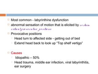
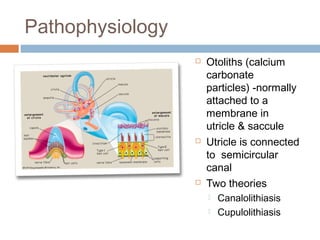
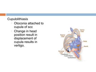
2. Most common - labyrinthine dysfunction
abnormal sensation of motion that is elicited by c e rta in
c ritic a l p ro vo c a tive p o s itio ns
Provocative positions
Head turn to affected side - getting out of bed
Extend head back to look up “Top shelf vertigo”
Causes
Idiopathic – 50%
Head trauma, middle ear infection, viral labyrinthitis,
ear surgery
3. Pathophysiology
Otoliths (calcium
carbonate
particles) -normally
attached to a
membrane in
utricle & saccule
Utricle is connected
to semicircular
canal
Two theories
Canalolithiasis
Cupulolithiasis
4. Canalolithiasis
Otoliths displaced from utricle
- enter the posterior
semicircular duct (most
dependent SCC )
Changing head position
relative to gravity causes the
free otoliths to gravitate
through the canal.
The concurrent flow of
endolymph stimulates the hair
cells of the affected
semicircular canal causing
vertigo.
5. Cupulolithiasis
Otoconia attached to
cupula of scc
Change in head
position result in
displacement of
cupula results in
vertigo.
6. Sixth decade
F>M
Clinical features
Sudden onset rotatory vertigo
Few secs
Triggered by provocative movements
No other aural symptoms
7. Dix-Hallpike maneuvre
Pt seats on the table
Pt’s head held, turned 45
deg to Rt & pt placed at
supine position – head
hangs 30 deg below
horizontal
Pt’s eyes observed for
nystagmus
Test repeated on Lt side
8. Comparision of positional nystagmus of BPPV
with lesions of the CNS
BPPV CNS
Latent period A few seconds nil
Distress Present nil
Direction of
Direction fixed – towards
nystagmus
the undermost ear
Direction changing
Duration of
nystagmus
Less than 30 sec Persists while
position
maintained
Fatiguablity Nystagmus stop with
repeated testing
Nystagmus
persists with
repeated testing
10. (S) Start: patient is seated
(1) Place head over end of table, 45 degrees to left.
(2) Keeping head tilted downward, rotate to 45 degrees
right.
(3) Rotate head and body until facing downward 135
degrees from supine.
(4) Keeping head turned right, bring patient to sitting
position.
(5) Turn head forward, chin down 20 degrees.
Pause at each position until nystagmus approaches
termination
11. Instructions following Epley’s maneuvre
Rest 10 min
Sleep in semi-recumbent
For at least 1 week
Use two pillows
Avoid bad side
No head turning far
up or down
12. Surgical mangement
Posterior canal wall
plugging
debris can no longer
move within the canal
Singular nerve
section
Section the nerve that
transmits information
from the posterior
semicircular canal
ampulla toward the
brain.
19. Prolonged – weeks to months
Late stage of vestibular neuritis, acute
labyrinthitis
Elderly patients
Drugs
Anticonvulsants, Gentamicin
Vestibular schwannoma
Functional
20. Examination
ENT
Nystagmus
Involuntary, rhythmical, oscillatory movement of
eyes
Slow / fast component – direction of the
nystagmus
Procedure
Examiner keeps finger about 30 cm from the
patients eyes in the central position & moves
it right or left
Do not exceed 30 degree from the centre
Enhanced with Frenzel glasses or in darkness
21. Otoscopic examination & Tuning fork test
Fistula test
Induce nystagmus - pressure changes in the
external ear which are then transmitted to labyrinth
pressure induced by
Intermittent pressure over the tragus
Siegel’s pneumatic speculum
22. Fistula test negative – normal
Fistula test positive
Labyrinthine fistula
Perilymph fistula
Post stapedectomy fistula
False negative fistula test
Cholesteatoma covering the fistula
False positive fistula test ( positive fistula test in
absence of fistula)
Meniere’s disease ( Hennebert’s sign)
23. Cranial nerves
Cerebellar function
Gait
Romberg’s test
Dysmetria
Dysdiadokokinesia
24. Management
Investigations
Audiomety
Caloric test
Induce nystagmus by thermal
stimulation of vestibular system
Bithermal caloric test
Supine, head tilted forward 30 deg
Ears irrigated with water
40 sec
Alternately with water at 30
& 44 deg C
25. Time taken from irrigation to end of nystagmus
charted on calorigram
Cold water – nystagmus to opposite side
Warm water – nystagmus to same side
(COWS)
Depending upon the response to caloric test
Canal paresis – depressed function of ipsilateral
labyrinth, vestibular nerve, vestibular nuclei
Directional preponderance – peripheral and central
lesion
26. Electronystagmography
Method of detecting & recording nystagmus
Rotational chair test
Computerized dynamic posturography
27. Treatment
Suppress vestibular symptoms
Wait for vestibular compensation
Treat the underlying cause
Medical
Surgical