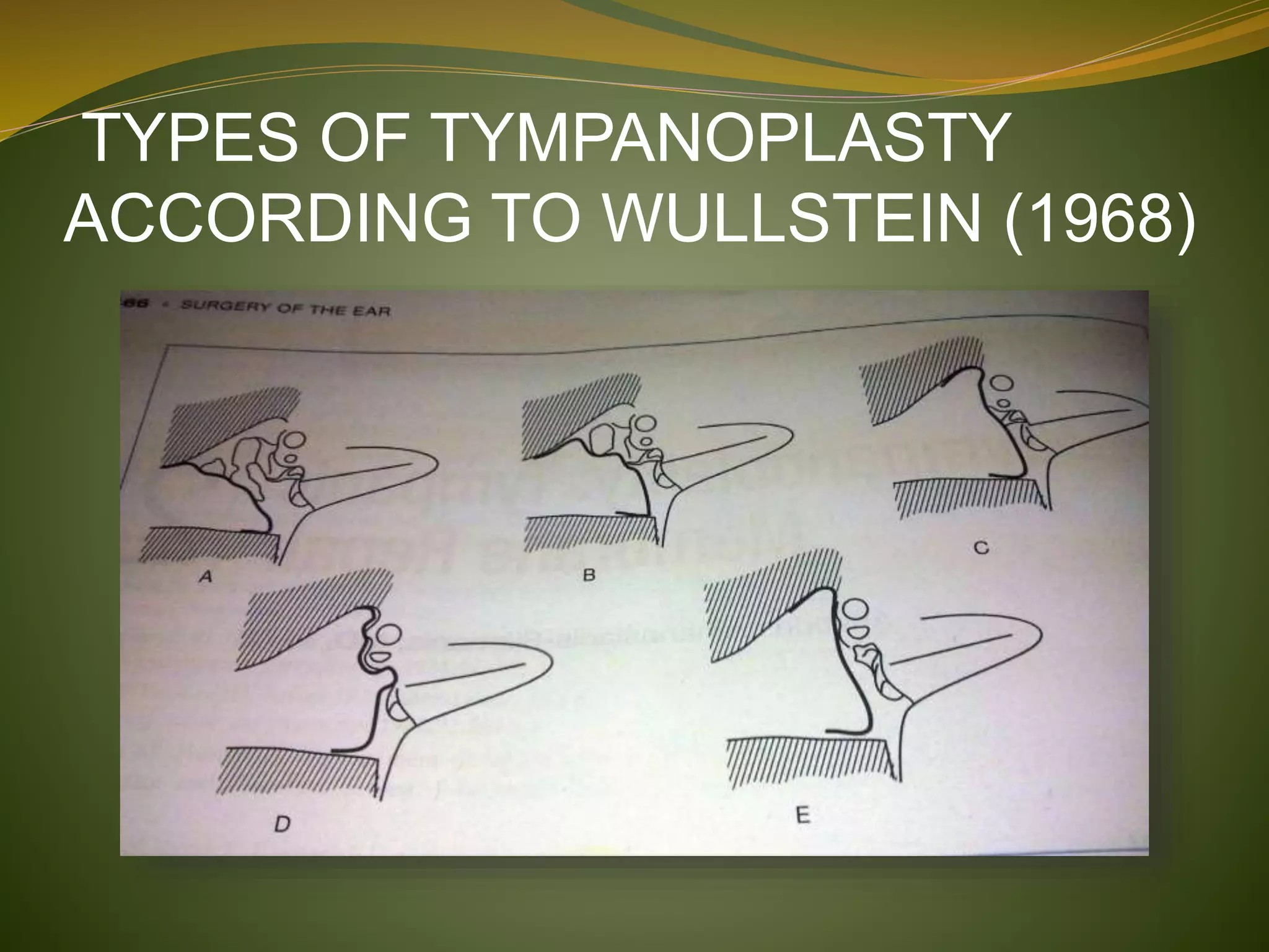
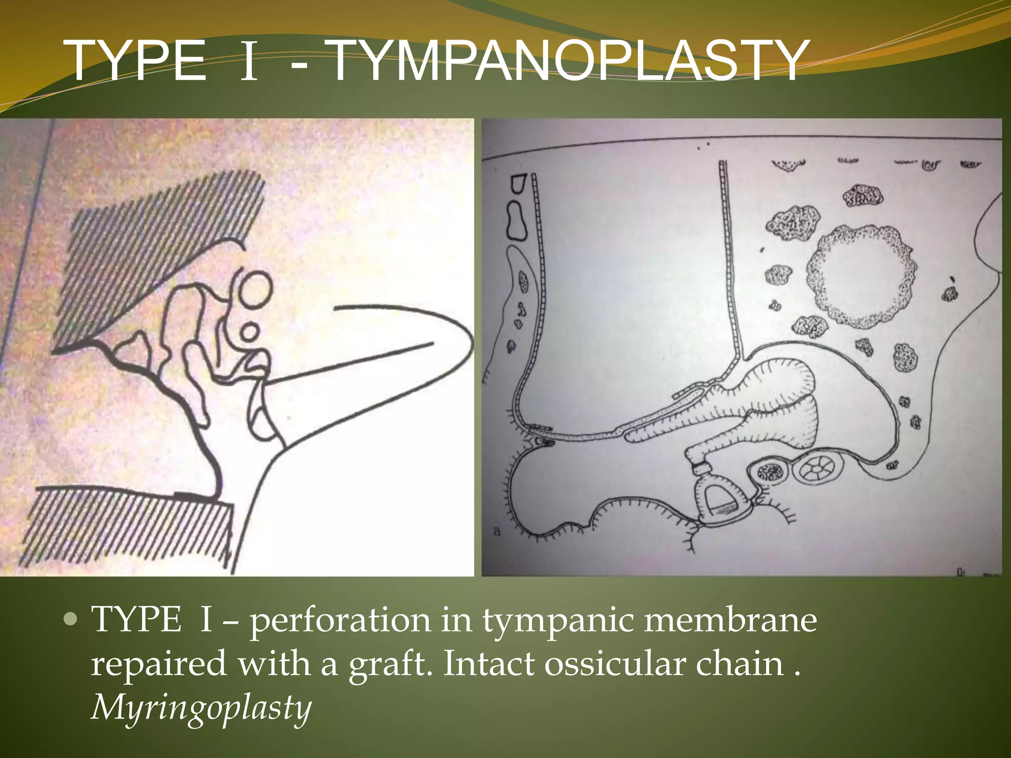
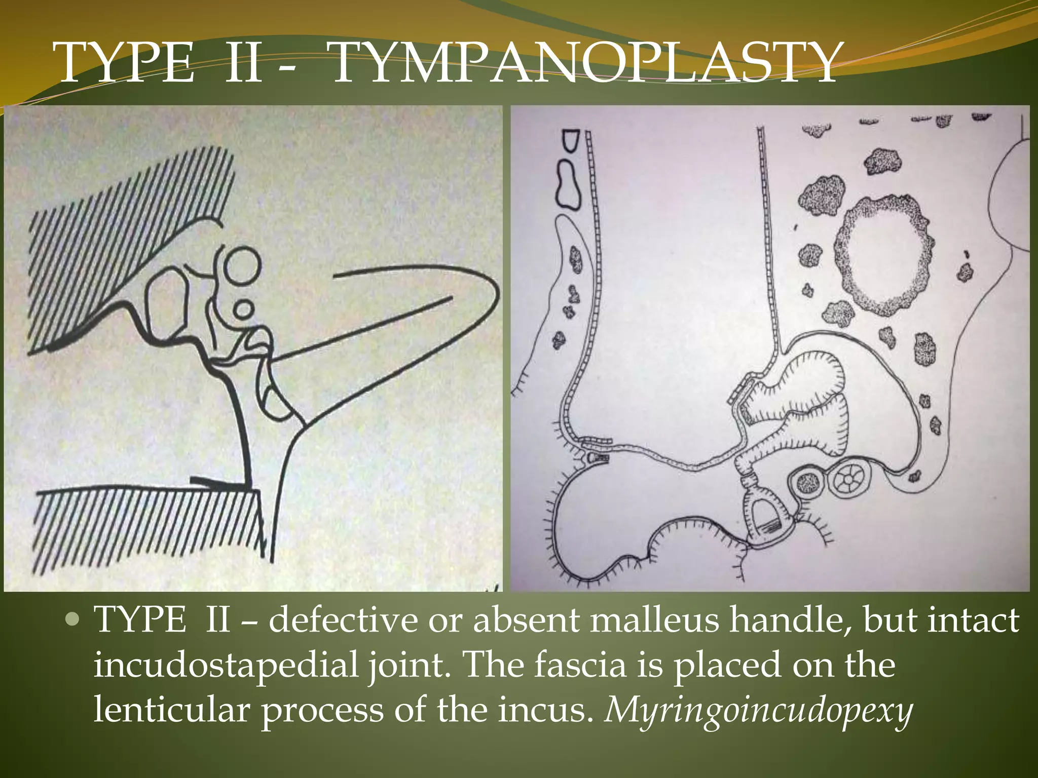
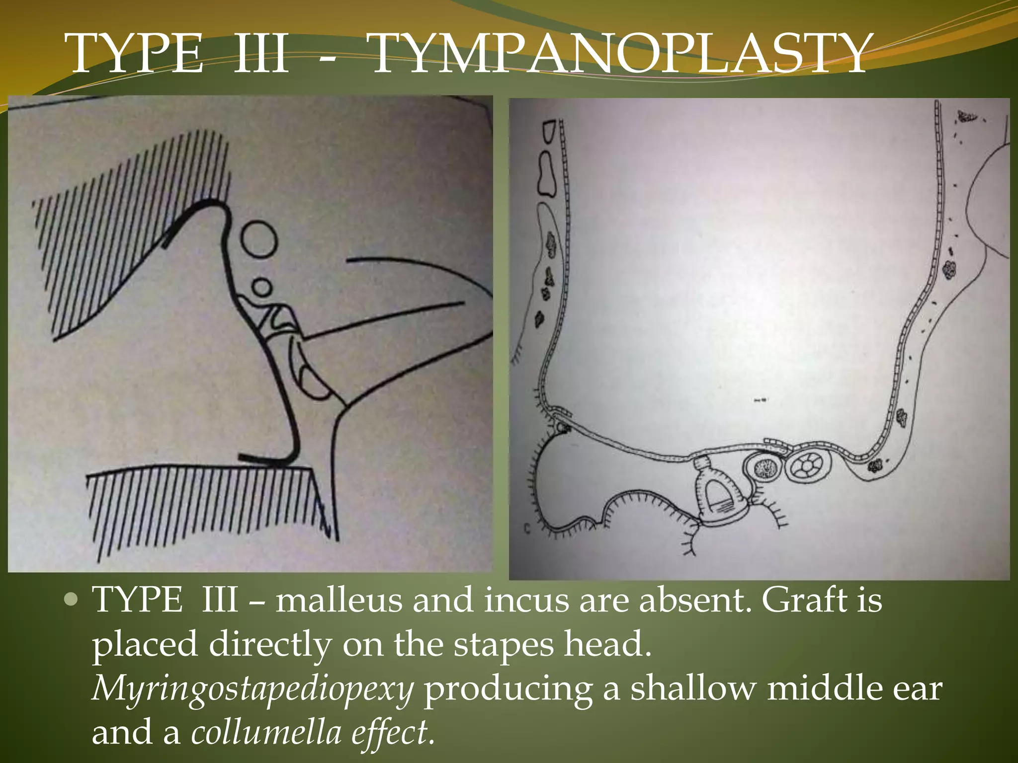
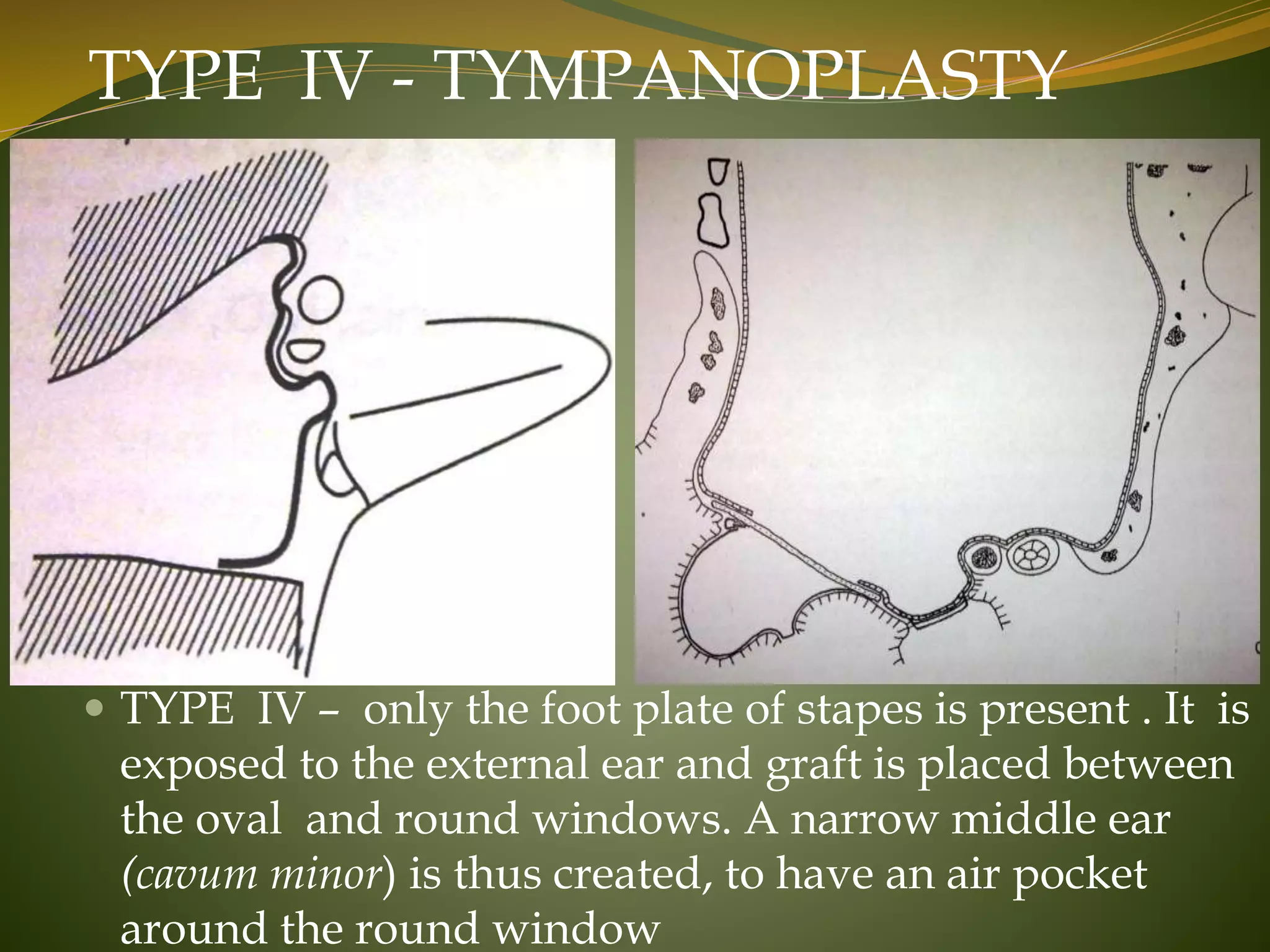
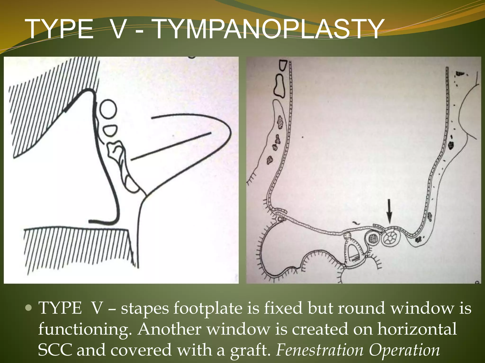
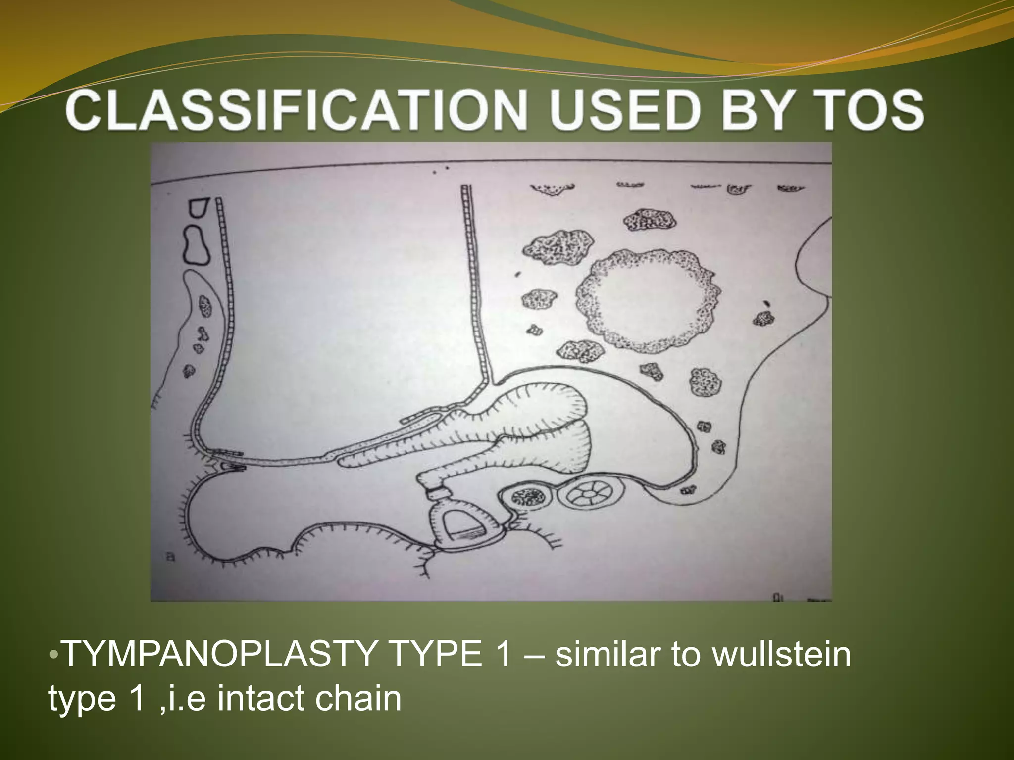
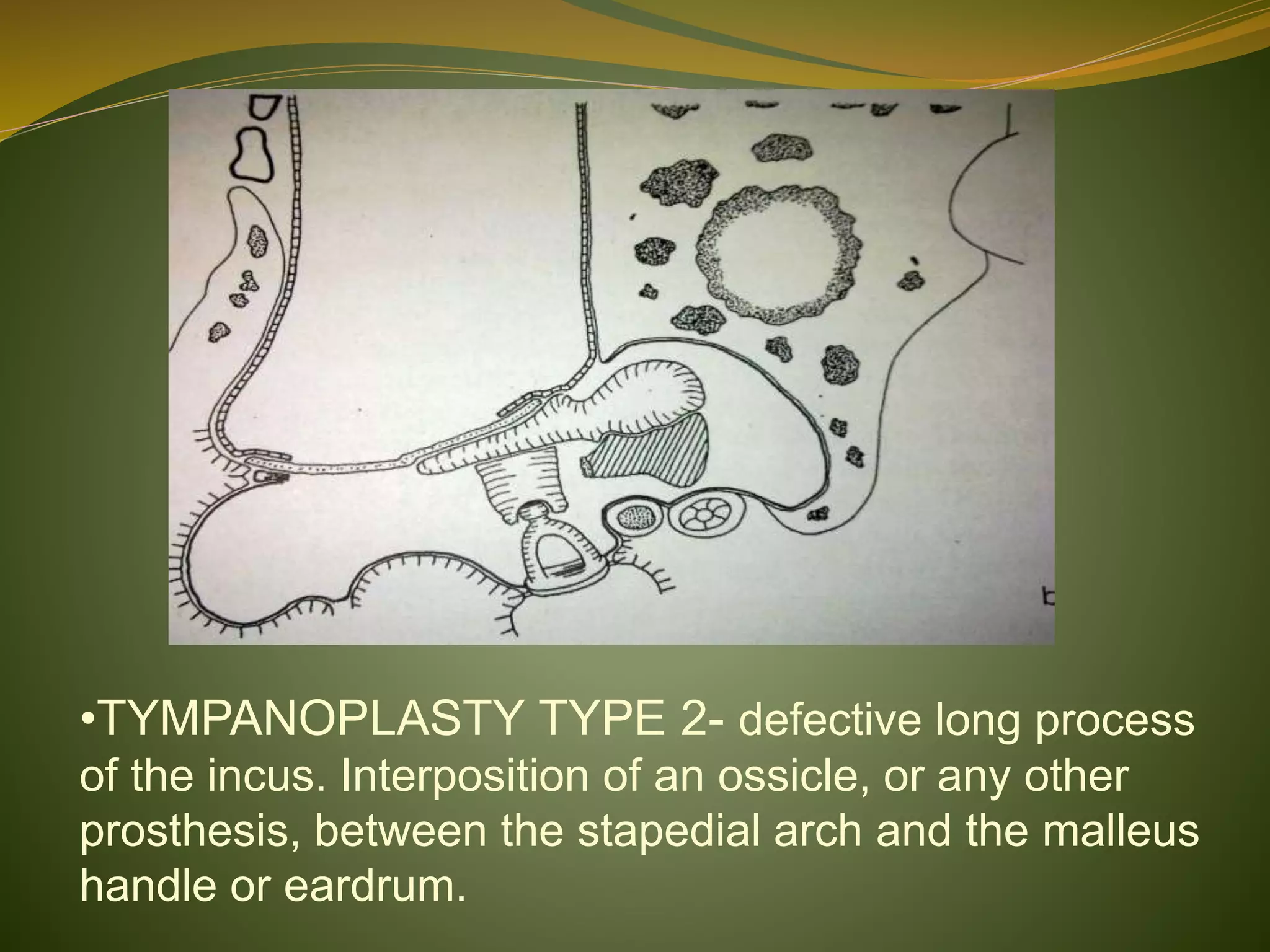
DR.PRASANNA DATTA
DEFINITION
TYMPANOPLASTY
 According to the American Academy of
Opthalmology and Otolaryngology Subcommittee on
Conversation of Hearing 1965 definition,
tympanoplasty is “ a procedure to eradicate
disease in the middle ear and to reconstruct the
hearing mechanism, with or without tympanic
membrane grafting “
TYMPANOPLASTY includes :
• Canalplasty (widening of bony part of the external
auditory canal)
• Myringoplasty (closure of the eardrum perforation in
cases with a normal ossicular chain and without any other
surgical procedures in the tympanic cavity or middle ear)
• Ossiculoplasty (reconstruction of ossicular chain)
HISTORY
 WULLSTEIN – in 1953 introduced the term
“TYMPANOPLASTY”
 KESSEL – in 1878 did stapes mobilization
 BERTHOLD – in 1878 plastic repair of tympanic
membrane
 SOURDILLE – tympanolabyrinthopexy for
otosclerosis
 MORITZ – in 1950 described use of pedicled flaps
to construct a closed middle ear cavity in cases of
chronic suppuration to provide sound shielding for
round window in preparation for later fenestration of
horizontal SCC
HISTORY
 WULLSTEIN – advocated free skin transplants rather
than the pedicled grafts used by MORITZ,
 ZOLLNER –soon after changed from pedicled to free
grafts as well. He replaced free distant skin graft with
meatal skin.
 SHEA AND TABB – in 1960 reported vein as grafting
material
 HEERMANN – 1961 described temporalis fascia as
grafting material
 STORRS – in 1963 introduced temporalis fascia as a
graft in the united states.
HISTORY
 HALL AND RYTZNER - in 1957 described ossicular
repositioning
 Homograft ossicles for reconstructing the ossicular
chain in tympanoplasty became popular in the early
1960s
 GLASSCOCK AND HOUSE – in 1968 reported the first
large series of homograft tympanic membrane
procedures
AIMS OF TYMPANOPLASTY
1. Eradication of disease
2. Restoration of tympanic membrane
3. Reconstruction of a sound transformer mechanism
OBJECTIVES OF
TYMPANOPLASTY
IN DECREASING ORDER OF PRIORITY
 elimination of disease to produce a safe and dry ear;
 alteration of anatomy to prevent recurrent disease,
and to optimize cleaning and otologic monitoring;
 reconstruction of the middle ear to achieve
serviceable and stable postoperative hearing
• The results of tympanoplasty are measured in terms of
success or failure of graft take and hearing improvement
• Individuals with bening perforations and simple ossicular
chain deficits have a very good to excelent chance of
obtaining a dry ear and hearing within normal range
•Such a patient may expect 93 to 97% chance of graft “take”
and an 85 to 90% chance for hearing gain to within 20dB of
bone level.
TYPES OF TYMPANOPLASTY
ACCORDING TO WULLSTEIN (1968)
TYPE I - TYMPANOPLASTY
 TYPE I – perforation in tympanic membrane
repaired with a graft. Intact ossicular chain .
Myringoplasty
TYPE II - TYMPANOPLASTY
 TYPE II – defective or absent malleus handle, but intact
incudostapedial joint. The fascia is placed on the
lenticular process of the incus. Myringoincudopexy
 TYPE III – malleus and incus are absent. Graft is
placed directly on the stapes head.
Myringostapediopexy producing a shallow middle ear
and a collumella effect.
TYPE III - TYMPANOPLASTY
TYPE IV - TYMPANOPLASTY
 TYPE IV – only the foot plate of stapes is present . It is
exposed to the external ear and graft is placed between
the oval and round windows. A narrow middle ear
(cavum minor) is thus created, to have an air pocket
around the round window
TYPE V - TYMPANOPLASTY
 TYPE V – stapes footplate is fixed but round window is
functioning. Another window is created on horizontal
SCC and covered with a graft. Fenestration Operation
•TYMPANOPLASTY TYPE 1 – similar to wullstein
type 1 ,i.e intact chain
•TYMPANOPLASTY TYPE 2- defective long process
of the incus. Interposition of an ossicle, or any other
prosthesis, between the stapedial arch and the malleus
handle or eardrum.
• TYMPANOPLASTY TYPE 3 – absent or severely
defective stapedial arch. Placement of a columella
between the footplate and the malleus handle or
eardrum
•TYMPANOPLASTY TYPE 4-sound protection of the round
window with a graft, and formation of an air space in the
hypotympanum. The footplate is covered by keratinized
epithelium.
• TYMPANOPLASTY TYPE 5A – fenestration of the lateral
SCC(arrow) in cases with no ossicles and a fixed footplate.
In such cases the stapedial arch is usually missing. The
round window is protected.
•TYMPANOPLASTY TYPE 5B – Platinectomy . The
oval window niche is filled with fatty or fibrous tissue
FARRIOR’S CLASSIFICATION
(1968)
 TYPE 1- cases with intact ossicular chain or
myringoplasty
 TYPE 2 – reconstruction of a new ear drum, placed in
contact with a normal, mobile incus in cases with a
missing malleus handle, similar to Wullstein type 2 –
myringoincudopexy
 TYPE 3 –interposition of a bone graft between the intact
stapes and the ear drum or the malleus handle,
corresponding to Tos type 2 classification.
 TYPE 4 – denotes cases with a missing stapedial arch,
reconstructed by a columella, corresponding to Tos type 3
classification.
 TYPE 5 – fenestration of the lateral SCC , same as
Wullstein’s type 5.
 TYPE 6 – myringoplasty in cases with no ossiculoplasty and
no restoration of the hearing, for instance in scar tissue,
tympanosclerosis around the windows, and disease of the
Eustachian tube.
OTHER CLASSIFICATIONS
 BELLUCCI’S modified Wullstein classification for
the prognosis of hearing improvement.
1. Type 1 : Intact ossicles
2. Type 2 : Minor ossicular defects
3. Type 3 : Severe ossicular defects but stapes arch
intact
4. Type 4 : Cavum minor
 KLEY’S CLASSIFICATION (1982)
(A) Type I. Repair of tympanic membrane (TM) with
temporalis fascia.
(B) Type III: minor columella. Ossicular strut or partial
ossicular replacement prosthesis (PORP) is placed
between stapes head and manubrium/TM.
(C) Type III: major columella. Total ossicular
replacement prosthesis (TORP) is placed from stapes
footplate to the manubrium/TM.
(D) Type III: stapes columella. Performed with canal wall-
down (CWD) mastoidectomy and obliteration of mastoid.
Thin cartilage disk and temporalis fascia are placed on
stapes head.
(E) Type IV. Round window is acoustically shielded by
thick cartilage and temporalis fascia while footplate is
covered with thin skin graft. Also performed with CWD
mastoidectomy
(F) Type V. Similar to type IV, except for total
stapedectomy and footplate replacement by an adipose
graft.
INDICATIONS FOR TYMPANOPLASTY
 Tympanic membrane perforations and associated
hearing loss with or without middle ear pathology
such as tympanosclerosis , small retraction pockets ,
and cholesteatomas.
CONTRAINDICATIONS
 ABSOLUTE –
1. Poor general health
2. Malignant tumours of outer / middle ear
3. Uncontrolled cholesteatoma
4. Unusual infections like malignant otitis externa
5. Complications of chronic ear disease such as
meningitis , brain abscess ,or lateral sinus
thrombosis
6. If it is the only or significantly better hearing ear.
CONTRAINDICATIONS
 RELATIVE –
1. Nonfunctioning eustachian tube
2. Nasal allergy
3. Chronic Otitis externa
4. Acute exacerbation of chronic otitis media ,
chronic mucoid discharge associated with allergic
rhinosinusitis
PREOPERATIVE EVALUATION
 Complete history and head and neck examination
 Otoscopic examination , best accomplished by
operating microscope
 Audiogram ,including PTA and air bone conduction
thresholds as well as speech discrimination scores.
ANESTHESIA
1. GENERAL ANESTHESIA –
• Extensive removal of tympanic cavity mucosa or tympanic
cavity cholesteatoma
• Any surgery in the anterior tympanon or tympanic orifice
of the Eustachian tube
• Cases requiring mastoidectomy or reconstruction of the ear
canal
• Children
• Uncooperative adults, apprehensive adults
• Patients who spontaneously prefer or request GA
• Any surgery lasting more than 1 ½ - 2 hours.
• Revision tympanoplasties where major pieces of temporal
muscle fascia have already been harvested previously
2. LOCAL ANESTHESIA –
• Limited to cooperative adults with dry, noninfected
ears and no evidence of mastoid disease.
POSITIONING OF THE PATIENT
 Patient is placed closed to the edge of the table,
 Patient’s body strapped on table with both arms
padded and tucked closed to body.
 Head turned approx 120 degrees away from surgeon
and is supported with a folded towel placed b/t
table and contralateral cheek.
 Operating table which can rotate along its long axis.
 Hydraulic chair.
INSTRUMENTS
 Bard parker handle and blade no 15, straight and curved scissors,
toothed and non toothed forceps, artery forceps, sponge holder
 Rosen aural speculum ,tumarkin slotted aural speculum
 Mollison and Wullstein self retaining retractors, Cottle double hook
retractor
 House graft press forceps
 Freer and Farabeuf periosteal elevators
 Wullstein needle
 Sickle knife
 Plestor first incision knife
 Circular cutting knife
 Rosen elevator
 House curette
 Micro aural crocodile forceps, micro aural cup forceps, micro aural
scissors
 Needle holder ( Kilner and micro fine)
SURGICAL APPROACHES
1. The transcanal approach
2. The endaural approach
3. The retroauricular approach
TRANSCANAL APPROACH
 Surgery is performed through an ear speculum in the ear canal.
 Mostly used for reparing acute truamatic perforations.
 Indicated when the external auditory canal is wide.
 Cannot be used when anterior margin of perforation is obscured by
overhanging canal wall.
TRANSCANAL APPROACH
 SURGICAL STEPS
 Local anesthesia
 Use of the ear speculum
 Fixation of the ear speculum
 Exposure of the traumatic perforation
 Outfolding of perforation margins (1.5mm,90 hook)
 Intratympanic fixation of perforation margins (gelfoam)
 Extratympanic fixation of perforation margins.
ENDAURAL APPROACH
 A small incision is made between the tragus and the helix.
 Selected for posterior perforations.
 A posterior overhang of bone can be eliminated with a burr.
 A more anterior surgical view than with the transcanal approach
 However , most anterior perforations are still obscured by the
anteroinferior overhang of the bony external canal
ENDAURAL APPROACH
 SURGICAL STEPS
 Local anesthesia
 Endaural incision
 Refreshing of perforation margins
 Elevation of tympanomeatal flap
 Anterior fascial underlay(fresh tragal perichondrium)
 Repositioning the tympanomeatal flap
 Fixation of underlaid fascia with intratympanic gelfoam
 Wound closure.
RETROAURICULAR APPROACH
 With this approach, the pinna and the attached retroauricular tissues are
reflected anteriorly.
 For anterior perforations whose margins cannot be seen entirely through the
intact external canal.
 The removal of the overhanging canal walls provides for complete exposure
of the anterior edge of the tympanic membrane
RETROAURICULAR APPROACH
 SURGICAL STEPS
 Anesthesia
 Retroauricular skin incision
 Temporalis fascia graft harvesting
 Periosteal flap raised
 Exposure of the external auditory canal
 Dissection of the perforation edge
 Elevation of tympanomeatal flap
 Checking the ossicular continuity
 Grafting and Repositioning the tympanomeatal flap
 Fixation of graft with gelfoam
 Wound closure.
TYMPANOTOMIES
 Opening the tympanic cavity by elevating a
tympanomeatal flap together with the fibrous annulus.
Tympanotomies can be divided into:
1. Posterior tympanotomy- 12–o’clock to 6-o’clock
posteriorly (rosen incision).
2. Inferior tympanotomy – 9-o’clock to 3-o’clock incision
inferiorly.
3. Anterior tympanotomy – 12-o’clock to the 6-o’clock
incision about 5mm lateral to annulus anteriorly.
4. Superior tympanotomy - 9-o’clock to 3-o’clock incision
about 5mm lateral to shrapnell’s membrane.
GRAFT MATERIALS
1) AUTOGRAFT (AUTOGENOUS GRAFT) – graft
from same person. These include:
 Temporalis muscle fascia
 Tragal perichondrium
 Conchal perichondrium
 Tragal or conchal cartilage
 Periosteum (mastoid process and temporal squama)
 Vein ( great saphenous vein, cubital vein)
 Fatty tissue (ear lobule)
 Subcutaneous tissue
 Fascia lata
 Ear canal skin
 Heterotropic skin ( skin harvested outside the ear canal)
2) ALLOGENOUS GRAFTS ( graft from another
person)
 Allogenous ear drum
 Lyophilized dura
3) XENOGENOUS GRAFTS (graft from animals)
 Bovine peritoneum
 Bovine drum
 Bovine jugular vein
GRAFTING TECHNIQUES
 OVERLAY TECHNIQUE
 INTERLAY TECHNIQUE
 UNDERLAY TECHNIQUE
OVERLAY TECHNIQUE
 This technique is used when there is no remnant of the tympanic
membrane.
 The graft rests over the anterior and the posterior tympanic sulcus
and underneath the malleus handle.
 The edges of the graft are covered by meatal skin.
UNDERLAY TECHNIQUE
 The presence of an anterior remnant of the tympanic membrane is required
for this type of fascial graft.
 The graft is placed under the anterior remanent of the tympanic membrane
and over the posterior tympanic sulcus.
 The graft lies under the malleus handle.
POSTOPERATIVE CARE
 PRECAUTIONS
• Do not drive home after discharged the next morning
• No air travel until 4 weeks after surgery
• Do not blow nose until ear is healed
• When sneezing, keep mouth open
• Avoid water entering ear canal
• Oral antibiotics
• First postoperative visit after one week
• The gelfoam over graft is gently suctioned away, if still
present , at the second visit 3 to 4 weeks later.
• Improvement in hearing can be noticed 6 to 8 weeks after
surgery, but maximum may take 4 to 6 months
COMPLICATIONS OF MYRINGOPLASTY
 Intraoperative Bleeding
 Facial Nerve Injury
 Wound infection / Perichondritis
 Wound Hematoma
 Chorda Tympani Nerve Injury
 Tympanoplasty Failure
 Recurrent / Residual Middle Ear Cholesteatoma
 Sensorineural Hearing Loss / Dizziness.
THANK YOU

Tympanoplasty; Indications, types, anesthesia, surgical procedure.

  • 1.
  • 2.
    DEFINITION TYMPANOPLASTY  According tothe American Academy of Opthalmology and Otolaryngology Subcommittee on Conversation of Hearing 1965 definition, tympanoplasty is “ a procedure to eradicate disease in the middle ear and to reconstruct the hearing mechanism, with or without tympanic membrane grafting “
  • 3.
    TYMPANOPLASTY includes : •Canalplasty (widening of bony part of the external auditory canal) • Myringoplasty (closure of the eardrum perforation in cases with a normal ossicular chain and without any other surgical procedures in the tympanic cavity or middle ear) • Ossiculoplasty (reconstruction of ossicular chain)
  • 4.
    HISTORY  WULLSTEIN –in 1953 introduced the term “TYMPANOPLASTY”  KESSEL – in 1878 did stapes mobilization  BERTHOLD – in 1878 plastic repair of tympanic membrane  SOURDILLE – tympanolabyrinthopexy for otosclerosis  MORITZ – in 1950 described use of pedicled flaps to construct a closed middle ear cavity in cases of chronic suppuration to provide sound shielding for round window in preparation for later fenestration of horizontal SCC
  • 5.
    HISTORY  WULLSTEIN –advocated free skin transplants rather than the pedicled grafts used by MORITZ,  ZOLLNER –soon after changed from pedicled to free grafts as well. He replaced free distant skin graft with meatal skin.  SHEA AND TABB – in 1960 reported vein as grafting material  HEERMANN – 1961 described temporalis fascia as grafting material  STORRS – in 1963 introduced temporalis fascia as a graft in the united states.
  • 6.
    HISTORY  HALL ANDRYTZNER - in 1957 described ossicular repositioning  Homograft ossicles for reconstructing the ossicular chain in tympanoplasty became popular in the early 1960s  GLASSCOCK AND HOUSE – in 1968 reported the first large series of homograft tympanic membrane procedures
  • 7.
    AIMS OF TYMPANOPLASTY 1.Eradication of disease 2. Restoration of tympanic membrane 3. Reconstruction of a sound transformer mechanism
  • 8.
    OBJECTIVES OF TYMPANOPLASTY IN DECREASINGORDER OF PRIORITY  elimination of disease to produce a safe and dry ear;  alteration of anatomy to prevent recurrent disease, and to optimize cleaning and otologic monitoring;  reconstruction of the middle ear to achieve serviceable and stable postoperative hearing
  • 9.
    • The resultsof tympanoplasty are measured in terms of success or failure of graft take and hearing improvement • Individuals with bening perforations and simple ossicular chain deficits have a very good to excelent chance of obtaining a dry ear and hearing within normal range •Such a patient may expect 93 to 97% chance of graft “take” and an 85 to 90% chance for hearing gain to within 20dB of bone level.
  • 10.
  • 11.
    TYPE I -TYMPANOPLASTY  TYPE I – perforation in tympanic membrane repaired with a graft. Intact ossicular chain . Myringoplasty
  • 12.
    TYPE II -TYMPANOPLASTY  TYPE II – defective or absent malleus handle, but intact incudostapedial joint. The fascia is placed on the lenticular process of the incus. Myringoincudopexy
  • 13.
     TYPE III– malleus and incus are absent. Graft is placed directly on the stapes head. Myringostapediopexy producing a shallow middle ear and a collumella effect. TYPE III - TYMPANOPLASTY
  • 14.
    TYPE IV -TYMPANOPLASTY  TYPE IV – only the foot plate of stapes is present . It is exposed to the external ear and graft is placed between the oval and round windows. A narrow middle ear (cavum minor) is thus created, to have an air pocket around the round window
  • 15.
    TYPE V -TYMPANOPLASTY  TYPE V – stapes footplate is fixed but round window is functioning. Another window is created on horizontal SCC and covered with a graft. Fenestration Operation
  • 16.
    •TYMPANOPLASTY TYPE 1– similar to wullstein type 1 ,i.e intact chain
  • 17.
    •TYMPANOPLASTY TYPE 2-defective long process of the incus. Interposition of an ossicle, or any other prosthesis, between the stapedial arch and the malleus handle or eardrum.
  • 18.
    • TYMPANOPLASTY TYPE3 – absent or severely defective stapedial arch. Placement of a columella between the footplate and the malleus handle or eardrum
  • 19.
    •TYMPANOPLASTY TYPE 4-soundprotection of the round window with a graft, and formation of an air space in the hypotympanum. The footplate is covered by keratinized epithelium.
  • 20.
    • TYMPANOPLASTY TYPE5A – fenestration of the lateral SCC(arrow) in cases with no ossicles and a fixed footplate. In such cases the stapedial arch is usually missing. The round window is protected.
  • 21.
    •TYMPANOPLASTY TYPE 5B– Platinectomy . The oval window niche is filled with fatty or fibrous tissue
  • 22.
    FARRIOR’S CLASSIFICATION (1968)  TYPE1- cases with intact ossicular chain or myringoplasty  TYPE 2 – reconstruction of a new ear drum, placed in contact with a normal, mobile incus in cases with a missing malleus handle, similar to Wullstein type 2 – myringoincudopexy  TYPE 3 –interposition of a bone graft between the intact stapes and the ear drum or the malleus handle, corresponding to Tos type 2 classification.
  • 23.
     TYPE 4– denotes cases with a missing stapedial arch, reconstructed by a columella, corresponding to Tos type 3 classification.  TYPE 5 – fenestration of the lateral SCC , same as Wullstein’s type 5.  TYPE 6 – myringoplasty in cases with no ossiculoplasty and no restoration of the hearing, for instance in scar tissue, tympanosclerosis around the windows, and disease of the Eustachian tube.
  • 24.
    OTHER CLASSIFICATIONS  BELLUCCI’Smodified Wullstein classification for the prognosis of hearing improvement. 1. Type 1 : Intact ossicles 2. Type 2 : Minor ossicular defects 3. Type 3 : Severe ossicular defects but stapes arch intact 4. Type 4 : Cavum minor  KLEY’S CLASSIFICATION (1982)
  • 25.
    (A) Type I.Repair of tympanic membrane (TM) with temporalis fascia.
  • 26.
    (B) Type III:minor columella. Ossicular strut or partial ossicular replacement prosthesis (PORP) is placed between stapes head and manubrium/TM.
  • 27.
    (C) Type III:major columella. Total ossicular replacement prosthesis (TORP) is placed from stapes footplate to the manubrium/TM.
  • 28.
    (D) Type III:stapes columella. Performed with canal wall- down (CWD) mastoidectomy and obliteration of mastoid. Thin cartilage disk and temporalis fascia are placed on stapes head.
  • 29.
    (E) Type IV.Round window is acoustically shielded by thick cartilage and temporalis fascia while footplate is covered with thin skin graft. Also performed with CWD mastoidectomy
  • 30.
    (F) Type V.Similar to type IV, except for total stapedectomy and footplate replacement by an adipose graft.
  • 32.
    INDICATIONS FOR TYMPANOPLASTY Tympanic membrane perforations and associated hearing loss with or without middle ear pathology such as tympanosclerosis , small retraction pockets , and cholesteatomas.
  • 33.
    CONTRAINDICATIONS  ABSOLUTE – 1.Poor general health 2. Malignant tumours of outer / middle ear 3. Uncontrolled cholesteatoma 4. Unusual infections like malignant otitis externa 5. Complications of chronic ear disease such as meningitis , brain abscess ,or lateral sinus thrombosis 6. If it is the only or significantly better hearing ear.
  • 34.
    CONTRAINDICATIONS  RELATIVE – 1.Nonfunctioning eustachian tube 2. Nasal allergy 3. Chronic Otitis externa 4. Acute exacerbation of chronic otitis media , chronic mucoid discharge associated with allergic rhinosinusitis
  • 35.
    PREOPERATIVE EVALUATION  Completehistory and head and neck examination  Otoscopic examination , best accomplished by operating microscope  Audiogram ,including PTA and air bone conduction thresholds as well as speech discrimination scores.
  • 36.
    ANESTHESIA 1. GENERAL ANESTHESIA– • Extensive removal of tympanic cavity mucosa or tympanic cavity cholesteatoma • Any surgery in the anterior tympanon or tympanic orifice of the Eustachian tube • Cases requiring mastoidectomy or reconstruction of the ear canal • Children
  • 37.
    • Uncooperative adults,apprehensive adults • Patients who spontaneously prefer or request GA • Any surgery lasting more than 1 ½ - 2 hours. • Revision tympanoplasties where major pieces of temporal muscle fascia have already been harvested previously 2. LOCAL ANESTHESIA – • Limited to cooperative adults with dry, noninfected ears and no evidence of mastoid disease.
  • 38.
    POSITIONING OF THEPATIENT  Patient is placed closed to the edge of the table,  Patient’s body strapped on table with both arms padded and tucked closed to body.  Head turned approx 120 degrees away from surgeon and is supported with a folded towel placed b/t table and contralateral cheek.  Operating table which can rotate along its long axis.  Hydraulic chair.
  • 39.
    INSTRUMENTS  Bard parkerhandle and blade no 15, straight and curved scissors, toothed and non toothed forceps, artery forceps, sponge holder  Rosen aural speculum ,tumarkin slotted aural speculum  Mollison and Wullstein self retaining retractors, Cottle double hook retractor  House graft press forceps  Freer and Farabeuf periosteal elevators  Wullstein needle  Sickle knife  Plestor first incision knife  Circular cutting knife  Rosen elevator  House curette  Micro aural crocodile forceps, micro aural cup forceps, micro aural scissors  Needle holder ( Kilner and micro fine)
  • 40.
    SURGICAL APPROACHES 1. Thetranscanal approach 2. The endaural approach 3. The retroauricular approach
  • 41.
    TRANSCANAL APPROACH  Surgeryis performed through an ear speculum in the ear canal.  Mostly used for reparing acute truamatic perforations.  Indicated when the external auditory canal is wide.  Cannot be used when anterior margin of perforation is obscured by overhanging canal wall.
  • 42.
    TRANSCANAL APPROACH  SURGICALSTEPS  Local anesthesia  Use of the ear speculum  Fixation of the ear speculum  Exposure of the traumatic perforation  Outfolding of perforation margins (1.5mm,90 hook)  Intratympanic fixation of perforation margins (gelfoam)  Extratympanic fixation of perforation margins.
  • 43.
    ENDAURAL APPROACH  Asmall incision is made between the tragus and the helix.  Selected for posterior perforations.  A posterior overhang of bone can be eliminated with a burr.  A more anterior surgical view than with the transcanal approach  However , most anterior perforations are still obscured by the anteroinferior overhang of the bony external canal
  • 44.
    ENDAURAL APPROACH  SURGICALSTEPS  Local anesthesia  Endaural incision  Refreshing of perforation margins  Elevation of tympanomeatal flap  Anterior fascial underlay(fresh tragal perichondrium)  Repositioning the tympanomeatal flap  Fixation of underlaid fascia with intratympanic gelfoam  Wound closure.
  • 45.
    RETROAURICULAR APPROACH  Withthis approach, the pinna and the attached retroauricular tissues are reflected anteriorly.  For anterior perforations whose margins cannot be seen entirely through the intact external canal.  The removal of the overhanging canal walls provides for complete exposure of the anterior edge of the tympanic membrane
  • 46.
    RETROAURICULAR APPROACH  SURGICALSTEPS  Anesthesia  Retroauricular skin incision  Temporalis fascia graft harvesting  Periosteal flap raised  Exposure of the external auditory canal  Dissection of the perforation edge  Elevation of tympanomeatal flap  Checking the ossicular continuity  Grafting and Repositioning the tympanomeatal flap  Fixation of graft with gelfoam  Wound closure.
  • 47.
    TYMPANOTOMIES  Opening thetympanic cavity by elevating a tympanomeatal flap together with the fibrous annulus. Tympanotomies can be divided into: 1. Posterior tympanotomy- 12–o’clock to 6-o’clock posteriorly (rosen incision). 2. Inferior tympanotomy – 9-o’clock to 3-o’clock incision inferiorly. 3. Anterior tympanotomy – 12-o’clock to the 6-o’clock incision about 5mm lateral to annulus anteriorly. 4. Superior tympanotomy - 9-o’clock to 3-o’clock incision about 5mm lateral to shrapnell’s membrane.
  • 48.
    GRAFT MATERIALS 1) AUTOGRAFT(AUTOGENOUS GRAFT) – graft from same person. These include:  Temporalis muscle fascia  Tragal perichondrium  Conchal perichondrium  Tragal or conchal cartilage  Periosteum (mastoid process and temporal squama)  Vein ( great saphenous vein, cubital vein)  Fatty tissue (ear lobule)  Subcutaneous tissue  Fascia lata  Ear canal skin  Heterotropic skin ( skin harvested outside the ear canal)
  • 49.
    2) ALLOGENOUS GRAFTS( graft from another person)  Allogenous ear drum  Lyophilized dura 3) XENOGENOUS GRAFTS (graft from animals)  Bovine peritoneum  Bovine drum  Bovine jugular vein
  • 50.
    GRAFTING TECHNIQUES  OVERLAYTECHNIQUE  INTERLAY TECHNIQUE  UNDERLAY TECHNIQUE
  • 51.
    OVERLAY TECHNIQUE  Thistechnique is used when there is no remnant of the tympanic membrane.  The graft rests over the anterior and the posterior tympanic sulcus and underneath the malleus handle.  The edges of the graft are covered by meatal skin.
  • 52.
    UNDERLAY TECHNIQUE  Thepresence of an anterior remnant of the tympanic membrane is required for this type of fascial graft.  The graft is placed under the anterior remanent of the tympanic membrane and over the posterior tympanic sulcus.  The graft lies under the malleus handle.
  • 53.
    POSTOPERATIVE CARE  PRECAUTIONS •Do not drive home after discharged the next morning • No air travel until 4 weeks after surgery • Do not blow nose until ear is healed • When sneezing, keep mouth open • Avoid water entering ear canal • Oral antibiotics • First postoperative visit after one week • The gelfoam over graft is gently suctioned away, if still present , at the second visit 3 to 4 weeks later. • Improvement in hearing can be noticed 6 to 8 weeks after surgery, but maximum may take 4 to 6 months
  • 54.
    COMPLICATIONS OF MYRINGOPLASTY Intraoperative Bleeding  Facial Nerve Injury  Wound infection / Perichondritis  Wound Hematoma  Chorda Tympani Nerve Injury  Tympanoplasty Failure  Recurrent / Residual Middle Ear Cholesteatoma  Sensorineural Hearing Loss / Dizziness.
  • 55.