SlideShare a Scribd company logo
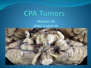
Mumtaz Ali
JPMC KARACHI
Outlines
 Anatomy of CPA
 Possible lesions of CPA
 Clinical features
 Differential diagnosis
 Radiology
 Diagnostic elements
CPA anatomy
CP angle:
 Irregularly shaped
 extra-axial
 potential space
 Between cerebellum & pons in posterior fossa
 lateral to pre-pontine cisterns
BORDERS
 Medial: lateral surface of the brainstem
 Lateral : petrous bone
 Superior : middle cerebellar peduncle & cerebellum
 Inferior : arachnoid tissue of lower cranial nerves
 Posterior : cerbellar peduncle
CONTENTS
 CSF
 arachnoid tissue
 cranial nerves
 vessels
POSSIBLE LESIONS OF CP ANGLE
 Nerve structure: schwanoma
 Meninges : Meningioma ,arachnoid cyst & metastasis
 Cisternal space :epidermoid cyst ,dermoid cyst &
lipoma
 Vascular : aneurysms , AVM & paraganglioma
 Cerebellar ventricles : epindymoma ,lymphoma &
glioma
 Skull base : chondroma ,pituitary adenoma &
cholestrol granuloma
Clinical features
 Chochlear CN-VIII : asymmetric sensorineural
hearing loss & tinnitus
 Vestibular CN-VIII : dysequilibrium & vertigo
 CN-VII : facial weakness
 CN-VI : lateral rectus palsy
 CN-IX : dysphagia
 CN-X : hoarsness & aspiration
 CN-XI : shoulder weakness
 Brainstem : headache ,visual loss & ataxia
Continue…
 Cerebellum:
 Nystagmus
 Finger-nose test
 Dysdiodokinesia
 Intentional Tremor
 Scanning Speech
 Wide gait
 Falling on ipsilateral side
 Romberg sign
Differential Diagnosis
 Accoustic neuroma/Vestibular schwanoma
 Meningioma
 Epidermoid
 Arachnoid cyst
 Dermoid cyst
 Trigeminal neuroma
 Metastasis
Accoustic neuroma
 In 1777, Eduard Sandidort
 Most common CPA tumor
 60-92% of CPA lesions
 benign & encapsulated tumor
 Schwann cells
 rubbery tissue with nodular surface
Continue….
 yellow and gray areas with interspersed foci of
hemorrhage and cyst
 Vestibular division of CN-VIII
 Inferior vestibular nerve > superior vestibular nerve
Histopathological types
 Antonie A:
spindle-shaped cells with rod-shaped nuclie,
dense reticulin arranged in fascicles
 Antonie B:
stellate cells
smaller hyperchromatic nuclei
Less reticulin
prominent cytoplasmic processes
large myxoid stroma
Radiology
 CT scan
 Non-contrast: isodense ,no calcification
 IV contrast : inhance homogenously
 Gas cisternogram : no longer used
 MRI
T1 : isointense to brain & hyperintense to CSF
T2 : hyperintense to brain & iso/hypointense to CSF
Gadolinium : enhancement of tumor on T1
Diagnostic elements
• Centered on Porus Acousticus
• "Ice cream on cone" pattern (intracanalicular
extension)
• Acute angles to petrous bone
• Often involves the IAC
• Homogeneous enhancement
• No dural tail
• No calcifications
 S-100 & vimentin
Auditory brain response(ABR)
 Stimulus =chochlear nerve + auditory pathway
 sensitivity = 92% to 98%
 Specificity = 80% to 90%
Meningioma
 2nd most common CPA tumor (3-7%)
 well-circumscribed
 globular or lobulated
 non-glial tumor
 arachnoidal cells:CN foramina , venous sinuses
Continue….
 clearly demarcated from the brain
 Usually arise : posterior surface of petrous
 Usually do not extend : IAC
 Vimentin & EMA
Diagnostic elements
 Arise : petrous bone (broad base)
 Obtuse angles to petrous bone
 Uncommonly involves the IAC
 "dural tail" sign (50-75%)
 Calcifications common (25%)
 Pial vessel flow voids
 Psammoma body
 Herniate into middle fossa (50%)
Continue…..
 Hyperstosis
 Homogenous signal
 Homogenous enhancement
 Extension:small ,internal auditory canal
Epidermoid cyst
 Accounts for 2-6 % of CPA masses
 Congenital lesions that present in adulthood
 Rests of ectodermal tissue containing stratified
squamous lining and keratin
 May arise within the temporal bone or in the CPA
Continue….
 Extra-axial lesions
 spread along the basal surface
 Rupture : aseptic meningitis
 Overwhelmingly benign
Diagnostic Elements
 Dumb bell : middle fossa or contralateral cistern
 Non-enhancing (25% mild peripheral enhancement)
 CT usually shows a mass hypodense to CSF
 Inhomogeneous lesion
 cauliflower surface appearance
 T1 - hypo-isointense
Continue….
 T2 - hyperintense
 (high protein,high signal=T1 & low signal=T2)
 FLAIR : iso-hyperintensity
 Diffusion weighting:characteristic moderate intensity
(restriction of diffusion)
Arachnoid cyst
 Loculated collections of CSF
 reduplication of arachnoidal membrane
 Erosion of the adjacent calvarium is often present
Diagnostic elements
 Avascular cystic mass
 Nonenhancing
 Smooth regular shape
 Homogeneous
 identical signal to CSF in all weighting
continue
 No calcifications
 FLAIR :intense signal suppresion
 Diffusion weighting : hypointensity (no restriction of
diffusion
Dermoid cyst
 Midline lesions
 rarely invade the CPA laterally
 elements from all layers of the skin
 FLAIR, CISS and DWI:diagnostic
 MR appearance depends : amount of fat
 increased signal : T1 & T2
Trigeminal neuroma
 Similar to vestibular neuoma
 arises from the trigeminal nerve
Thank You

More Related Content

What's hot

Surgical approaches to skull base
Surgical approaches to skull base Surgical approaches to skull base
Surgical approaches to skull base
Ajay Mourya
 
Cerebellopontine angle tumours
Cerebellopontine angle tumoursCerebellopontine angle tumours
Cerebellopontine angle tumours
Dr Safika Zaman
 
Anatomy of cerebral veins
Anatomy of cerebral veinsAnatomy of cerebral veins
Anatomy of cerebral veins
Amar Patil
 
CP ANGLE TUMORS MANAGEMENT
CP ANGLE TUMORS MANAGEMENT CP ANGLE TUMORS MANAGEMENT
CP ANGLE TUMORS MANAGEMENT
Vamsi Reloaded
 
Cp angle tumours
Cp angle tumoursCp angle tumours
Cp angle tumours
Khalidmsayed
 
Presentation2.pptx. posterior fossa tumour
Presentation2.pptx. posterior fossa tumourPresentation2.pptx. posterior fossa tumour
Presentation2.pptx. posterior fossa tumourAbdellah Nazeer
 
Cisterns of brain
Cisterns of brainCisterns of brain
Cisterns of brain
suresh Bishokarma
 
CP ANGLE TUMOR.pptx
CP ANGLE TUMOR.pptxCP ANGLE TUMOR.pptx
CP ANGLE TUMOR.pptx
SujanPandey11
 
Third ventricular surgical approaches
Third ventricular surgical approachesThird ventricular surgical approaches
Third ventricular surgical approaches
suresh Bishokarma
 
Pterional craniotomy
Pterional craniotomyPterional craniotomy
Pterional craniotomy
Dr Fakir Mohan Sahu
 
Accustic neuroma 1
Accustic neuroma 1Accustic neuroma 1
Accustic neuroma 1Verdah Sabih
 
Frontotemporal FTOZ craniotomy
Frontotemporal FTOZ craniotomyFrontotemporal FTOZ craniotomy
Frontotemporal FTOZ craniotomy
Dr. Shahnawaz Alam
 
Endoscopic pituitary surgery
Endoscopic pituitary surgeryEndoscopic pituitary surgery
Endoscopic pituitary surgery
Ajay Mourya
 
Paragangliomas of head and neck
Paragangliomas of head and neckParagangliomas of head and neck
Paragangliomas of head and neck
Mamoon Ameen
 
Cavernous sinus-ANATOMY AND SURGICAL APPROACHES
Cavernous sinus-ANATOMY AND SURGICAL APPROACHESCavernous sinus-ANATOMY AND SURGICAL APPROACHES
Cavernous sinus-ANATOMY AND SURGICAL APPROACHES
Sanjeev Sreenivasan
 
The temporal bone and transtemporal approaches
The temporal bone and transtemporal approachesThe temporal bone and transtemporal approaches
The temporal bone and transtemporal approaches
Dr. Shahnawaz Alam
 

What's hot (20)

10 triangles 360°
10 triangles 360°10 triangles 360°
10 triangles 360°
 
Surgical approaches to skull base
Surgical approaches to skull base Surgical approaches to skull base
Surgical approaches to skull base
 
Cerebellopontine angle tumours
Cerebellopontine angle tumoursCerebellopontine angle tumours
Cerebellopontine angle tumours
 
Anatomy of cerebral veins
Anatomy of cerebral veinsAnatomy of cerebral veins
Anatomy of cerebral veins
 
Petrous apex and skull base
Petrous apex and skull basePetrous apex and skull base
Petrous apex and skull base
 
CP ANGLE TUMORS MANAGEMENT
CP ANGLE TUMORS MANAGEMENT CP ANGLE TUMORS MANAGEMENT
CP ANGLE TUMORS MANAGEMENT
 
CP angle 360°
CP angle 360°CP angle 360°
CP angle 360°
 
Cp angle tumours
Cp angle tumoursCp angle tumours
Cp angle tumours
 
Presentation2.pptx. posterior fossa tumour
Presentation2.pptx. posterior fossa tumourPresentation2.pptx. posterior fossa tumour
Presentation2.pptx. posterior fossa tumour
 
Cisterns of brain
Cisterns of brainCisterns of brain
Cisterns of brain
 
CP ANGLE TUMOR.pptx
CP ANGLE TUMOR.pptxCP ANGLE TUMOR.pptx
CP ANGLE TUMOR.pptx
 
Third ventricular surgical approaches
Third ventricular surgical approachesThird ventricular surgical approaches
Third ventricular surgical approaches
 
Pterional craniotomy
Pterional craniotomyPterional craniotomy
Pterional craniotomy
 
Accustic neuroma 1
Accustic neuroma 1Accustic neuroma 1
Accustic neuroma 1
 
Frontotemporal FTOZ craniotomy
Frontotemporal FTOZ craniotomyFrontotemporal FTOZ craniotomy
Frontotemporal FTOZ craniotomy
 
CSF cisterns
CSF cisternsCSF cisterns
CSF cisterns
 
Endoscopic pituitary surgery
Endoscopic pituitary surgeryEndoscopic pituitary surgery
Endoscopic pituitary surgery
 
Paragangliomas of head and neck
Paragangliomas of head and neckParagangliomas of head and neck
Paragangliomas of head and neck
 
Cavernous sinus-ANATOMY AND SURGICAL APPROACHES
Cavernous sinus-ANATOMY AND SURGICAL APPROACHESCavernous sinus-ANATOMY AND SURGICAL APPROACHES
Cavernous sinus-ANATOMY AND SURGICAL APPROACHES
 
The temporal bone and transtemporal approaches
The temporal bone and transtemporal approachesThe temporal bone and transtemporal approaches
The temporal bone and transtemporal approaches
 

Similar to Cp angle tumors

Cpa sol radio discussion
Cpa sol radio discussion Cpa sol radio discussion
Cpa sol radio discussion
Dr. Shahnawaz Alam
 
Tumors of middle and inner ear dr.sithanandha kumar 14.03.2016
Tumors of middle and inner ear  dr.sithanandha kumar 14.03.2016Tumors of middle and inner ear  dr.sithanandha kumar 14.03.2016
Tumors of middle and inner ear dr.sithanandha kumar 14.03.2016
ophthalmgmcri
 
Diagnostic Neuroradiology
Diagnostic NeuroradiologyDiagnostic Neuroradiology
Diagnostic NeuroradiologySumit Prajapati
 
skullbase imaging.pptx
skullbase imaging.pptxskullbase imaging.pptx
skullbase imaging.pptx
ssuser0aca5c
 
Vascular brain lesions for radiology by Dr Soumitra Halder
Vascular brain lesions for radiology by Dr Soumitra HalderVascular brain lesions for radiology by Dr Soumitra Halder
Vascular brain lesions for radiology by Dr Soumitra Halder
Soumitra Halder
 
Vestibular schwanoma
Vestibular schwanoma Vestibular schwanoma
Vestibular schwanoma
MD Sayad Zaman
 
CNS Rapid Review of Radiology
CNS Rapid Review of RadiologyCNS Rapid Review of Radiology
CNS Rapid Review of Radiology
Double M
 
Acoustic neuroma
Acoustic neuromaAcoustic neuroma
Acoustic neuroma
Ajay Manickam
 
Imaging of the temporal bone
Imaging of the temporal bone Imaging of the temporal bone
Imaging of the temporal bone
Kanu Saha
 
Fourth ventricular tumors
Fourth ventricular tumorsFourth ventricular tumors
Fourth ventricular tumors
Neurosurgeon Mumtaz Ali Narejo
 
thyriod gland imaging part 3 (benign malignant thyriod nodule) Dr Ahmed Esawy
thyriod gland imaging part 3 (benign malignant thyriod nodule)  Dr Ahmed Esawythyriod gland imaging part 3 (benign malignant thyriod nodule)  Dr Ahmed Esawy
thyriod gland imaging part 3 (benign malignant thyriod nodule) Dr Ahmed Esawy
AHMED ESAWY
 
VESTIBULAR SCHWANNOMA.pptx
VESTIBULAR SCHWANNOMA.pptxVESTIBULAR SCHWANNOMA.pptx
VESTIBULAR SCHWANNOMA.pptx
KarishmaMishra13
 
Head and neck
Head and neckHead and neck
Head and neck
Dalia El Said
 
Congenital CNS lesions
Congenital CNS lesionsCongenital CNS lesions
Congenital CNS lesions
Milan Silwal
 
extra axial Meningioma
extra axial Meningiomaextra axial Meningioma
extra axial Meningioma
Hassan Alqarni
 
Presentation1.pptx, radiological imaging of cerebello pontine angle mass lesi...
Presentation1.pptx, radiological imaging of cerebello pontine angle mass lesi...Presentation1.pptx, radiological imaging of cerebello pontine angle mass lesi...
Presentation1.pptx, radiological imaging of cerebello pontine angle mass lesi...Abdellah Nazeer
 
Cranial usg final.pptx
Cranial usg final.pptxCranial usg final.pptx
Cranial usg final.pptx
Nishitha Ashok
 
Head and Neck Imaging
Head and Neck ImagingHead and Neck Imaging
Head and Neck Imaging
Marco Alberto Brion
 
IMAGING OF INFRATENTORIAL BRAIN TUMORS.pptx
IMAGING OF INFRATENTORIAL BRAIN TUMORS.pptxIMAGING OF INFRATENTORIAL BRAIN TUMORS.pptx
IMAGING OF INFRATENTORIAL BRAIN TUMORS.pptx
Arya Anish
 
Basic approach to brain tumor
Basic approach to brain tumorBasic approach to brain tumor
Basic approach to brain tumor
Khon Kaen university
 

Similar to Cp angle tumors (20)

Cpa sol radio discussion
Cpa sol radio discussion Cpa sol radio discussion
Cpa sol radio discussion
 
Tumors of middle and inner ear dr.sithanandha kumar 14.03.2016
Tumors of middle and inner ear  dr.sithanandha kumar 14.03.2016Tumors of middle and inner ear  dr.sithanandha kumar 14.03.2016
Tumors of middle and inner ear dr.sithanandha kumar 14.03.2016
 
Diagnostic Neuroradiology
Diagnostic NeuroradiologyDiagnostic Neuroradiology
Diagnostic Neuroradiology
 
skullbase imaging.pptx
skullbase imaging.pptxskullbase imaging.pptx
skullbase imaging.pptx
 
Vascular brain lesions for radiology by Dr Soumitra Halder
Vascular brain lesions for radiology by Dr Soumitra HalderVascular brain lesions for radiology by Dr Soumitra Halder
Vascular brain lesions for radiology by Dr Soumitra Halder
 
Vestibular schwanoma
Vestibular schwanoma Vestibular schwanoma
Vestibular schwanoma
 
CNS Rapid Review of Radiology
CNS Rapid Review of RadiologyCNS Rapid Review of Radiology
CNS Rapid Review of Radiology
 
Acoustic neuroma
Acoustic neuromaAcoustic neuroma
Acoustic neuroma
 
Imaging of the temporal bone
Imaging of the temporal bone Imaging of the temporal bone
Imaging of the temporal bone
 
Fourth ventricular tumors
Fourth ventricular tumorsFourth ventricular tumors
Fourth ventricular tumors
 
thyriod gland imaging part 3 (benign malignant thyriod nodule) Dr Ahmed Esawy
thyriod gland imaging part 3 (benign malignant thyriod nodule)  Dr Ahmed Esawythyriod gland imaging part 3 (benign malignant thyriod nodule)  Dr Ahmed Esawy
thyriod gland imaging part 3 (benign malignant thyriod nodule) Dr Ahmed Esawy
 
VESTIBULAR SCHWANNOMA.pptx
VESTIBULAR SCHWANNOMA.pptxVESTIBULAR SCHWANNOMA.pptx
VESTIBULAR SCHWANNOMA.pptx
 
Head and neck
Head and neckHead and neck
Head and neck
 
Congenital CNS lesions
Congenital CNS lesionsCongenital CNS lesions
Congenital CNS lesions
 
extra axial Meningioma
extra axial Meningiomaextra axial Meningioma
extra axial Meningioma
 
Presentation1.pptx, radiological imaging of cerebello pontine angle mass lesi...
Presentation1.pptx, radiological imaging of cerebello pontine angle mass lesi...Presentation1.pptx, radiological imaging of cerebello pontine angle mass lesi...
Presentation1.pptx, radiological imaging of cerebello pontine angle mass lesi...
 
Cranial usg final.pptx
Cranial usg final.pptxCranial usg final.pptx
Cranial usg final.pptx
 
Head and Neck Imaging
Head and Neck ImagingHead and Neck Imaging
Head and Neck Imaging
 
IMAGING OF INFRATENTORIAL BRAIN TUMORS.pptx
IMAGING OF INFRATENTORIAL BRAIN TUMORS.pptxIMAGING OF INFRATENTORIAL BRAIN TUMORS.pptx
IMAGING OF INFRATENTORIAL BRAIN TUMORS.pptx
 
Basic approach to brain tumor
Basic approach to brain tumorBasic approach to brain tumor
Basic approach to brain tumor
 

More from Neurosurgeon Mumtaz Ali Narejo

Meningioma presentation by dr,Mumtaz Ali narejo
Meningioma presentation by dr,Mumtaz Ali narejoMeningioma presentation by dr,Mumtaz Ali narejo
Meningioma presentation by dr,Mumtaz Ali narejo
Neurosurgeon Mumtaz Ali Narejo
 
Blue Printing in medical education by Dr.Mumtaz Ali.pptx
Blue Printing in medical education by Dr.Mumtaz Ali.pptxBlue Printing in medical education by Dr.Mumtaz Ali.pptx
Blue Printing in medical education by Dr.Mumtaz Ali.pptx
Neurosurgeon Mumtaz Ali Narejo
 
Endoscopic 3rd Ventriculostomy-DR.MUMTAZ ALI NAREJO.pptx
Endoscopic 3rd Ventriculostomy-DR.MUMTAZ ALI NAREJO.pptxEndoscopic 3rd Ventriculostomy-DR.MUMTAZ ALI NAREJO.pptx
Endoscopic 3rd Ventriculostomy-DR.MUMTAZ ALI NAREJO.pptx
Neurosurgeon Mumtaz Ali Narejo
 
head injury according to ATLS BY DR MUMTAZ ALI.pptx
head injury according to ATLS BY DR MUMTAZ ALI.pptxhead injury according to ATLS BY DR MUMTAZ ALI.pptx
head injury according to ATLS BY DR MUMTAZ ALI.pptx
Neurosurgeon Mumtaz Ali Narejo
 
Spine injury according to atls by dr mumtaz ali narejo.pptx
Spine injury according to atls by dr mumtaz ali narejo.pptxSpine injury according to atls by dr mumtaz ali narejo.pptx
Spine injury according to atls by dr mumtaz ali narejo.pptx
Neurosurgeon Mumtaz Ali Narejo
 
Statistical software packages ,their layout & applications
Statistical software packages ,their layout & applicationsStatistical software packages ,their layout & applications
Statistical software packages ,their layout & applications
Neurosurgeon Mumtaz Ali Narejo
 
Neurofibromatosis Type.pptx
Neurofibromatosis Type.pptxNeurofibromatosis Type.pptx
Neurofibromatosis Type.pptx
Neurosurgeon Mumtaz Ali Narejo
 
Protein microarray.pptx
Protein microarray.pptxProtein microarray.pptx
Protein microarray.pptx
Neurosurgeon Mumtaz Ali Narejo
 
TRAUMATIC BRAIN INJURY - Copy.pptx
TRAUMATIC BRAIN INJURY - Copy.pptxTRAUMATIC BRAIN INJURY - Copy.pptx
TRAUMATIC BRAIN INJURY - Copy.pptx
Neurosurgeon Mumtaz Ali Narejo
 
Road Safty Presentation (2) (1).ppt
Road Safty Presentation (2) (1).pptRoad Safty Presentation (2) (1).ppt
Road Safty Presentation (2) (1).ppt
Neurosurgeon Mumtaz Ali Narejo
 
PRESENTATION OF SPINE INJURY BY DR.MUMTAZ ALI.ppt
PRESENTATION OF SPINE INJURY BY DR.MUMTAZ ALI.pptPRESENTATION OF SPINE INJURY BY DR.MUMTAZ ALI.ppt
PRESENTATION OF SPINE INJURY BY DR.MUMTAZ ALI.ppt
Neurosurgeon Mumtaz Ali Narejo
 
Spine lecture for Final year MBBS by Dr.Mumtaz Ali.pptx
Spine lecture for Final year MBBS by Dr.Mumtaz Ali.pptxSpine lecture for Final year MBBS by Dr.Mumtaz Ali.pptx
Spine lecture for Final year MBBS by Dr.Mumtaz Ali.pptx
Neurosurgeon Mumtaz Ali Narejo
 
Skeletal muscle excitation by dr mumtaz ali
Skeletal muscle excitation by dr mumtaz aliSkeletal muscle excitation by dr mumtaz ali
Skeletal muscle excitation by dr mumtaz ali
Neurosurgeon Mumtaz Ali Narejo
 
Skeletal muscle contraction by dr.mumtaz ali
Skeletal muscle contraction by dr.mumtaz aliSkeletal muscle contraction by dr.mumtaz ali
Skeletal muscle contraction by dr.mumtaz ali
Neurosurgeon Mumtaz Ali Narejo
 
Vestibular scwanomma (case pressentation)dr.mumtaz ali
Vestibular scwanomma (case pressentation)dr.mumtaz aliVestibular scwanomma (case pressentation)dr.mumtaz ali
Vestibular scwanomma (case pressentation)dr.mumtaz ali
Neurosurgeon Mumtaz Ali Narejo
 
Spinal schwannoma(case presentation) dr.mumtaz ali
Spinal schwannoma(case presentation) dr.mumtaz aliSpinal schwannoma(case presentation) dr.mumtaz ali
Spinal schwannoma(case presentation) dr.mumtaz ali
Neurosurgeon Mumtaz Ali Narejo
 
Spinal ependymoma (case presentation)dr.mumtaz ali
Spinal ependymoma (case presentation)dr.mumtaz aliSpinal ependymoma (case presentation)dr.mumtaz ali
Spinal ependymoma (case presentation)dr.mumtaz ali
Neurosurgeon Mumtaz Ali Narejo
 
Spinal meningioma (case presentation)dr.mumtaz ali
Spinal  meningioma (case presentation)dr.mumtaz aliSpinal  meningioma (case presentation)dr.mumtaz ali
Spinal meningioma (case presentation)dr.mumtaz ali
Neurosurgeon Mumtaz Ali Narejo
 
Right sphenoid wing meningioma (case presentation)dr.mumtaz a li
Right sphenoid wing meningioma (case presentation)dr.mumtaz a liRight sphenoid wing meningioma (case presentation)dr.mumtaz a li
Right sphenoid wing meningioma (case presentation)dr.mumtaz a li
Neurosurgeon Mumtaz Ali Narejo
 
Orbital tumor (case presentation)dr.mumtaz ali
Orbital tumor (case presentation)dr.mumtaz aliOrbital tumor (case presentation)dr.mumtaz ali
Orbital tumor (case presentation)dr.mumtaz ali
Neurosurgeon Mumtaz Ali Narejo
 

More from Neurosurgeon Mumtaz Ali Narejo (20)

Meningioma presentation by dr,Mumtaz Ali narejo
Meningioma presentation by dr,Mumtaz Ali narejoMeningioma presentation by dr,Mumtaz Ali narejo
Meningioma presentation by dr,Mumtaz Ali narejo
 
Blue Printing in medical education by Dr.Mumtaz Ali.pptx
Blue Printing in medical education by Dr.Mumtaz Ali.pptxBlue Printing in medical education by Dr.Mumtaz Ali.pptx
Blue Printing in medical education by Dr.Mumtaz Ali.pptx
 
Endoscopic 3rd Ventriculostomy-DR.MUMTAZ ALI NAREJO.pptx
Endoscopic 3rd Ventriculostomy-DR.MUMTAZ ALI NAREJO.pptxEndoscopic 3rd Ventriculostomy-DR.MUMTAZ ALI NAREJO.pptx
Endoscopic 3rd Ventriculostomy-DR.MUMTAZ ALI NAREJO.pptx
 
head injury according to ATLS BY DR MUMTAZ ALI.pptx
head injury according to ATLS BY DR MUMTAZ ALI.pptxhead injury according to ATLS BY DR MUMTAZ ALI.pptx
head injury according to ATLS BY DR MUMTAZ ALI.pptx
 
Spine injury according to atls by dr mumtaz ali narejo.pptx
Spine injury according to atls by dr mumtaz ali narejo.pptxSpine injury according to atls by dr mumtaz ali narejo.pptx
Spine injury according to atls by dr mumtaz ali narejo.pptx
 
Statistical software packages ,their layout & applications
Statistical software packages ,their layout & applicationsStatistical software packages ,their layout & applications
Statistical software packages ,their layout & applications
 
Neurofibromatosis Type.pptx
Neurofibromatosis Type.pptxNeurofibromatosis Type.pptx
Neurofibromatosis Type.pptx
 
Protein microarray.pptx
Protein microarray.pptxProtein microarray.pptx
Protein microarray.pptx
 
TRAUMATIC BRAIN INJURY - Copy.pptx
TRAUMATIC BRAIN INJURY - Copy.pptxTRAUMATIC BRAIN INJURY - Copy.pptx
TRAUMATIC BRAIN INJURY - Copy.pptx
 
Road Safty Presentation (2) (1).ppt
Road Safty Presentation (2) (1).pptRoad Safty Presentation (2) (1).ppt
Road Safty Presentation (2) (1).ppt
 
PRESENTATION OF SPINE INJURY BY DR.MUMTAZ ALI.ppt
PRESENTATION OF SPINE INJURY BY DR.MUMTAZ ALI.pptPRESENTATION OF SPINE INJURY BY DR.MUMTAZ ALI.ppt
PRESENTATION OF SPINE INJURY BY DR.MUMTAZ ALI.ppt
 
Spine lecture for Final year MBBS by Dr.Mumtaz Ali.pptx
Spine lecture for Final year MBBS by Dr.Mumtaz Ali.pptxSpine lecture for Final year MBBS by Dr.Mumtaz Ali.pptx
Spine lecture for Final year MBBS by Dr.Mumtaz Ali.pptx
 
Skeletal muscle excitation by dr mumtaz ali
Skeletal muscle excitation by dr mumtaz aliSkeletal muscle excitation by dr mumtaz ali
Skeletal muscle excitation by dr mumtaz ali
 
Skeletal muscle contraction by dr.mumtaz ali
Skeletal muscle contraction by dr.mumtaz aliSkeletal muscle contraction by dr.mumtaz ali
Skeletal muscle contraction by dr.mumtaz ali
 
Vestibular scwanomma (case pressentation)dr.mumtaz ali
Vestibular scwanomma (case pressentation)dr.mumtaz aliVestibular scwanomma (case pressentation)dr.mumtaz ali
Vestibular scwanomma (case pressentation)dr.mumtaz ali
 
Spinal schwannoma(case presentation) dr.mumtaz ali
Spinal schwannoma(case presentation) dr.mumtaz aliSpinal schwannoma(case presentation) dr.mumtaz ali
Spinal schwannoma(case presentation) dr.mumtaz ali
 
Spinal ependymoma (case presentation)dr.mumtaz ali
Spinal ependymoma (case presentation)dr.mumtaz aliSpinal ependymoma (case presentation)dr.mumtaz ali
Spinal ependymoma (case presentation)dr.mumtaz ali
 
Spinal meningioma (case presentation)dr.mumtaz ali
Spinal  meningioma (case presentation)dr.mumtaz aliSpinal  meningioma (case presentation)dr.mumtaz ali
Spinal meningioma (case presentation)dr.mumtaz ali
 
Right sphenoid wing meningioma (case presentation)dr.mumtaz a li
Right sphenoid wing meningioma (case presentation)dr.mumtaz a liRight sphenoid wing meningioma (case presentation)dr.mumtaz a li
Right sphenoid wing meningioma (case presentation)dr.mumtaz a li
 
Orbital tumor (case presentation)dr.mumtaz ali
Orbital tumor (case presentation)dr.mumtaz aliOrbital tumor (case presentation)dr.mumtaz ali
Orbital tumor (case presentation)dr.mumtaz ali
 

Recently uploaded

Eureka, I found it! - Special Libraries Association 2021 Presentation
Eureka, I found it! - Special Libraries Association 2021 PresentationEureka, I found it! - Special Libraries Association 2021 Presentation
Eureka, I found it! - Special Libraries Association 2021 Presentation
Access Innovations, Inc.
 
Announcement of 18th IEEE International Conference on Software Testing, Verif...
Announcement of 18th IEEE International Conference on Software Testing, Verif...Announcement of 18th IEEE International Conference on Software Testing, Verif...
Announcement of 18th IEEE International Conference on Software Testing, Verif...
Sebastiano Panichella
 
Acorn Recovery: Restore IT infra within minutes
Acorn Recovery: Restore IT infra within minutesAcorn Recovery: Restore IT infra within minutes
Acorn Recovery: Restore IT infra within minutes
IP ServerOne
 
0x01 - Newton's Third Law: Static vs. Dynamic Abusers
0x01 - Newton's Third Law:  Static vs. Dynamic Abusers0x01 - Newton's Third Law:  Static vs. Dynamic Abusers
0x01 - Newton's Third Law: Static vs. Dynamic Abusers
OWASP Beja
 
Media as a Mind Controlling Strategy In Old and Modern Era
Media as a Mind Controlling Strategy In Old and Modern EraMedia as a Mind Controlling Strategy In Old and Modern Era
Media as a Mind Controlling Strategy In Old and Modern Era
faizulhassanfaiz1670
 
Competition and Regulation in Professional Services – KLEINER – June 2024 OEC...
Competition and Regulation in Professional Services – KLEINER – June 2024 OEC...Competition and Regulation in Professional Services – KLEINER – June 2024 OEC...
Competition and Regulation in Professional Services – KLEINER – June 2024 OEC...
OECD Directorate for Financial and Enterprise Affairs
 
Doctoral Symposium at the 17th IEEE International Conference on Software Test...
Doctoral Symposium at the 17th IEEE International Conference on Software Test...Doctoral Symposium at the 17th IEEE International Conference on Software Test...
Doctoral Symposium at the 17th IEEE International Conference on Software Test...
Sebastiano Panichella
 
International Workshop on Artificial Intelligence in Software Testing
International Workshop on Artificial Intelligence in Software TestingInternational Workshop on Artificial Intelligence in Software Testing
International Workshop on Artificial Intelligence in Software Testing
Sebastiano Panichella
 
Getting started with Amazon Bedrock Studio and Control Tower
Getting started with Amazon Bedrock Studio and Control TowerGetting started with Amazon Bedrock Studio and Control Tower
Getting started with Amazon Bedrock Studio and Control Tower
Vladimir Samoylov
 
María Carolina Martínez - eCommerce Day Colombia 2024
María Carolina Martínez - eCommerce Day Colombia 2024María Carolina Martínez - eCommerce Day Colombia 2024
María Carolina Martínez - eCommerce Day Colombia 2024
eCommerce Institute
 
Bonzo subscription_hjjjjjjjj5hhhhhhh_2024.pdf
Bonzo subscription_hjjjjjjjj5hhhhhhh_2024.pdfBonzo subscription_hjjjjjjjj5hhhhhhh_2024.pdf
Bonzo subscription_hjjjjjjjj5hhhhhhh_2024.pdf
khadija278284
 
Obesity causes and management and associated medical conditions
Obesity causes and management and associated medical conditionsObesity causes and management and associated medical conditions
Obesity causes and management and associated medical conditions
Faculty of Medicine And Health Sciences
 
Bitcoin Lightning wallet and tic-tac-toe game XOXO
Bitcoin Lightning wallet and tic-tac-toe game XOXOBitcoin Lightning wallet and tic-tac-toe game XOXO
Bitcoin Lightning wallet and tic-tac-toe game XOXO
Matjaž Lipuš
 
somanykidsbutsofewfathers-140705000023-phpapp02.pptx
somanykidsbutsofewfathers-140705000023-phpapp02.pptxsomanykidsbutsofewfathers-140705000023-phpapp02.pptx
somanykidsbutsofewfathers-140705000023-phpapp02.pptx
Howard Spence
 
Sharpen existing tools or get a new toolbox? Contemporary cluster initiatives...
Sharpen existing tools or get a new toolbox? Contemporary cluster initiatives...Sharpen existing tools or get a new toolbox? Contemporary cluster initiatives...
Sharpen existing tools or get a new toolbox? Contemporary cluster initiatives...
Orkestra
 
Supercharge your AI - SSP Industry Breakout Session 2024-v2_1.pdf
Supercharge your AI - SSP Industry Breakout Session 2024-v2_1.pdfSupercharge your AI - SSP Industry Breakout Session 2024-v2_1.pdf
Supercharge your AI - SSP Industry Breakout Session 2024-v2_1.pdf
Access Innovations, Inc.
 

Recently uploaded (16)

Eureka, I found it! - Special Libraries Association 2021 Presentation
Eureka, I found it! - Special Libraries Association 2021 PresentationEureka, I found it! - Special Libraries Association 2021 Presentation
Eureka, I found it! - Special Libraries Association 2021 Presentation
 
Announcement of 18th IEEE International Conference on Software Testing, Verif...
Announcement of 18th IEEE International Conference on Software Testing, Verif...Announcement of 18th IEEE International Conference on Software Testing, Verif...
Announcement of 18th IEEE International Conference on Software Testing, Verif...
 
Acorn Recovery: Restore IT infra within minutes
Acorn Recovery: Restore IT infra within minutesAcorn Recovery: Restore IT infra within minutes
Acorn Recovery: Restore IT infra within minutes
 
0x01 - Newton's Third Law: Static vs. Dynamic Abusers
0x01 - Newton's Third Law:  Static vs. Dynamic Abusers0x01 - Newton's Third Law:  Static vs. Dynamic Abusers
0x01 - Newton's Third Law: Static vs. Dynamic Abusers
 
Media as a Mind Controlling Strategy In Old and Modern Era
Media as a Mind Controlling Strategy In Old and Modern EraMedia as a Mind Controlling Strategy In Old and Modern Era
Media as a Mind Controlling Strategy In Old and Modern Era
 
Competition and Regulation in Professional Services – KLEINER – June 2024 OEC...
Competition and Regulation in Professional Services – KLEINER – June 2024 OEC...Competition and Regulation in Professional Services – KLEINER – June 2024 OEC...
Competition and Regulation in Professional Services – KLEINER – June 2024 OEC...
 
Doctoral Symposium at the 17th IEEE International Conference on Software Test...
Doctoral Symposium at the 17th IEEE International Conference on Software Test...Doctoral Symposium at the 17th IEEE International Conference on Software Test...
Doctoral Symposium at the 17th IEEE International Conference on Software Test...
 
International Workshop on Artificial Intelligence in Software Testing
International Workshop on Artificial Intelligence in Software TestingInternational Workshop on Artificial Intelligence in Software Testing
International Workshop on Artificial Intelligence in Software Testing
 
Getting started with Amazon Bedrock Studio and Control Tower
Getting started with Amazon Bedrock Studio and Control TowerGetting started with Amazon Bedrock Studio and Control Tower
Getting started with Amazon Bedrock Studio and Control Tower
 
María Carolina Martínez - eCommerce Day Colombia 2024
María Carolina Martínez - eCommerce Day Colombia 2024María Carolina Martínez - eCommerce Day Colombia 2024
María Carolina Martínez - eCommerce Day Colombia 2024
 
Bonzo subscription_hjjjjjjjj5hhhhhhh_2024.pdf
Bonzo subscription_hjjjjjjjj5hhhhhhh_2024.pdfBonzo subscription_hjjjjjjjj5hhhhhhh_2024.pdf
Bonzo subscription_hjjjjjjjj5hhhhhhh_2024.pdf
 
Obesity causes and management and associated medical conditions
Obesity causes and management and associated medical conditionsObesity causes and management and associated medical conditions
Obesity causes and management and associated medical conditions
 
Bitcoin Lightning wallet and tic-tac-toe game XOXO
Bitcoin Lightning wallet and tic-tac-toe game XOXOBitcoin Lightning wallet and tic-tac-toe game XOXO
Bitcoin Lightning wallet and tic-tac-toe game XOXO
 
somanykidsbutsofewfathers-140705000023-phpapp02.pptx
somanykidsbutsofewfathers-140705000023-phpapp02.pptxsomanykidsbutsofewfathers-140705000023-phpapp02.pptx
somanykidsbutsofewfathers-140705000023-phpapp02.pptx
 
Sharpen existing tools or get a new toolbox? Contemporary cluster initiatives...
Sharpen existing tools or get a new toolbox? Contemporary cluster initiatives...Sharpen existing tools or get a new toolbox? Contemporary cluster initiatives...
Sharpen existing tools or get a new toolbox? Contemporary cluster initiatives...
 
Supercharge your AI - SSP Industry Breakout Session 2024-v2_1.pdf
Supercharge your AI - SSP Industry Breakout Session 2024-v2_1.pdfSupercharge your AI - SSP Industry Breakout Session 2024-v2_1.pdf
Supercharge your AI - SSP Industry Breakout Session 2024-v2_1.pdf
 

Cp angle tumors

  • 2. Outlines  Anatomy of CPA  Possible lesions of CPA  Clinical features  Differential diagnosis  Radiology  Diagnostic elements
  • 3. CPA anatomy CP angle:  Irregularly shaped  extra-axial  potential space  Between cerebellum & pons in posterior fossa  lateral to pre-pontine cisterns
  • 4. BORDERS  Medial: lateral surface of the brainstem  Lateral : petrous bone  Superior : middle cerebellar peduncle & cerebellum  Inferior : arachnoid tissue of lower cranial nerves  Posterior : cerbellar peduncle
  • 5. CONTENTS  CSF  arachnoid tissue  cranial nerves  vessels
  • 6.
  • 7.
  • 8. POSSIBLE LESIONS OF CP ANGLE  Nerve structure: schwanoma  Meninges : Meningioma ,arachnoid cyst & metastasis  Cisternal space :epidermoid cyst ,dermoid cyst & lipoma  Vascular : aneurysms , AVM & paraganglioma  Cerebellar ventricles : epindymoma ,lymphoma & glioma  Skull base : chondroma ,pituitary adenoma & cholestrol granuloma
  • 9. Clinical features  Chochlear CN-VIII : asymmetric sensorineural hearing loss & tinnitus  Vestibular CN-VIII : dysequilibrium & vertigo  CN-VII : facial weakness  CN-VI : lateral rectus palsy  CN-IX : dysphagia  CN-X : hoarsness & aspiration  CN-XI : shoulder weakness  Brainstem : headache ,visual loss & ataxia
  • 10. Continue…  Cerebellum:  Nystagmus  Finger-nose test  Dysdiodokinesia  Intentional Tremor  Scanning Speech  Wide gait  Falling on ipsilateral side  Romberg sign
  • 11. Differential Diagnosis  Accoustic neuroma/Vestibular schwanoma  Meningioma  Epidermoid  Arachnoid cyst  Dermoid cyst  Trigeminal neuroma  Metastasis
  • 12. Accoustic neuroma  In 1777, Eduard Sandidort  Most common CPA tumor  60-92% of CPA lesions  benign & encapsulated tumor  Schwann cells  rubbery tissue with nodular surface
  • 13. Continue….  yellow and gray areas with interspersed foci of hemorrhage and cyst  Vestibular division of CN-VIII  Inferior vestibular nerve > superior vestibular nerve
  • 14. Histopathological types  Antonie A: spindle-shaped cells with rod-shaped nuclie, dense reticulin arranged in fascicles  Antonie B: stellate cells smaller hyperchromatic nuclei Less reticulin prominent cytoplasmic processes large myxoid stroma
  • 15.
  • 16.
  • 17. Radiology  CT scan  Non-contrast: isodense ,no calcification  IV contrast : inhance homogenously  Gas cisternogram : no longer used  MRI T1 : isointense to brain & hyperintense to CSF T2 : hyperintense to brain & iso/hypointense to CSF Gadolinium : enhancement of tumor on T1
  • 18. Diagnostic elements • Centered on Porus Acousticus • "Ice cream on cone" pattern (intracanalicular extension) • Acute angles to petrous bone • Often involves the IAC • Homogeneous enhancement • No dural tail • No calcifications  S-100 & vimentin
  • 19.
  • 20.
  • 21.
  • 22. Auditory brain response(ABR)  Stimulus =chochlear nerve + auditory pathway  sensitivity = 92% to 98%  Specificity = 80% to 90%
  • 23.
  • 24. Meningioma  2nd most common CPA tumor (3-7%)  well-circumscribed  globular or lobulated  non-glial tumor  arachnoidal cells:CN foramina , venous sinuses
  • 25. Continue….  clearly demarcated from the brain  Usually arise : posterior surface of petrous  Usually do not extend : IAC  Vimentin & EMA
  • 26. Diagnostic elements  Arise : petrous bone (broad base)  Obtuse angles to petrous bone  Uncommonly involves the IAC  "dural tail" sign (50-75%)  Calcifications common (25%)  Pial vessel flow voids  Psammoma body  Herniate into middle fossa (50%)
  • 27. Continue…..  Hyperstosis  Homogenous signal  Homogenous enhancement  Extension:small ,internal auditory canal
  • 28.
  • 29.
  • 30. Epidermoid cyst  Accounts for 2-6 % of CPA masses  Congenital lesions that present in adulthood  Rests of ectodermal tissue containing stratified squamous lining and keratin  May arise within the temporal bone or in the CPA
  • 31. Continue….  Extra-axial lesions  spread along the basal surface  Rupture : aseptic meningitis  Overwhelmingly benign
  • 32. Diagnostic Elements  Dumb bell : middle fossa or contralateral cistern  Non-enhancing (25% mild peripheral enhancement)  CT usually shows a mass hypodense to CSF  Inhomogeneous lesion  cauliflower surface appearance  T1 - hypo-isointense
  • 33. Continue….  T2 - hyperintense  (high protein,high signal=T1 & low signal=T2)  FLAIR : iso-hyperintensity  Diffusion weighting:characteristic moderate intensity (restriction of diffusion)
  • 34.
  • 35. Arachnoid cyst  Loculated collections of CSF  reduplication of arachnoidal membrane  Erosion of the adjacent calvarium is often present
  • 36. Diagnostic elements  Avascular cystic mass  Nonenhancing  Smooth regular shape  Homogeneous  identical signal to CSF in all weighting
  • 37. continue  No calcifications  FLAIR :intense signal suppresion  Diffusion weighting : hypointensity (no restriction of diffusion
  • 38.
  • 39. Dermoid cyst  Midline lesions  rarely invade the CPA laterally  elements from all layers of the skin  FLAIR, CISS and DWI:diagnostic  MR appearance depends : amount of fat  increased signal : T1 & T2
  • 40.
  • 41. Trigeminal neuroma  Similar to vestibular neuoma  arises from the trigeminal nerve
  • 42.