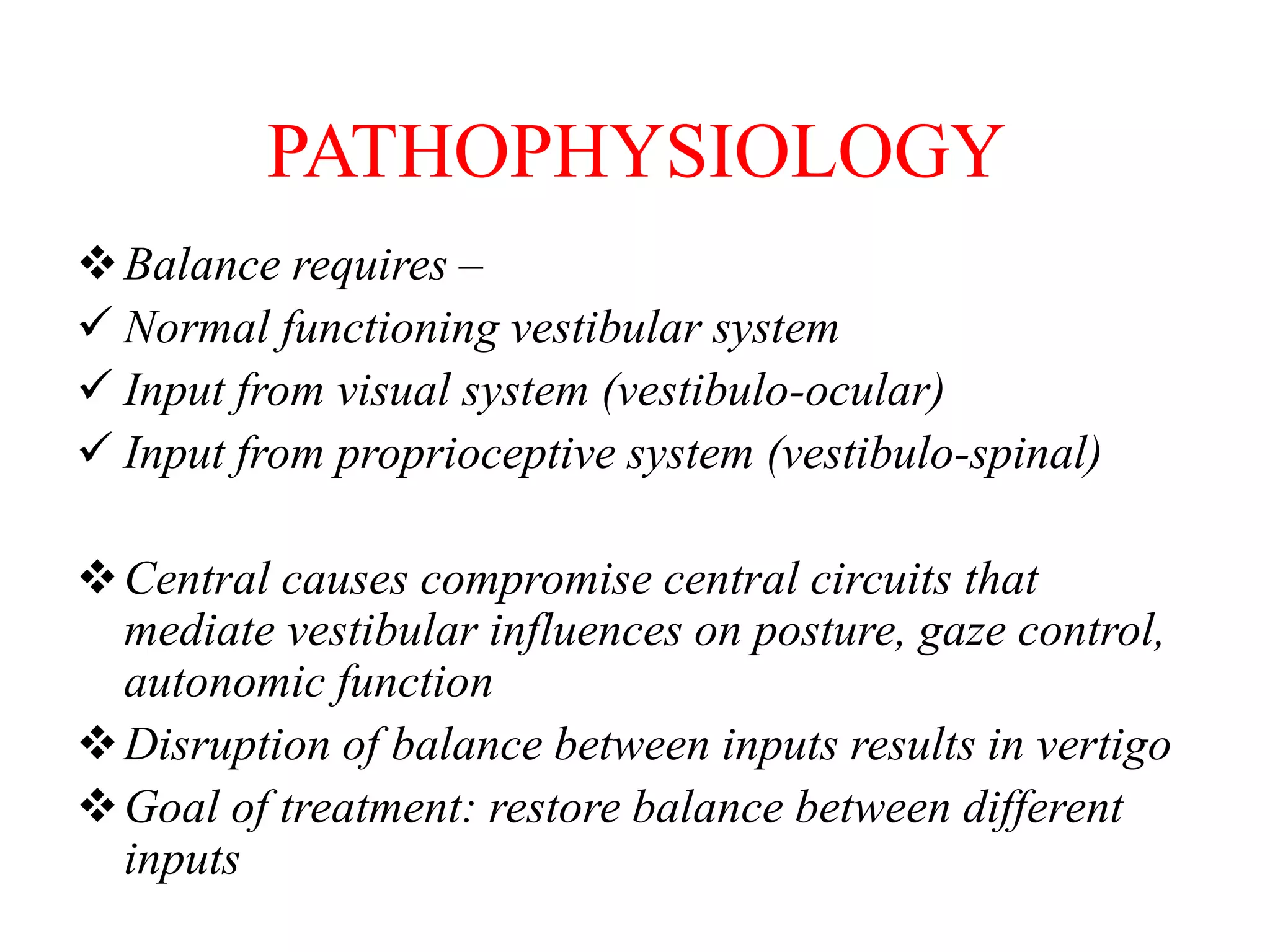
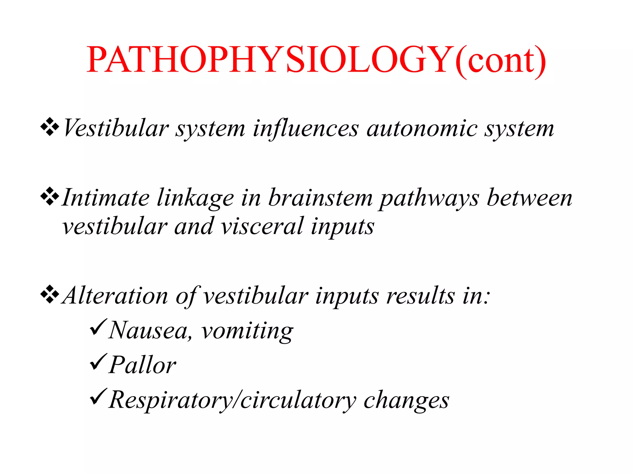
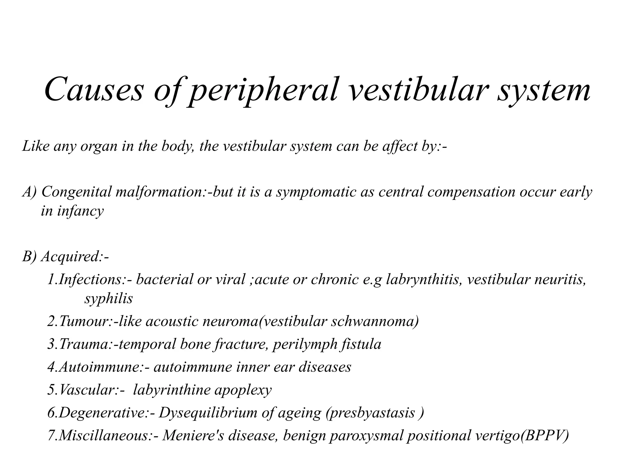
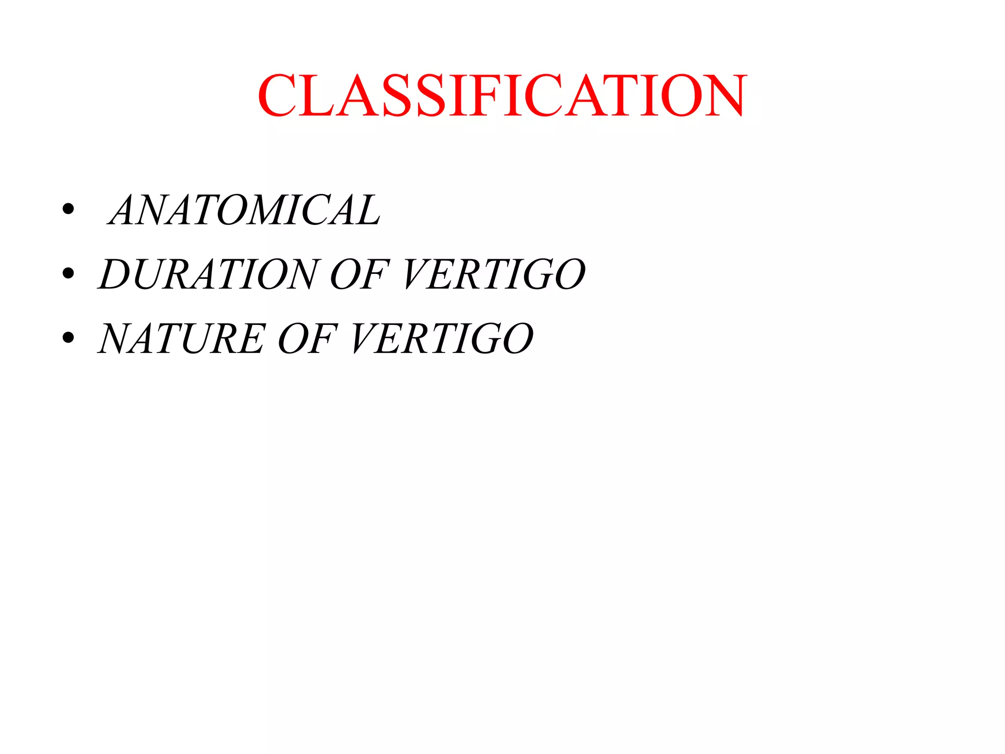
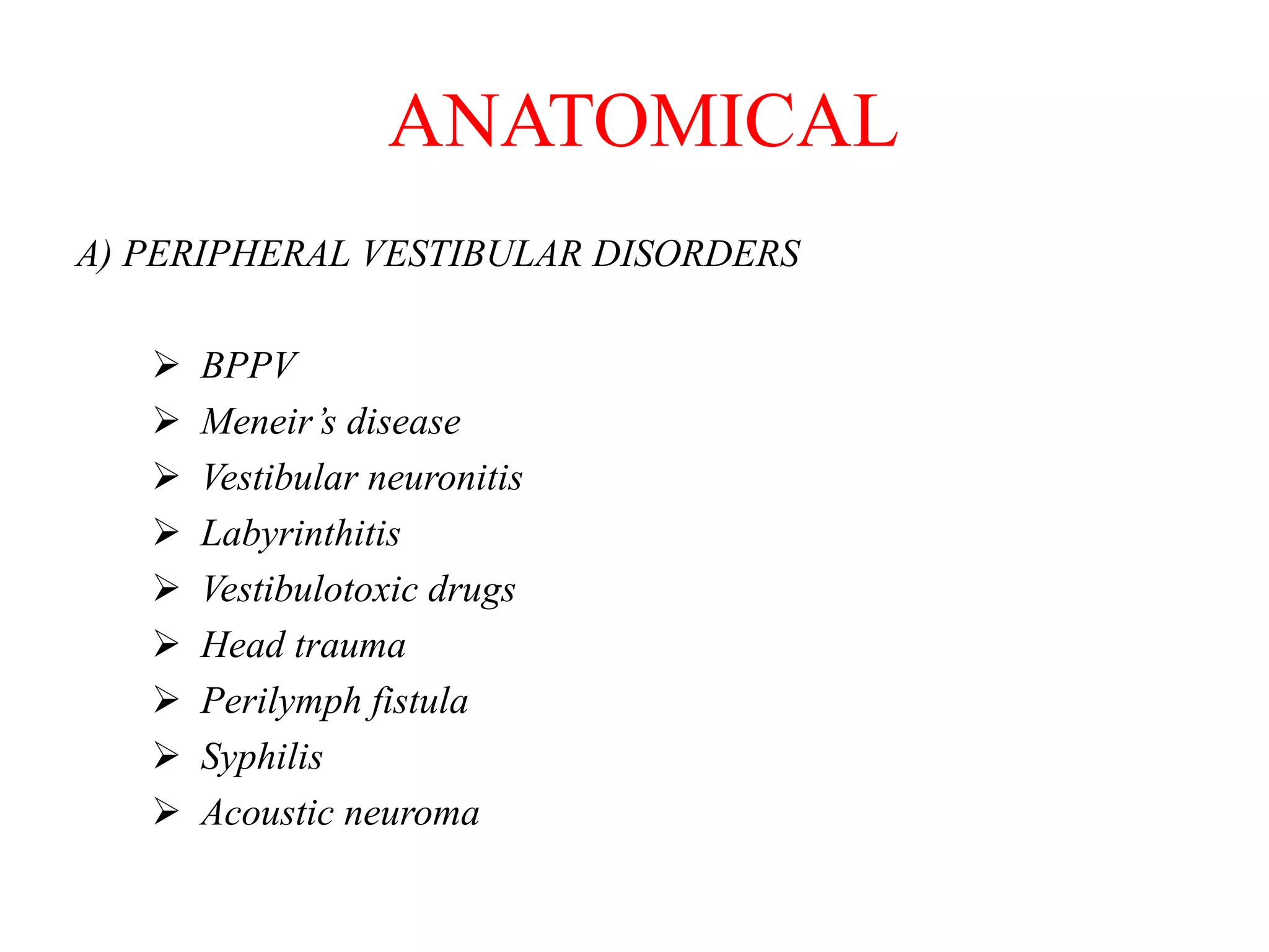
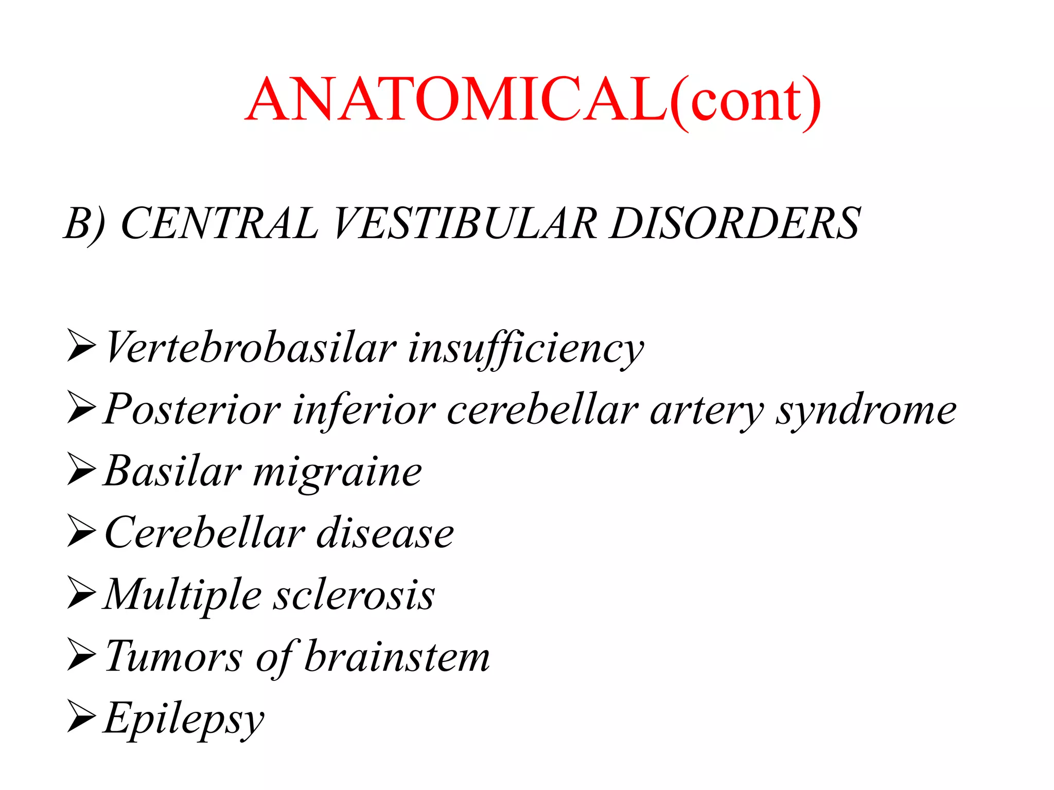
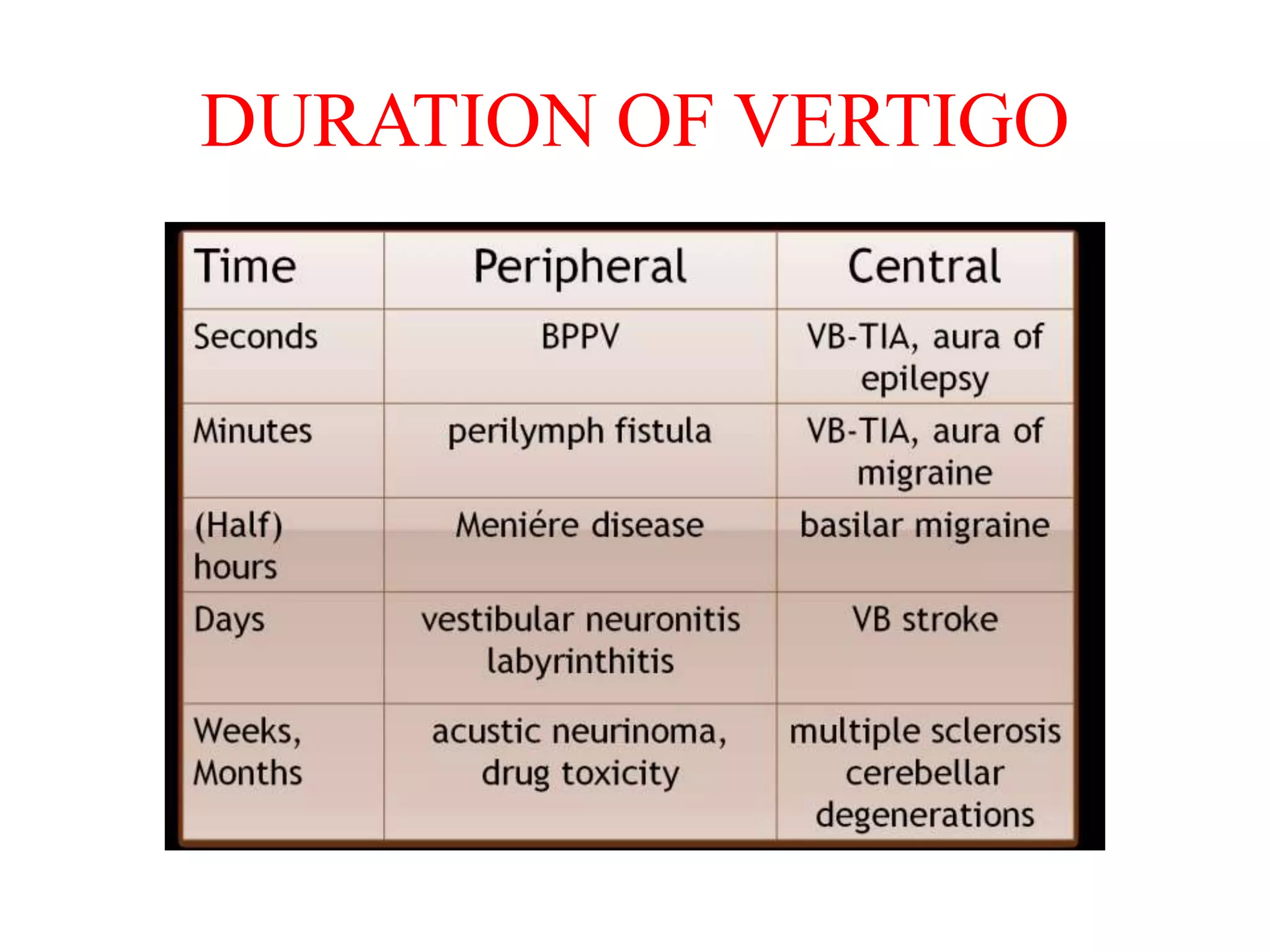
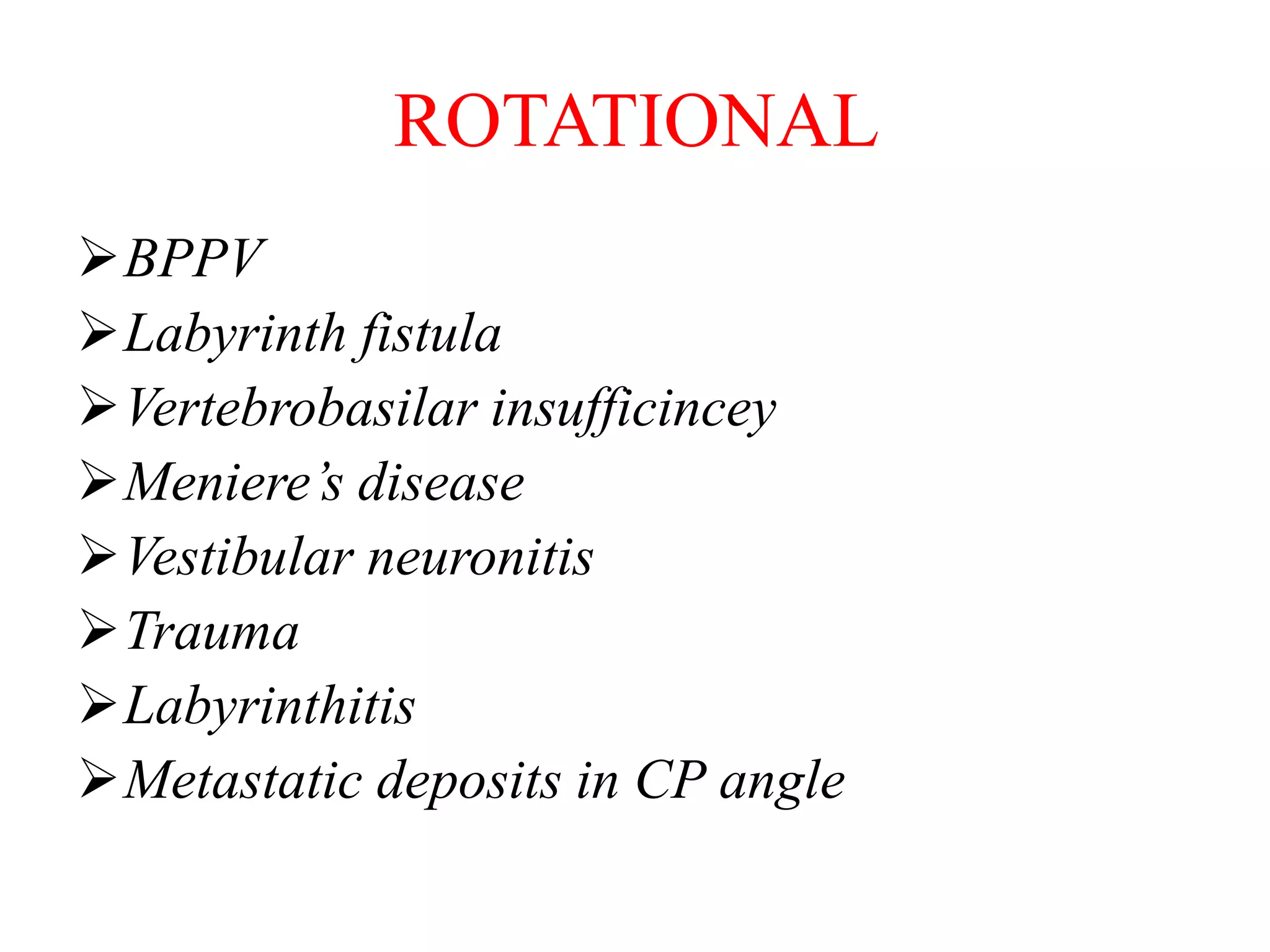
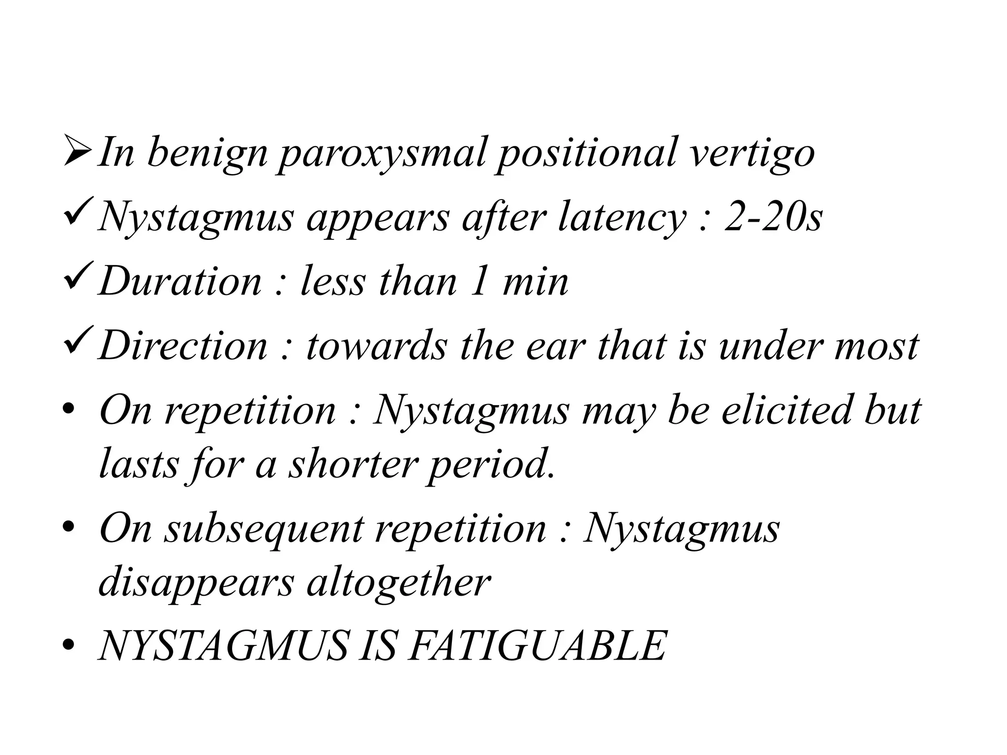
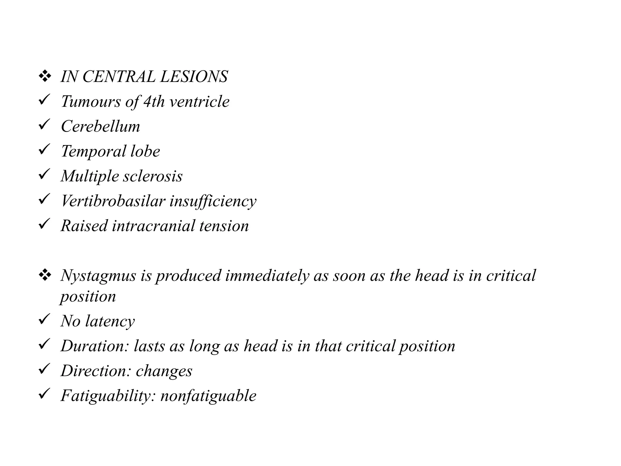
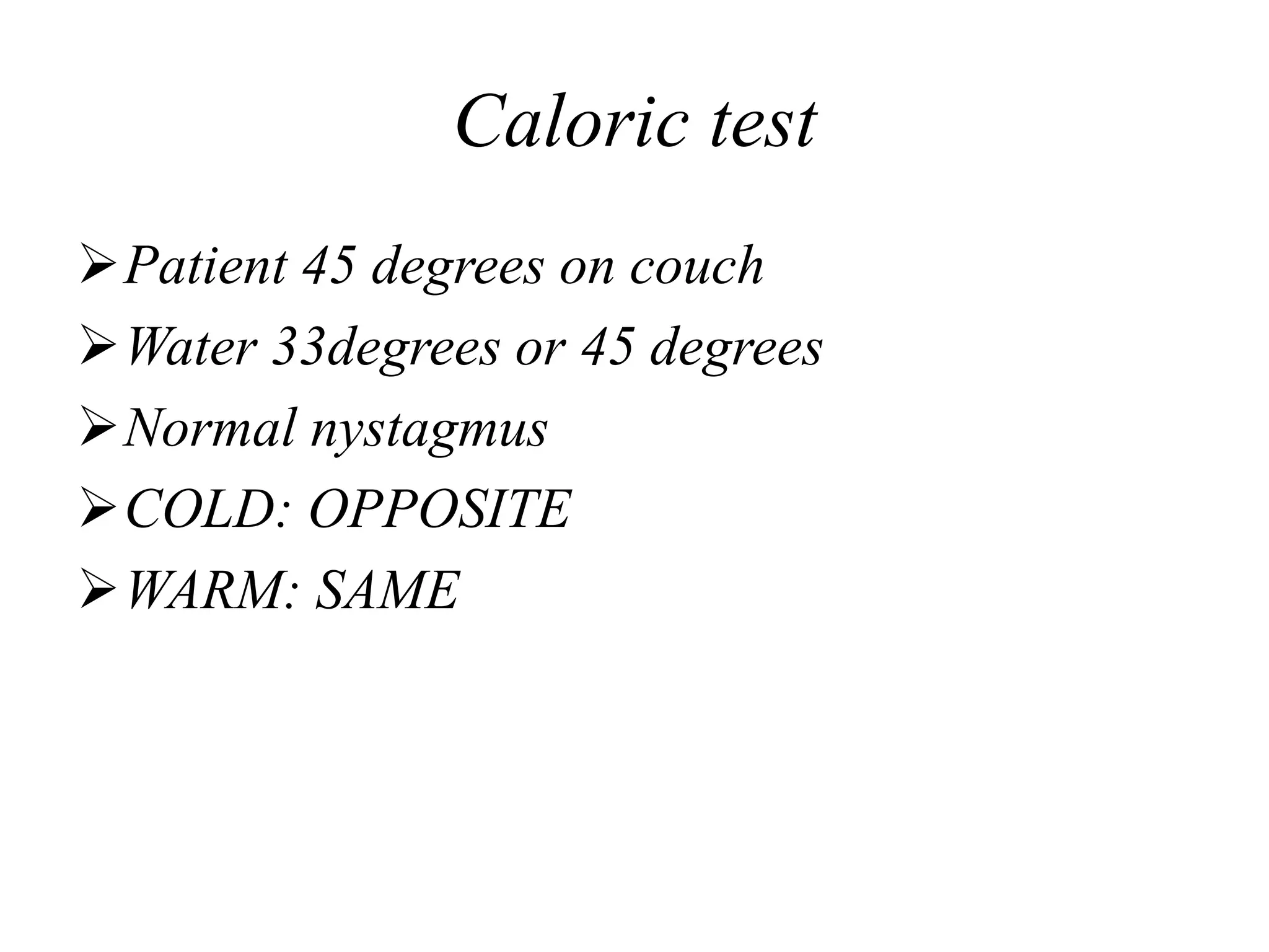
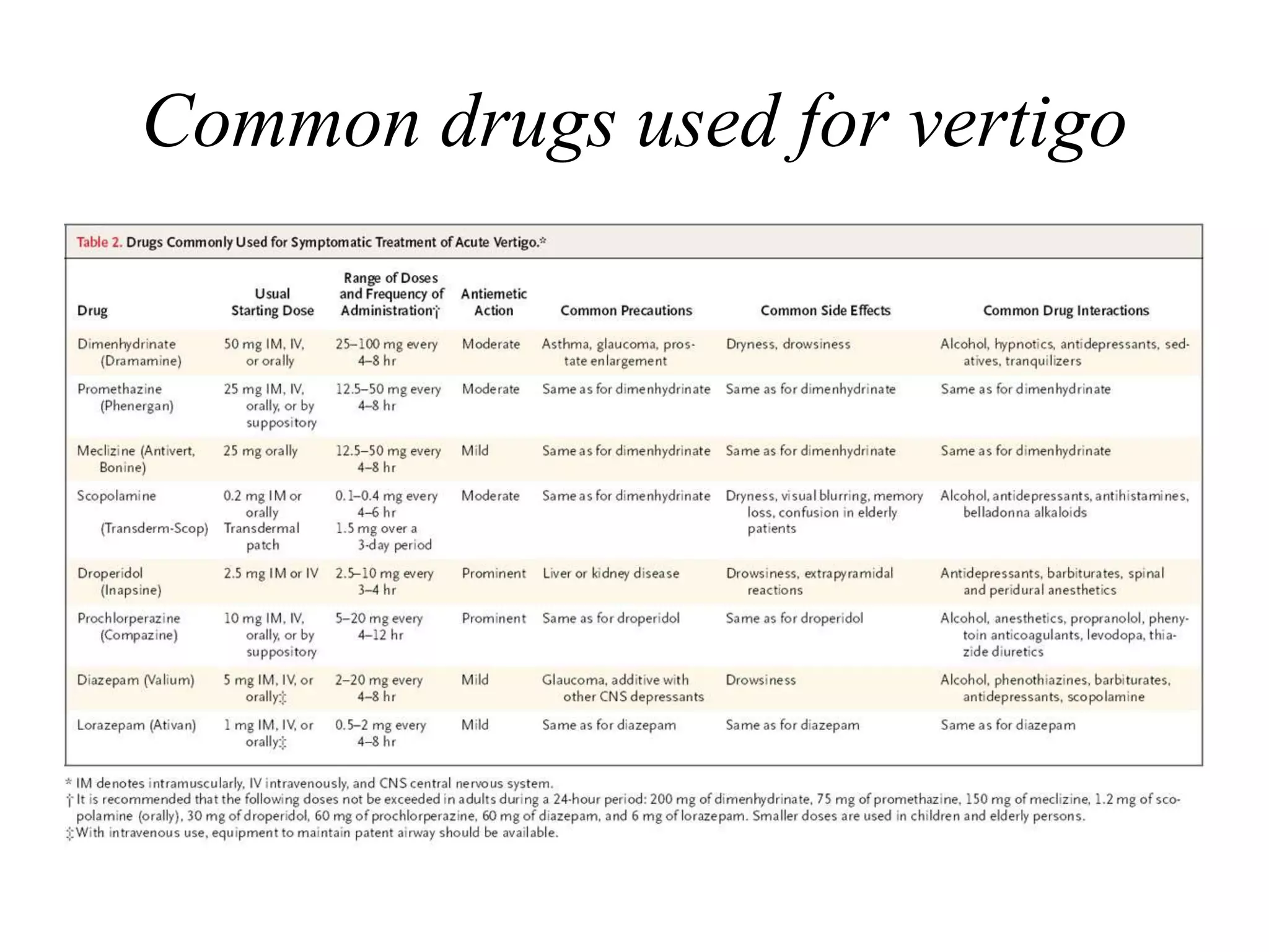
2. DEFINITION
SUBJECTIVE SENSE OF IMBALANCE
“Sensation as if the external world is
revolving around the patient or as if he
himself is revolving in space”
3. VESTIBULAR SYSTEM
Peripheral vestibular system:-include the vestibular apparatus( 3 SCC ,
Utricle & Saccule) and vestibular nerve
Five Vestibular Receptor organs are present in the Vestibular Labyrinth.
o Two Maculae in utricle and saccule (otolith organs) --- Linear
Acceleration.
o Three Cristae Ampullares of SSC --- Angular Accleration.
Central vestibular system(within CNS):starts from vestibular nuclei in the
Pons and its central connection(including cerebellum)
4. PATHOPHYSIOLOGY
Balance requires –
Normal functioning vestibular system
Input from visual system (vestibulo-ocular)
Input from proprioceptive system (vestibulo-spinal)
Central causes compromise central circuits that
mediate vestibular influences on posture, gaze control,
autonomic function
Disruption of balance between inputs results in vertigo
Goal of treatment: restore balance between different
inputs
5. PATHOPHYSIOLOGY(cont)
Vestibular system influences autonomic system
Intimate linkage in brainstem pathways between
vestibular and visceral inputs
Alteration of vestibular inputs results in:
Nausea, vomiting
Pallor
Respiratory/circulatory changes
6. Causes of peripheral vestibular system
Like any organ in the body, the vestibular system can be affect by:-
A) Congenital malformation:-but it is a symptomatic as central compensation occur early
in infancy
B) Acquired:-
1.Infections:- bacterial or viral ;acute or chronic e.g labrynthitis, vestibular neuritis,
syphilis
2.Tumour:-like acoustic neuroma(vestibular schwannoma)
3.Trauma:-temporal bone fracture, perilymph fistula
4.Autoimmune:- autoimmune inner ear diseases
5.Vascular:- labyrinthine apoplexy
6.Degenerative:- Dysequilibrium of ageing (presbyastasis )
7.Miscillaneous:- Meniere's disease, benign paroxysmal positional vertigo(BPPV)
14. BENIGN PAROXYSMAL POSITIONAL
VERTIGO (BPPV)
Most common cause
Dysfunction of posterior SCC
Cupulolithiasis vs. Canalithiasis
Cupulolithiasis
Calcium deposits embedded on cupula
PSCC becomes dependent on gravity
Canalithiasis
Calcium debris (otoconia) displaced into PSCC
Does not adhere to cupula
15. BPPV(cont)
Head movements
Looking up
Lying down
Rolling onto affected ear
Result in displacement of “sludge” / otoconia
Vertigo lasting a few seconds
Treatment approaches
Laboratory maneuvers
Particle repositioning
Habituation exercises
16. MENIERE’S DISEASE
Is an inner ear disease characterized by :-
• Episodic vertigo ,hearing loss ,tinnitus and
ear fullness lasting for minutes –days.
• The disease affect cochlea and SCC and
vestibule(whole membranous labyrinth)
which reflect its symptoms
17. MENIERE’S DISEASE (cont)
Pathogenesis
Distension of the membranous labyrinth or
endolymphatic hydrops is postulated as a
cause of this condition either due to excessive
production or impair absorption of endolymph
The attack occurs due to small ruptures in
Reissner membrane leading to mixing of the
endolymph and perilymph.
18. MENIERE’S DISEASE (cont)
Clinical features
The duration of the vertigo last usually for 30 minutes
to 4 hours
The vertigo is often disabling and very acute in onset
Associated with nausea and vomiting
The patient feels tired for the next few days after the
attack.
19. MENIERE’S DISEASE (cont)
Hearing loss is sensorineural in type and in early stage of the disease affects the
lower frequencies and return to normal after the attack (fluctuating hearing loss )
Tinnitus and a feeling of fullness or pressure in the affected ear may precedes the
attack
Over the course of the disease hearing loss and tinnitus become permanent
The disease is usually unilateral initially but can become bilateral
Sign:- spontaneous rotatory nystagmus
Investigations:- P.T.A. , brain MRI may be needed to role out central causes
20. ACOUSTIC NEUROMA
Vestibular symptoms are variable and tinnitus
is common.
Diagnosis is made on the basis of radiological
investigations.
Treatment depends upon size of tumour.
Various modalities are surgery, steriotactic surgery.
21. VESTIBULAR NEURITIS
Etiology
Most likely due to viral infection supported by polymerase chain reaction to detect viral DNA
In postmortem study mostly Herpes simplex virus type one
Symptoms
Acute spontaneous vertigo associated with nausea and vomiting lasting from days –weeks,
No cochlear & CNS involvement, no
The symptoms subside over the following days but many patients have residual imbalance that
last for months .
Hearing is normal
Sign
Acute attack there is nystagmus (horizontal towards the unaffected side )
After acute stage passed, nystagmus disappear but patient characteristically rotate towards the
affected side when attempt to march on a spot with their eyes closed positive Unterberger test
PTA ,brain MRI may be needed if central vertigo cannot be excluded easily
22. TRAUMATIC INNER EAR DISEASE
Non-Operative: Labyrinthine concussion or fracture of temporal bone.
Post-Operative: A perilymph fistula may occur after ear surgery esp.
`stapedectomy
Temporal bone fractures
Longitudinal fractures 80 -90 % , Usually spare the labyrinth and facial
nerve
Transverse fractures 10- 20% , Transverse fracture usually involve the
labyrinth and thus lead to sensorineural hearing loss with profound
vertigo. This vertigo will settle with time as central compensation occurs .
23. TRAUMATIC INNER EAR DISEASE(cont)
Management
For fracture itself--conservative plus broad spectrum antibiotic
Vertigo---vestibular sedative
Haemotympanum----conservative treatment usually resolves within 3-4 weeks
Tympanic membrane perforation----usually heals within 3 months if persists then myringoplasty
Facial nerve paralysis:
a. Immediate paralysis treated by surgical exploration with attempt to repair the nerve.
b. Delayed paralysis : usually treated conservatively
CSF leak :
Medical treatment : bed rest ,head elevation, stool softener , lumbar drain and antibiotics to
prevent meningitis
Surgical closure of the defect if medical treatment fail
24. LABYRINTHITIS
Inflammation of labyrinth due to any cause.
May be viral or bacterial.
Viral may occur during course of an
exanthematous disease like mumps/measles
or influenza type illness.
Bacterial labyrinthitis may be circumscribed,
serous or suppurative in a case of otorrhoea.
It may also occur during course of meningitis.
25. SUPERIOR SEMICIRCULAR CANAL
DEHISCENCE SYNDROME
Syndrome of vertigo and oscillopsia induced by
loud noises or by stimuli that change middle
ear or intracranial pressure
Tullio phenomenon - eye movement - loud noise
Hennebert's sign
“Third mobile window
26. MIGRAINE
Concomitant vertigo and disequilibrium
Headache control improves vertigo
Diagnostic criteria
Personal/family history
Motion intolerance
Vestibular symptoms - do not fit other causes
Theories - vascular origin, abnormal neural
activity (brainstem), abnormal voltage-gated
calcium channel genes.
27. VERTEBROBASILAR INSUFFICIENCY
Vertigo, diplopia, dysarthria, gait ataxia and
bilateral sensory & motor disturbance
Transient ischemia - low stroke risk
Antiplatelet therapy - aspirin 325mg qD
Ticlid
Platelet aggregate inhibitor
Risk of life-threatening neutropenia
Only in patients unable to tolerate aspirin
28. CNS NEOPLASM
Tumors involving brainstem, cerebellum or
midbrain
Other signs of intracranial disease are found
On ENG nystagmus is found to be irregular
and enhanced on eye opening.
29. POSTERIOR INFERIOR CEREBELLAR ARTERY
THROMBOSIS (LATERAL MEDULLARY SYNDROME
Onset is with severe vertigo with contralateral
hemianalgesia.
31. HISTORY
DESCRIPTION
Ask the patient to describe the problem
True rotatory vertigo or dizziness.
Severity
Number of attacks
Temporal pattern (continuous vs. episodic / short vs. prolonged)
If associated with turning the head, lying supine, or sitting upright.
Vestibular & cochlear symptoms (hearing loss either fluctuating or progressive, tinnitus, ear
pressure, nausea and vomiting)
Degree of impairment during the attack
Syncope:Transient loss of consciousness with loss of postural tone
Presyncope: Lightheadedness-an impending loss of consciousness
Psychiatric dizziness: Dizziness not related to vestibular dysfunction
Disequilibrium: Feeling of unsteadiness, imbalance or sensation of “floating” while walking
33. PREVIOUS HISTORY
Injuries:
Head trauma in the past (post traumatic hydrops)
History Of prior ear surgery (labyrinthine fistula,
perilymphatic fistula.)
Drugs : Aminoglycosides, cisplatin, miocycline
Stress situations
Family illness
Systemic Diseases:
History of DM (causes visual, proprioceptive, vascular
problems) HTN, cardiovascular and cerebrovascular
diseases
34. GPE
Cardiovascular, BP (including orthostatic) in both
arms, pulse
Neurologic
ENT HEAD AND NECK EXAMINATION
Detailed ENT Examination
Tympanic membrane for retraction, perforation,
Infection, cholesteatoma,valsalva
Assess hearing on both sides
Cranial nerves
Bruits in the neck
35. SPECIFIC VESTIBULAR SYSTEM EXAMINATION
(Balance tests need not be performed in acute vertigo)
Nystagmus
Corneal test
Fistula test
Romberg test
Gait
Past-pointing and falling
Hallpike-manoeuvre (positional test)
Caloric tests
Electronystagmography
Test of cerebellar dysfunction
36. Nystagmus
Defined as involuntary, rhythmical, oscillatory
movement of eyes
It is an important sign in evaluation of vestibular
system
It can be either horizontal /vertical/rotatory nystagmus
It has 2 components : SLOW & FAST
The direction of fast component indicates the direction
of the nystagmus
Slow component usually ipsilateral to diseased
structure
Fast component due to cortical correction
37. Intensity of nystagmus is indicated by its degree.
AS PER ALEXANDER’S LAW
1st DEGREE
It is weak nystagmus and is present when patient looks in the direction of fast
component
2nd DEGREE
It is stronger than 1st degree and is present when patient looks straight ahead
3rd DEGREE
It is stronger than the 2nd degree and is present when the patient looks in the direction
of the slow component
This law may not hold true in case of nystagmus of central region
38. PRESENCE of spontaneous nystagmus is indicative of ORGANIC LESIONS - Tone
of imbalance of vestibulo-ocular reflux
Peripheral nystagmus – is suppressed by optic fixation
Enhanced by darkness and use of FRENZEL GLASS
Central nystagmus is not supressed by optic fixation
TORSIONAL NYSTAGMUS – Indicates lesion of brainstem/vestibular nuclei e.g.
SYRINGOMYELIA
VERTICAL DOWNBEAT NYSTAGMUS – Lesion is at cranio-cervical region e.g.
Arnold-chiari malformation/degenerative lesion of cerebellum
VERTICAL UPBEAT NYSTAGMUS – Lesion at the junction of pons and
medulla/pons and midbrain
PENDULAR NYSTAGMUS – congenital/acquired e.g. Multiple sclerosis
May also be disconjugate Via., vertical in one eye and horizontal in other.
39. Central Peripheral
Spontaneous nystagmus that cannot be Suppressed by fixation suppressed by fixation.
Changes direction with gaze Doesn’t change direction
with gaze.
Purely vertical, Horizontal, rotatory,
horizontal, or torsional Never vertical
Paroxysmal but Not fatigable Paroxysmal but fatigable in
in Dix-hallpike test, Dix-hallpike test, has latency,
no latency, Lasting lasts less than a minute,
longer than 60 sec. and doesn't change direction with
often vertical, may different head positions.
change direction with
different head positions.
41. FISTULA TEST
Apply intermittent pressure on tragus OR By using Siegel's speculum
IN NORMAL PERSON: NEGATIVE
Because pressure changes in external auditory canal can’t be transmitted to
labyrinth
ABNORMALITY: POSITIVE
Erosion of horizontal semi-circular canal- cholesteatoma
Surgically created window in horizontal canal- fenestration operation
Abnormal opening in oval window- post stapedectomy fistula
Abnormal opening in round window- rupture of round window membrane
ALSO INDICATES THAT LABYRINTH IS STILL FUNCTIONAL
42. FALSE NEGATIVE FISTULA TEST :
IN CHOLESTEATOMA: it covers the site of fistula and it
doesn’t allow pressure changes to be transmitted to the
labyrinth
IN LABYRINTH DEAD
FALSE POSITIVE FISTULA TEST :
Means +ve test without presence of fistula
It is seen in two conditions : 1.congenital syphilis
2.Meniere’s disease.
Congenital syphilis: stapes footplate is hypermobile
Meniere’s disease: due to fibrous bands connecting
utricular macula to the stapes footplate.
43. ROMBERG TEST
Patient is asked to stand with feet together and arms by side with
eyes first open and then closed.
With eyes open : patient can still compensates the balance
With eyes closed : patient cant compensate –Here vestibular system
is at more disadvantage
Central: instability
Peripheral: Patient sways to side of lesion
If patient perform this test without sway then SHARPENED
ROMBERG TEST is performed.
• Patient is asked to stand with one heel in front of toes and arms
folded across the chest.
• Inability to perform this test indicates vestibular impairment
44. GAIT
Patient walks along a straight line to a fixed
point first with eyes opened and then closed.
In the case of uncompensated lesion of
peripheral vestibular system, with eyes closed
Patient deviates to affected side
45. HALLPIKE MANOEUVRE (POSITIONAL
TEST)
USES
When patient complains of vertigo in head position
Helps to differentiate a peripheral from a central lesion.
METHOD
Patient sits in the couch
Examiner holds the patient’s head, turns it 45˚ to the right and then places the
patient in a supine position so that his head hangs 30˚ below the horizontal.
Patient’s eyes are observed for nystagmus
The test is repeated with head turned to left and then again in straight head-
hanging position .
Four parameters are observed:
1. Latency
2. Duration
3. Direction
4. Fatiguability
48. In benign paroxysmal positional vertigo
Nystagmus appears after latency : 2-20s
Duration : less than 1 min
Direction : towards the ear that is under most
• On repetition : Nystagmus may be elicited but
lasts for a shorter period.
• On subsequent repetition : Nystagmus
disappears altogether
• NYSTAGMUS IS FATIGUABLE
49. IN CENTRAL LESIONS
Tumours of 4th ventricle
Cerebellum
Temporal lobe
Multiple sclerosis
Vertibrobasilar insufficiency
Raised intracranial tension
Nystagmus is produced immediately as soon as the head is in critical
position
No latency
Duration: lasts as long as head is in that critical position
Direction: changes
Fatiguability: nonfatiguable
50. TEST OF CEREBELLAR
DYSFUNCTION For cerebellar diseases – all cases of giddiness should be tested.
CEREBELLAR HEMISPHERE CAUSES:
1. Asynergia(abn finger-nose test)
2. Dysmetria(inability to control range of motion)
3. Adiadochokinesia (inability to perform rapid alternating movements)
4. Rebound phenomenon (inability to control movement of extremity when
opposing forceful restraint is suddenly released)
MIDLINE DISEASE OF CEREBELLUM CAUSES:
1. Wide base gait
2. Falling in any direction
3. Inability to make sudden turns while walking
4. Truncal ataxia
51. Nystagmus observed in cerebellar diseases
either in hemisphere or midline diseases
include
GAZE
EVOKED NYSTAGMUS
REBOUND NYSTAGMUS
ABNORMAL OPTOKINETIC NYSTAGMUS
52. Caloric test
Patient 45 degrees on couch
Water 33degrees or 45 degrees
Normal nystagmus
COLD: OPPOSITE
WARM: SAME
53. 1.CANAL PARESIS:
Decreased duration of nystagmus
Both hot and cold
2.DEAD LABYRINTH:
No nystagmoid response
3.DIRECTION PREPONDERANCE:
BY Both Hot and Cold
CENTRAL/PERIPHERAL lesion
59. Some drugs of the antihistamine class are
useful for symptomatic control of vertigo
Have anti-motion sickness properties in large
part due to inhibition of vestibular system H1
histaminergic neurotransmitters
Examples include dimenhydrinate
(Dramamine) and promethazine (Phenergan)
Also suppress the vomiting center
62. Vestibular Neuritis
Acute stage
• Bed rest , vestibular sedative, steroids can be used to
shorten the course of the disease
• Antiviral??
Resolution stage
After vertigo subsides (vestibular rehabilitation
exercise) will enhance central compensation(as it is a
paretic pathology)
63. Meniere's ‘s disease
In acute phase :- treatment consists of
vestibular sedative(stugerone, anti-emetics),
Betahistine(betaserc) ,diuretics
In long term :-avoidance of caffeine and salt
and reassurance can reduce the number of the
attacks and increase the patient ability to cope
with the attacks
64. Surgical Treatment:- rarely needed
A. Endolymphatic sac surgery: By transmastoid
approachch the endolymaphatic sac either
decompressed or a shunt is placed in the sac
that communicates with the subarachnoid
space or mastoid cavity
B. Vestibular nerve section:- in patients with
serviceable hearing
C. Labyrinthectomy : - in patients with
unilateral Meniere’s disease and poor
hearing
65. Ablation therapy
• Gentamycin is given through the tympanic
membrane will be absorbed through the round
window and selectively damage the vestibular
cells relative to the cochlear cells
67. The Epley and Semont Maneuvers
Are both intended to move debris out of the sensitive
part of the ear (posterior canal) to a less sensitive
location. Each maneuver takes about 15 minutes to
complete.
Semont maneuver: It involves a procedure whereby the
patient is rapidly moved from lying on one side to lying
on the other.
Epley Maneuver: It involves sequential movement of
the head into four positions, staying in each position for
roughly 30 seconds
70. Home Treatment Of BPPV:
Brandt-Daroff Exercises
The Brandt-Daroff Exercises is a method of treating BPPV, usually used
when the office treatment fails. They succeed in 95% of cases.
71. Selective posterior canal plugging offers a
reasonable surgical approach to intractable
symptoms.
Singular neurectomy, an older procedure, is
less popular because it produces hearing loss
in 7 to 17% of patients and fails in 8 to 12%.
72. Otosyphilis
Penicillin established treatment
IM and IV routes acceptable
IM - 2.4 million units benzathine PCN weekly
x 3 consecutive weeks is minimal treatment
(some advocate up to 1 year)
IV - 10 million units PCN G qD in divided
doses x 10 days, followed by 2.4 million units
benzathine PCN x 2 weeks
73. Vertebrobasilar insufficiency
Vertigo, diplopia, dysarthria, gait ataxia and bilateral sensory &
motor disturbance
Transient ischemia - low stroke risk
Antiplatelet therapy - aspirin
Ticlid
Platelet aggregate inhibitor
Risk of life-threatening neutropenia
Only in patients unable to tolerate aspirin
74. Migraine
Treatment
Modifying risk factors
Exercise and diet
Avoid nicotine, caffeine, red wine and chocolate
Abortive medical therapy
Ergots
Sumatriptin
Midrin
Prophylactic medical therapy
B blockers, Ca channel blockers, NSAIDs, amitryptiline, and lithium
76. Cawthorne Cooksey Exercises:
Sitting
Eye movements and head movements
Shoulder shrugging and circling
Bending forward and picking up objects from the ground
Standing
Eye, head and shoulder movements as before
Changing form sitting to standing position with eyes open and shut
Throwing a small ball from hand to hand (above eye level)
Throwing a ball from hand to hand under knee
Changing from sitting to standing and turning around in between
Moving about (in class)
Circle around center person who will throw a large ball and to whom it will be
returned
Walk across room with eyes open and then closed
77. Habituation of pathologic responses
Postural control exercises
Visual-vestibular interaction
Conditioning activities
B.I.D., most improve after 4-6 weeks
VRT - Elderly
Multifactorial causes of balance difficulty
Need 2 of 3 systems functional : vestibular, visual,
proprioceptive
78. Conclusions
Vestibular complaints common to ENT
Thorough evaluation and understanding
Dx and treat acute symptoms
Wean vestibular suppressants
Specific pharmacotherapy instituted
Chronic, uncompensated disease benefits from
early VRT