Downloaded 150 times


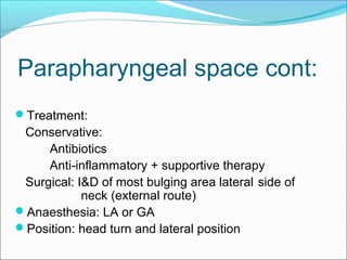


The document discusses several clinically important deep neck spaces, including the peritonsillar space, submaxillary space, retropharyngeal space, and parapharyngeal space. Infections in these spaces can spread rapidly to dangerous areas if not treated properly. The peritonsillar space is prone to infections leading to peritonsillar abscesses. The submaxillary space infection is known as Ludwig's angina. Retropharyngeal space infections present with pain, fever, and difficulty swallowing. Both conservative and surgical treatments are discussed for infections in these deep neck spaces.


