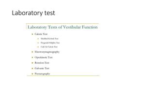
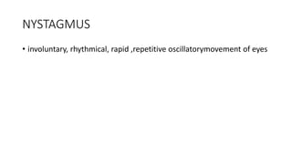
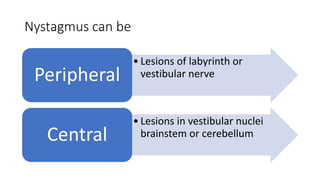
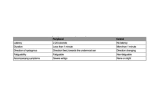
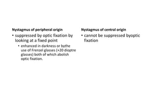
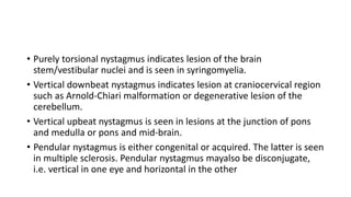
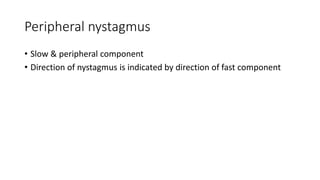
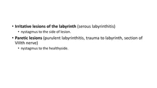
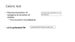
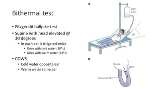
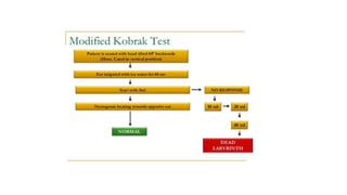
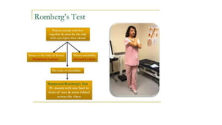
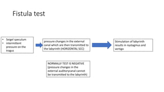
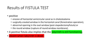
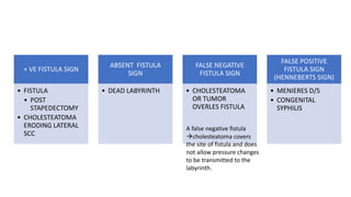
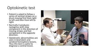
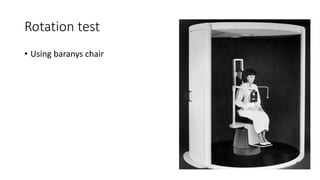
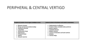
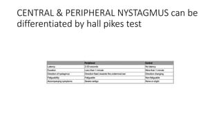
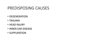
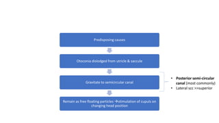
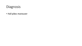
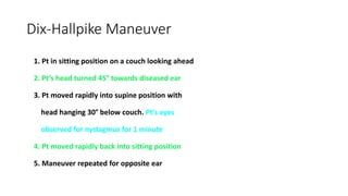
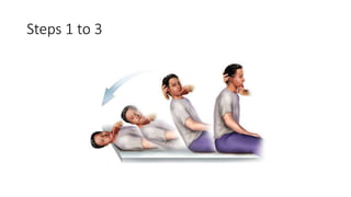
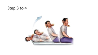
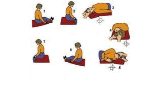
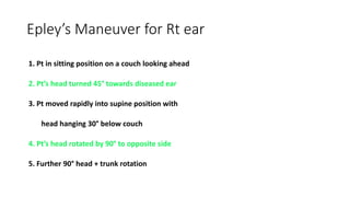
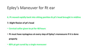
6. Nystagmus
• Stimulation of horizontal scc
Horizontal
• Stimulation of vertical (posterior)scc
Vertical
• Stimulation of superior (anterior)semicircular cana
Rotatory
7. Nystagmus can be
• Lesions of labyrinth or
vestibular nervePeripheral
• Lesions in vestibular nuclei
brainstem or cerebellumCentral
8.
9. Nystagmus of peripheral origin
• suppressed by optic fixation by
looking at a fixed point
• enhanced in darkness or bythe
use of Frenzel glasses (+20 dioptre
glasses) both of which abolish
optic fixation.
Nystagmus of central origin
• cannot be suppressed byoptic
fixation
10. • Purely torsional nystagmus indicates lesion of the brain
stem/vestibular nuclei and is seen in syringomyelia.
• Vertical downbeat nystagmus indicates lesion at craniocervical region
such as Arnold-Chiari malformation or degenerative lesion of the
cerebellum.
• Vertical upbeat nystagmus is seen in lesions at the junction of pons
and medulla or pons and mid-brain.
• Pendular nystagmus is either congenital or acquired. The latter is seen
in multiple sclerosis. Pendular nystagmus mayalso be disconjugate,
i.e. vertical in one eye and horizontal in the other
11. Peripheral nystagmus
• Slow & peripheral component
• Direction of nystagmus is indicated by direction of fast component
12. • Irritative lesions of the labyrinth (serous labyrinthitis)
• nystagmus to the side of lesion.
• Paretic lesions (purulent labyrinthitis, trauma to labyrinth, section of
VIIIth nerve)
• nystagmus to the healthyside.
13. Caloric test
• Thermal stimulation
nystagmus & sensation of
rotation
• Thermal gradient along lateral scc
• c/I in perforated TM
By caloric test only lateral scc can
be assessed
In perforated TM cold air caloric test
15. Bithermal test
• Fitzgerald hallpike test
• Supine with head elevated @
30 degrees
• In each ear is irrigated twice
• Once with cold water (30*c)
• Once with warm water (44*C)
• COWS
• Cold water opposite ear
• Warm water same ear
16.
17.
18. Fistula test
• Seigel speculum
• intermittent
pressure on the
tragus
pressure changes in the external
canal which are then transmitted to
the labyrinth (HORIZONTAL SCC)
Stimulation of labyrinth
results in nystagmus and
vertigo
NORMALLY TEST IS NEGATIVE
(pressure changes in the
external auditorycanal cannot
be transmitted to the labyrinth)
19. Results of FISTULA TEST
• positive
• erosion of horizontal semicircular canal as in cholesteatoma
• surgically-created window in the horizontal canal (fenestration operation),
• abnormal opening in the oval window (post-stapedectomyfistula) or
• the round window (rupture of round window membrane).
• A positive fistula also implies that the labyrinth is still functioning;
• absent when labyrinth is dead.
20. + VE FISTULA SIGN
• FISTULA
• POST
STAPEDECTOMY
• CHOLESTEATOMA
ERODING LATERAL
SCC
ABSENT FISTULA
SIGN
• DEAD LABYRINTH
FALSE NEGATIVE
FISTULA SIGN
• CHOLESTEATOMA
OR TUMOR
OVERLES FISTULA
FALSE POSITIVE
FISTULA SIGN
(HENNEBERTS SIGN)
• MENIERES D/S
• CONGENITAL
SYPHILIS
A false negative fistula
cholesteatoma covers
the site of fistula and does
not allow pressure changes
to be transmitted to the
labyrinth.
21. HENNEBERT SIGN
• A false positive fistula test
• In congenital syphilis, stapes footplate is hypermobile
• in Meniere's disease it is due to the fibrous bands connecting utricular macula
to the stapes footplate.
• In both these conditions, movements of stapes result in stimulation of
the utricular macula.
23. USES OF SEIGEL SPECULUM
• TO TEST MOBILITY OF TYMPANIC MEMBRANE
• MAGNIFIED VIEW OF SMALL PERFORATION
• INTRODUCE MEDICINE INTO M/E
• PERFORM FISTULA TEST
24. Optokinetic test
• Patient is asked to follow a
series of vertical stripes on a
drum moving first from right
to left and then from left to
right.
• Normally it produces
nystagmus with slow
component in the direction of
moving stripes and fast
component in the opposite
direction.
• Optokinetic abnormalities are
seen in brainstem and
cerebral hemisphere lesions.
Thus this test is useful to
diagnose a central lesion.
26. Galvanic Test
• only vestibular test which helps in differentiating an end organ
lesion from that of vestibular nerve.
• Patient stands with his feet together, eyes closed and arms
outstretched and then a current of 1 mAis passed to one ear.
Normally, person sways towards the side of anodal current. Body
sway can be studied bya special platform
32. Predisposing causes
Otoconia dislodged from utricle & saccule
Gravitate to semicircular canal
Remain as free floating particles stimulation of cupuls on
changing head position
• Posterior semi-circular
canal (most commonly)
• Lateral scc >>superior
34. Dix-Hallpike Maneuver
1. Pt in sitting position on a couch looking ahead
2. Pt’s head turned 45° towards diseased ear
3. Pt moved rapidly into supine position with
head hanging 30° below couch. Pt’s eyes
observed for nystagmus for 1 minute
4. Pt moved rapidly back into sitting position
5. Maneuver repeated for opposite ear
40. Epley’s Maneuver for Rt ear
1. Pt in sitting position on a couch looking ahead
2. Pt’s head turned 45° towards diseased ear
3. Pt moved rapidly into supine position with
head hanging 30° below couch
4. Pt’s head rotated by 90° to opposite side
5. Further 90° head + trunk rotation
41. Epley’s Maneuver for Rt ear
6. Pt moved rapidly back into sitting position & pt’s head brought in midline
7. Slight flexion of pt’s head
• Cervical collar given to pt for 48 hours
• Pt must have nystagmus at every step of Epley’s manoeuvre if it is done
properly
• 80% pt get cured by a single maneuver
42. Surgical treatment
Considered when Epley maneuver, Semont maneuver + Brandt-Daroff
exercises have failed and diagnosis of BPPV is clear
1. Posterior semicircular canal plugging (Parnes)
2. Singular neurectomy (Gacek)
45. Vestibular neuronitis
• 2nd most common cause of peripheral vertigo after BPPV
• Following URTI a/c onset vertigo
• Hearing is unaffected (unlike in a/c labyrinthitis )
46. Cogan syndrome
• Interstitial keratitis
• Audiovestibular involvement
• SNHL
• Vertigo
• Tinnitus
• Auto Ab against enothelium
vasculitis affecting aorta renal
arteries & coronary arteries