Downloaded 184 times


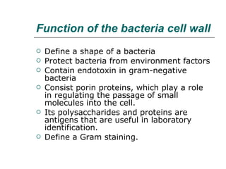


The document provides an overview of microbiology, including the structure and morphology of microorganisms such as bacteria, fungi, protozoa, and viruses. It discusses topics like bacterial cell structure, flagella and pili, endospores, capsules, inclusion bodies, and the contributions of Anton van Leeuwenhoek, who is considered the father of microbiology.


![06_Kingdom_Prokaryotae769[1].pptx](https://cdn.slidesharecdn.com/ss_thumbnails/06kingdomprokaryotae7691-240127144445-3a0d06ed-thumbnail.jpg?width=640&height=640&fit=bounds)


