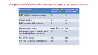
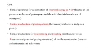
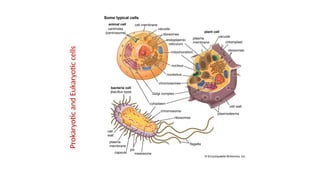
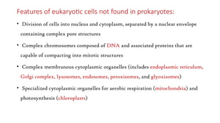
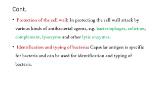
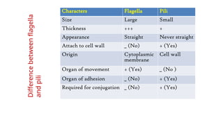
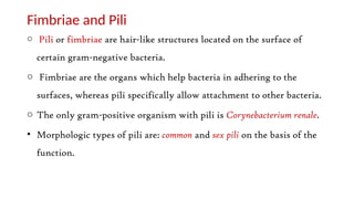
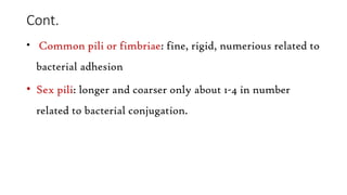
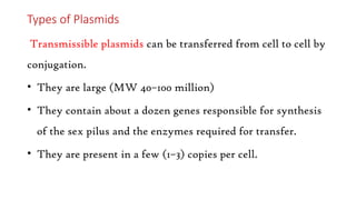
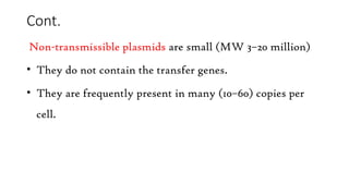
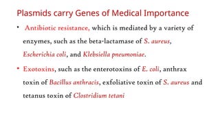
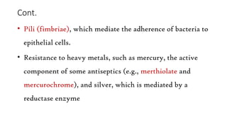
South University of Medicine, Science and Technology
Course: General Microbiology Class: 4th
year
Lecture 3 Date: 31/01/ 2025
Academic year 2024/2025
Faculty of Medicine and Health Science
Topics
• Bacterial cell -essential and non-essential components:
• Cell envelope, cytoplasm
• Cytoplasmic inclusions
• Capsules, flagella, fimbriae, spores, etc.
Important features of microorganisms
• The distinction between microorganisms (bacteria, fungi,
viruses and protozoa /helminthes) are based on three
criteria, viz:
• Structure of the cell.
• Methods of replication.
• Nature of nucleic acids.
Bacteria cell
• Structure, bacteria cells have a nucleoid (nuclear material), which
contains DNA; this is surrounded by cytoplasm, within which
proteins are synthesized and energy is generated.
• Where as viruses have an inner core of genetic material (either DNA
or RNA) but not both also no cytoplasm, and so they depend on host
cells to provide the machinery for protein synthesis and energy
generation.
Methods of Replication in Microorganisms
• Method of Replication. Cells replicate either by binary fission or by
mitosis, during which one parent cell divides to make two progeny
cells.
• Prokaryotic cells (e.g., bacteria) replicate by binary fission, whereas
eukaryotic cells replicate by mitosis.
• In contrast, viruses disassemble, produce many copies of their nucleic
acid and protein, and then reassemble into multiple progeny viruses.
note: viruses must replicate within host cells.
Types of cells found in Microorganisms
• Generally, cells have evolved into two fundamentally
different types:
- Eukaryotic cells
- Prokaryotic cells
Fungal (yeast ) cell Protozoan cell
Eukaryotic
cells
Structure of bacterium cell
Prokaryotic
cell
Characteristics Eukaryotic cell
(human cell)
Prokaryotic cell
(bacterial cell)
DNA within a nuclear membrane Yes No
Mitotic division Yes No
DNA associate with histones Yes No
Chromosome number More than one One
Membrane bound organelles such
as mitochondria and lysosomes
Yes No
Size of ribosomes 80S 70S
Cell wall containing peptidoglycan No Yes
Comparison of characteristics between prokaryotic and eukaryotic cells
Features held in common by the two types of cells
(Prokaryotic and Eukaryotic)
• Plasma membrane of similar construction
• Genetic information encoded in DNA using identical genetic code
• Similar mechanisms for transcription and translation of genetic
information, including similar ribosomes
• Shared metabolic pathways (e.g., glycolysis and TCA cycle)
Cont.
• Similar apparatus for conservation of chemical energy as ATP (located in the
plasma membrane of prokaryotes and the mitochondrial membrane of
eukaryotes)
• Similar mechanism of photosynthesis (between cyanobacteria and green
plants)
• Similar mechanism for synthesizing and inserting membrane proteins
• Proteasomes (protein digesting structures) of similar construction (between
archaebacteria and eukaryotes
Prokaryotic
and
Eukaryotic
cells
Features of eukaryotic cells not found in prokaryotes:
• Division of cells into nucleus and cytoplasm, separated by a nuclear envelope
containing complex pore structures
• Complex chromosomes composed of DNA and associated proteins that are
capable of compacting into mitotic structures
• Complex membranous cytoplasmic organelles (includes endoplasmic reticulum,
Golgi complex, lysosomes, endosomes, peroxisomes, and glyoxisomes)
• Specialized cytoplasmic organelles for aerobic respiration (mitochondria) and
photosynthesis (chloroplasts)
Cont.
• Complex cytoskeletal system (including microfilaments,
intermediate filaments, and microtubules) and associated motor
proteins
• Complex flagella and cilia
• Ability to ingest fluid and particulate material by enclosure within
plasma membrane vesicles (endocytosis and phagocytosis)
• Cellulose-containing cell walls (in plants).
Cont.
• Cell division using a microtubule-containing mitotic spindle that
separates chromosomes
• Presence of two copies of genes per cell (diploidy), one from each
parent
• Presence of three different RNA synthesizing enzymes (RNA
polymerases)
• Sexual reproduction requiring meiosis and fertilization
Bacteria are classified by shape into
three basic groups:
• Spheroidal (cocci)
• Cylindrical (bacilli or Rods)
• Spirilla (spirochetes)
Shapes and Sizes of Bacteria
Complex Shape
• In addition, Some bacteria are variable in shape and are
referred to as pleomorphic (many-shaped) e.g.
Actinomycetes.
• Actinomycetes, are branching filamentous bacteria
resembling fungi. They possess a rigid cell wall.
Sizes of Bacteria cell
Bacteria are microscopic and very small in size. The size of
bacteria is measured in units of length called microns.
• A micron (micrometer, m) is the unit of measurement in
bacteriology.
• 1 micron (m) = 1/1000 millimeter (mm).
• 1 nanometer (nm) = 1/1000 micron (m).
• 1 Angstrom unit (Å) =1/10 nm (nanometer).
Bacteria of medical importance measure 2 - 5µm (length) x
0.2 -1.5 µm (width).
Cont.
• Cocci are true spheres with diameter ranging between 0.75 to 1.25 μm
(and average of 1 μm).
• Bacilli vary in length from 2-10 times their width.
• Coccobacilli are very short bacilli
• Filaments are long threads of bacilli which have not separated into
single cells.
• Curved bacterial rods vary from small, coma shaped or mildly helical
shaped organisms with only one curve such as Vibrio cholerae.
Cocci may occur in
pairs e.g. Diplococci
Chain e.g. streptococci
Grapelike cluster e.g.
staphylococcus
Tetrads e.g. Micrococci
Arrangement of cocci cells
Streptococcus agalactiae
cocci in chains
Staphylococcus aureus
cocci in clusters
Bacillus megaterium
rods in chains
Arrangement of Bacilli (Rods)
Are rod shaped and can be arranged or exist in:
• Coccobacilli, length of the bacteria is approximately the
same as its width, e.g., Brucella
• Streptobacilli, cells are arranged in chains, e.g.,
Streptobacillus.
Arrangement of Bacilli
• Comma shaped, their cells exhibit curved
appearance, e.g., Vibrio.
• Fusiform Bacilli
Vibrio cholerae – comma shaped
Arrangement of Spirochetes
Spirochetes
• They exhibit rigid spiral forms, e.g., Spirillum).
• Relaxed coil (e.g., Borrelia).
• Tightly coiled (e.g., Treponema).
Structure of Bacterial cell
Bacterial capsule
• Use of capsule-swelling reaction: This phenomenon is seen in and
allows rapid identification of capsular serotypes of Streptococcus
pneumoniae, Neisseria meningitidis, several groups of streptococci,
Klebsiella, Haemophilus influenzae, Yersinia and Bacillus
Function of bacterial capsule
• Usually weakly antigenic
• Not necessary for viability of the bacterium
• Enhances virulence
• Protects from phagocytosis
• Plays a role in the adherence of bacteria
Cont.
• Capsulated strains are invariably non-motile
• Visualized by:
• - negative staining (India ink)
• - special capsular staining
• Detected by quellung reaction (swelling reaction)
Capsule as a virulence factor
• Capsules often act as a virulence factor by protecting the bacterium
from ingestion by phagocytosis.
• where non-capsulate mutant of these bacteria are nonvirulent.
• Repeated subcultures in vitro lead to the loss of capsule and also of
virulence.
Cont.
• Protection of the cell wall: In protecting the cell wall attack by
various kinds of antibacterial agents, e.g. bacteriophages, colicines,
complement, lysozyme and other lytic enzymes.
• Identification and typing of bacteria: Capsular antigen is specific
for bacteria and can be used for identification and typing of
bacteria.
Characters Flagella Pili
Size Large Small
Thickness +++ +
Appearance Straight Never straight
Attach to cell wall _ (No) + (Yes)
Origin Cytoplasmic
membrane
Cell wall
Organ of movement + (Yes) _ (No )
Organ of adhesion _ (No) + (Yes)
Required for conjugation _ (No) + (Yes)
Difference
between
flagella
and
pili
Fimbriae and Pili
o Pili or fimbriae are hair-like structures located on the surface of
certain gram-negative bacteria.
o Fimbriae are the organs which help bacteria in adhering to the
surfaces, whereas pili specifically allow attachment to other bacteria.
o The only gram-positive organism with pili is Corynebacterium renale.
• Morphologic types of pili are: common and sex pili on the basis of the
function.
Cont.
• Common pili or fimbriae: fine, rigid, numerious related to
bacterial adhesion
• Sex pili: longer and coarser only about 1-4 in number
related to bacterial conjugation.
Functions of Fimbriae
• Organ of adhesion.
• Hemagglutination
• Conjugation tube through which genetic material is transferred
from the donor to recipient cell.
• They are antigenic.
• Agglutination and pellicle formation
Flagella
• These are long, sinnous contractile filamentous appendages.
• These are organs of locomotion, e.g. Escherichia coli, salmonella, vibrio,
pseudomonas, etc.
• The number of flagella varies up to 10 to 20 per cells according to species of bacteria.
Escherichia coli has a motility of about 30 µ and that of Vibrio cholerae about 60 µ per
second.
• These are extremely thin 12 to 30 nm, helical shaped structure of uniform diameter
throughout their length.
• These are 3 to 20 µ long. Each flagellum consists of hook and basal body
Flagellated
bacteria
Parts and composition
Each flagellum consists of three parts;
i. Filament
ii. Hook
iii. Basal body.
• Filament: The filament is the longest and most obvious portion
which extends from the cell surface to the tip.
Cont.
• Hook: The hook is a short, curved segment which links the
filament to its basal body and functions as universal joint between
the basal body and the filament.
• Basal body: The basal body is embedded in the cell (cytoplasmic
membrane). In the gram-negative bacteria, the basal body has four
rings connected to a central rod (L, P, S and M).
Cont.
• The outer L and P rings associated with the lipopolysaccharide and
peptidoglycan layers respectively. S ring is located just above the
cytoplasmic membrane and the inner M ring contacts the
cytoplasmic membrane.
• Gram-positive bacteria have only two basal body rings, an inner
ring connected to the cytoplasmic membrane and an outer one
probably attached to peptidoglycan.
Features of flagella
• Flagella antibodies are not protective, but may help in
serodiagnosis.
• Though different genera of bacteria have flagella of same
chemical composition, they are antigenically different.
• Polar flagellated bacteria move the fastest, e.g. darting
mobility in Vibrio cholerae, may be as fast as 200 μm per
second.
Arrangement of Bacterial flagella
Flagella have characteristic arrangement in different bacteria
and this is helpful in classification of certain bacteria.
• Monotrichous, bacteria which have one polar flagellum, e.g.
Vibrio cholerae.
• Lophotrichous, bacteria with a tuft of several polar flagella,
e.g. in Spirilla.
Cont.
• Amphitrichous, bacteria with flagella at both the ends,
e.g. in some campylobacter sps.
• Peritrichous, bacteria with flagella distributed all over the
surface of the bacterium, e.g. in Salmonella typhi.
• Atrichous, bacteria which do not possess any flagellum,
e.g. in shigella sps.
Characteristic arrangements of flagella in bacteria
Medically importance of flagella
Flagella are medically important for two reasons:
• Some species of motile bacteria (e.g., E. coli and Proteus
species) are common causes of urinary tract infections.
Hence flagella play a role in pathogenesis.
• Some species of bacteria (e.g., Salmonella species) are
identified in the clinical laboratory by the use of specific
antibodies against flagella proteins.
The plasmids
• Plasmids are extrachromosomal, double-stranded, circular
DNA molecules that are capable of replicating
independently of the bacterial chromosome.
• Plasmids occur in both gram-positive and gram-negative
bacteria,
Types of Plasmids
Transmissible plasmids can be transferred from cell to cell by
conjugation.
• They are large (MW 40–100 million)
• They contain about a dozen genes responsible for synthesis
of the sex pilus and the enzymes required for transfer.
• They are present in a few (1–3) copies per cell.
Cont.
Non-transmissible plasmids are small (MW 3–20 million)
• They do not contain the transfer genes.
• They are frequently present in many (10–60) copies per
cell.
Plasmids carry Genes of Medical Importance
• Antibiotic resistance, which is mediated by a variety of
enzymes, such as the beta-lactamase of S. aureus,
Escherichia coli, and Klebsiella pneumoniae.
• Exotoxins, such as the enterotoxins of E. coli, anthrax
toxin of Bacillus anthracis, exfoliative toxin of S. aureus and
tetanus toxin of Clostridium tetani
Cont.
• Pili (fimbriae), which mediate the adherence of bacteria to
epithelial cells.
• Resistance to heavy metals, such as mercury, the active
component of some antiseptics (e.g., merthiolate and
mercurochrome), and silver, which is mediated by a
reductase enzyme
Cont.
• Resistance to ultraviolet light, which is mediated by DNA
repair enzymes.
• Bacteriocins are toxic proteins produced by certain
bacteria that are lethal for other bacteria
Bacterial spores
• These highly resistant structures are formed in response to
adverse conditions by two genera of medically important.
• Gram-positive rods: the genus Bacillus, which includes the
agent of anthrax and the genus Clostridium, which
includes the agents of tetanus and botulism.
Bacterial Spore formation
• Spore formation (sporulation) occurs when nutrients, such
as sources of carbon and nitrogen, are depleted
• The bacterial endospore is not a reproductive structure.
One cell forms one spore under adverse conditions in a
process called sporulation.
Cont.
• Bacterial endospores or spores, are small, dehydrated, metabolically
quiescent forms that are produced by some bacteria in response to
nutrient limitation or a related sign other dangers.
• Some spore-forming bacteria are of great importance in medicine,
causing such diseases as anthrax, gas gangrene, tetanus, and
botulism.
• All medically important spore formers are gram-positive rods.
Stages
of
bacterial
spore
formation
Structure
of
bacteria
spore
Properties of endospores
• Core: The fully developed spore has the core which is the spore
protoplast containing the normal cell structures but is metabolically
inactive.
• Spore wall: The innermost layer surrounding the inner spore
membrane is called the spore wall. It contains normal peptidoglycan
and becomes the cell wall of the germinating vegetative cell
Cont.
• Cortex: The cortex is the thickest layer of the spore envelope.
Cortex peptidoglycan is extremely sensitive to lysozyme, and its
autolysins plays a role in spore germination.
• Spore coat: Cortex, in turn, is enclosed by fairly thick spore coat.
• Exosporium: Spores of some species have an additional, apparently
rather loose covering known as the exosporium, which may have
distinctive ridges and grooves
Shape and Position of Spores
The shape and position of the spore and its size relative to the parent cell are
species characteristics.
• Spores may be central (equatorial), subterminal (close to one end), or terminal.
• The appearance may be spherical, ovoid or elongated, and being narrower than
the cell, or broader and bulging it.
• The diameter of spore may be same or less than the width of bacteria
(Bacillus), or may be wider than the bacillary body producing a distension or
bulge in the cell (Clostridium).
Types of spores
• Central, bulging
• Central, not bulging
• Subterminal, bulging
• Subterminal, not bulging
• Terminal, spherical
• Terminal, oval
Resistance
• Bacterial spores constitute some of the most resistant forms of life.
• they may remain viable for centuries.
• spores of all medically important species are destroyed by autoclaving at
121°C for 15 minutes.
• Methods of sterilization and disinfection should ensure that spores also
are destroyed.
• Sporulation helps bacteria survive for long periods under unfavorable
environments.
Endospore
• Endospore heat resistance probably is due to several factors:
calcium-dipicolinate and acid-soluble protein stabilization of DNA,
protoplast dehydration, the spore coat, DNA repair, the greater
stability of the cell proteins in bacteria adapted to growth at high
temperatures and others
Clinical relevance of bacteria spore
• Clinical importance of bacterial spores lies in their extraordinary
resistance to heat and chemicals.
• this property of bacterial spores is exploited when they are used for
evaluating the sterilization efficacy of autoclaves. steam heating
under pressure (autoclaving) at 121°C, for at least 15
minutes, is required to ensure the sterility of products for
medical use.
Character Gram + ve
bacteria
Gram – ve bacteria Acid fast bacteria
Peptidoglycan Thick layer Thin layer Small amount
Lipids Very little Lipopolysaccharide
(major)
Mycolic acid and
other waxes
Outer membrane Absent Present Absent
Periplasmic space Absent Present Absent
Cell shape Always rigid Rigid or flexible Rigid or flexible
Result of enzyme
digestion
Protoplast Spheroplast Difficult to digest
Sensitivity to dyes
and antibiotics
+++ ++ +
Characteristics
of
cell
walls
of
Gram-positive,
Gram
–
negative
and
acid
fast
bacteria
Characteristics Bacteria Fungi Protozoa and
helminthes
Viruses
Cell Yes Yes Yes No
Nucleic acid Both DNA
and RNA
Both DNA and
RNA
Both DNA and
RNA
Either DNA or
RNA
Type of nucleus Prokaryotic Eukaryotic Eukaryotic None
Ribosomes 70S 80S 80S Absent
Mitochondria Absent Present Present Absent
Motility Some None Most None
Method of
replication
Binary
fission
Budding and
mitosis
Mitosis Not binary
fission
Approximate
diameter in (µM)
1-5 3-10 (yeasts) 15-25
(trophozoites)
0.02- 0.2
Comparison of medically important microbes and
viruses

MEDICAL MICROBIOLGY......................................................

  • 1.
    South University ofMedicine, Science and Technology Course: General Microbiology Class: 4th year Lecture 3 Date: 31/01/ 2025 Academic year 2024/2025 Faculty of Medicine and Health Science
  • 2.
    Topics • Bacterial cell-essential and non-essential components: • Cell envelope, cytoplasm • Cytoplasmic inclusions • Capsules, flagella, fimbriae, spores, etc.
  • 3.
    Important features ofmicroorganisms • The distinction between microorganisms (bacteria, fungi, viruses and protozoa /helminthes) are based on three criteria, viz: • Structure of the cell. • Methods of replication. • Nature of nucleic acids.
  • 4.
    Bacteria cell • Structure,bacteria cells have a nucleoid (nuclear material), which contains DNA; this is surrounded by cytoplasm, within which proteins are synthesized and energy is generated. • Where as viruses have an inner core of genetic material (either DNA or RNA) but not both also no cytoplasm, and so they depend on host cells to provide the machinery for protein synthesis and energy generation.
  • 5.
    Methods of Replicationin Microorganisms • Method of Replication. Cells replicate either by binary fission or by mitosis, during which one parent cell divides to make two progeny cells. • Prokaryotic cells (e.g., bacteria) replicate by binary fission, whereas eukaryotic cells replicate by mitosis. • In contrast, viruses disassemble, produce many copies of their nucleic acid and protein, and then reassemble into multiple progeny viruses. note: viruses must replicate within host cells.
  • 6.
    Types of cellsfound in Microorganisms • Generally, cells have evolved into two fundamentally different types: - Eukaryotic cells - Prokaryotic cells
  • 7.
    Fungal (yeast )cell Protozoan cell Eukaryotic cells
  • 8.
    Structure of bacteriumcell Prokaryotic cell
  • 9.
    Characteristics Eukaryotic cell (humancell) Prokaryotic cell (bacterial cell) DNA within a nuclear membrane Yes No Mitotic division Yes No DNA associate with histones Yes No Chromosome number More than one One Membrane bound organelles such as mitochondria and lysosomes Yes No Size of ribosomes 80S 70S Cell wall containing peptidoglycan No Yes Comparison of characteristics between prokaryotic and eukaryotic cells
  • 10.
    Features held incommon by the two types of cells (Prokaryotic and Eukaryotic) • Plasma membrane of similar construction • Genetic information encoded in DNA using identical genetic code • Similar mechanisms for transcription and translation of genetic information, including similar ribosomes • Shared metabolic pathways (e.g., glycolysis and TCA cycle)
  • 11.
    Cont. • Similar apparatusfor conservation of chemical energy as ATP (located in the plasma membrane of prokaryotes and the mitochondrial membrane of eukaryotes) • Similar mechanism of photosynthesis (between cyanobacteria and green plants) • Similar mechanism for synthesizing and inserting membrane proteins • Proteasomes (protein digesting structures) of similar construction (between archaebacteria and eukaryotes
  • 12.
  • 13.
    Features of eukaryoticcells not found in prokaryotes: • Division of cells into nucleus and cytoplasm, separated by a nuclear envelope containing complex pore structures • Complex chromosomes composed of DNA and associated proteins that are capable of compacting into mitotic structures • Complex membranous cytoplasmic organelles (includes endoplasmic reticulum, Golgi complex, lysosomes, endosomes, peroxisomes, and glyoxisomes) • Specialized cytoplasmic organelles for aerobic respiration (mitochondria) and photosynthesis (chloroplasts)
  • 14.
    Cont. • Complex cytoskeletalsystem (including microfilaments, intermediate filaments, and microtubules) and associated motor proteins • Complex flagella and cilia • Ability to ingest fluid and particulate material by enclosure within plasma membrane vesicles (endocytosis and phagocytosis) • Cellulose-containing cell walls (in plants).
  • 15.
    Cont. • Cell divisionusing a microtubule-containing mitotic spindle that separates chromosomes • Presence of two copies of genes per cell (diploidy), one from each parent • Presence of three different RNA synthesizing enzymes (RNA polymerases) • Sexual reproduction requiring meiosis and fertilization
  • 16.
    Bacteria are classifiedby shape into three basic groups: • Spheroidal (cocci) • Cylindrical (bacilli or Rods) • Spirilla (spirochetes) Shapes and Sizes of Bacteria
  • 17.
    Complex Shape • Inaddition, Some bacteria are variable in shape and are referred to as pleomorphic (many-shaped) e.g. Actinomycetes. • Actinomycetes, are branching filamentous bacteria resembling fungi. They possess a rigid cell wall.
  • 18.
    Sizes of Bacteriacell Bacteria are microscopic and very small in size. The size of bacteria is measured in units of length called microns. • A micron (micrometer, m) is the unit of measurement in bacteriology. • 1 micron (m) = 1/1000 millimeter (mm). • 1 nanometer (nm) = 1/1000 micron (m). • 1 Angstrom unit (Å) =1/10 nm (nanometer). Bacteria of medical importance measure 2 - 5µm (length) x 0.2 -1.5 µm (width).
  • 19.
    Cont. • Cocci aretrue spheres with diameter ranging between 0.75 to 1.25 μm (and average of 1 μm). • Bacilli vary in length from 2-10 times their width. • Coccobacilli are very short bacilli • Filaments are long threads of bacilli which have not separated into single cells. • Curved bacterial rods vary from small, coma shaped or mildly helical shaped organisms with only one curve such as Vibrio cholerae.
  • 20.
    Cocci may occurin pairs e.g. Diplococci Chain e.g. streptococci Grapelike cluster e.g. staphylococcus Tetrads e.g. Micrococci Arrangement of cocci cells
  • 21.
    Streptococcus agalactiae cocci inchains Staphylococcus aureus cocci in clusters Bacillus megaterium rods in chains
  • 22.
    Arrangement of Bacilli(Rods) Are rod shaped and can be arranged or exist in: • Coccobacilli, length of the bacteria is approximately the same as its width, e.g., Brucella • Streptobacilli, cells are arranged in chains, e.g., Streptobacillus.
  • 23.
    Arrangement of Bacilli •Comma shaped, their cells exhibit curved appearance, e.g., Vibrio. • Fusiform Bacilli Vibrio cholerae – comma shaped
  • 24.
    Arrangement of Spirochetes Spirochetes •They exhibit rigid spiral forms, e.g., Spirillum). • Relaxed coil (e.g., Borrelia). • Tightly coiled (e.g., Treponema).
  • 25.
  • 26.
    Bacterial capsule • Useof capsule-swelling reaction: This phenomenon is seen in and allows rapid identification of capsular serotypes of Streptococcus pneumoniae, Neisseria meningitidis, several groups of streptococci, Klebsiella, Haemophilus influenzae, Yersinia and Bacillus
  • 27.
    Function of bacterialcapsule • Usually weakly antigenic • Not necessary for viability of the bacterium • Enhances virulence • Protects from phagocytosis • Plays a role in the adherence of bacteria
  • 28.
    Cont. • Capsulated strainsare invariably non-motile • Visualized by: • - negative staining (India ink) • - special capsular staining • Detected by quellung reaction (swelling reaction)
  • 29.
    Capsule as avirulence factor • Capsules often act as a virulence factor by protecting the bacterium from ingestion by phagocytosis. • where non-capsulate mutant of these bacteria are nonvirulent. • Repeated subcultures in vitro lead to the loss of capsule and also of virulence.
  • 30.
    Cont. • Protection ofthe cell wall: In protecting the cell wall attack by various kinds of antibacterial agents, e.g. bacteriophages, colicines, complement, lysozyme and other lytic enzymes. • Identification and typing of bacteria: Capsular antigen is specific for bacteria and can be used for identification and typing of bacteria.
  • 31.
    Characters Flagella Pili SizeLarge Small Thickness +++ + Appearance Straight Never straight Attach to cell wall _ (No) + (Yes) Origin Cytoplasmic membrane Cell wall Organ of movement + (Yes) _ (No ) Organ of adhesion _ (No) + (Yes) Required for conjugation _ (No) + (Yes) Difference between flagella and pili
  • 32.
    Fimbriae and Pili oPili or fimbriae are hair-like structures located on the surface of certain gram-negative bacteria. o Fimbriae are the organs which help bacteria in adhering to the surfaces, whereas pili specifically allow attachment to other bacteria. o The only gram-positive organism with pili is Corynebacterium renale. • Morphologic types of pili are: common and sex pili on the basis of the function.
  • 33.
    Cont. • Common pilior fimbriae: fine, rigid, numerious related to bacterial adhesion • Sex pili: longer and coarser only about 1-4 in number related to bacterial conjugation.
  • 34.
    Functions of Fimbriae •Organ of adhesion. • Hemagglutination • Conjugation tube through which genetic material is transferred from the donor to recipient cell. • They are antigenic. • Agglutination and pellicle formation
  • 35.
    Flagella • These arelong, sinnous contractile filamentous appendages. • These are organs of locomotion, e.g. Escherichia coli, salmonella, vibrio, pseudomonas, etc. • The number of flagella varies up to 10 to 20 per cells according to species of bacteria. Escherichia coli has a motility of about 30 µ and that of Vibrio cholerae about 60 µ per second. • These are extremely thin 12 to 30 nm, helical shaped structure of uniform diameter throughout their length. • These are 3 to 20 µ long. Each flagellum consists of hook and basal body
  • 36.
  • 37.
    Parts and composition Eachflagellum consists of three parts; i. Filament ii. Hook iii. Basal body. • Filament: The filament is the longest and most obvious portion which extends from the cell surface to the tip.
  • 38.
    Cont. • Hook: Thehook is a short, curved segment which links the filament to its basal body and functions as universal joint between the basal body and the filament. • Basal body: The basal body is embedded in the cell (cytoplasmic membrane). In the gram-negative bacteria, the basal body has four rings connected to a central rod (L, P, S and M).
  • 39.
    Cont. • The outerL and P rings associated with the lipopolysaccharide and peptidoglycan layers respectively. S ring is located just above the cytoplasmic membrane and the inner M ring contacts the cytoplasmic membrane. • Gram-positive bacteria have only two basal body rings, an inner ring connected to the cytoplasmic membrane and an outer one probably attached to peptidoglycan.
  • 40.
    Features of flagella •Flagella antibodies are not protective, but may help in serodiagnosis. • Though different genera of bacteria have flagella of same chemical composition, they are antigenically different. • Polar flagellated bacteria move the fastest, e.g. darting mobility in Vibrio cholerae, may be as fast as 200 μm per second.
  • 41.
    Arrangement of Bacterialflagella Flagella have characteristic arrangement in different bacteria and this is helpful in classification of certain bacteria. • Monotrichous, bacteria which have one polar flagellum, e.g. Vibrio cholerae. • Lophotrichous, bacteria with a tuft of several polar flagella, e.g. in Spirilla.
  • 42.
    Cont. • Amphitrichous, bacteriawith flagella at both the ends, e.g. in some campylobacter sps. • Peritrichous, bacteria with flagella distributed all over the surface of the bacterium, e.g. in Salmonella typhi. • Atrichous, bacteria which do not possess any flagellum, e.g. in shigella sps.
  • 43.
    Characteristic arrangements offlagella in bacteria
  • 44.
    Medically importance offlagella Flagella are medically important for two reasons: • Some species of motile bacteria (e.g., E. coli and Proteus species) are common causes of urinary tract infections. Hence flagella play a role in pathogenesis. • Some species of bacteria (e.g., Salmonella species) are identified in the clinical laboratory by the use of specific antibodies against flagella proteins.
  • 45.
    The plasmids • Plasmidsare extrachromosomal, double-stranded, circular DNA molecules that are capable of replicating independently of the bacterial chromosome. • Plasmids occur in both gram-positive and gram-negative bacteria,
  • 46.
    Types of Plasmids Transmissibleplasmids can be transferred from cell to cell by conjugation. • They are large (MW 40–100 million) • They contain about a dozen genes responsible for synthesis of the sex pilus and the enzymes required for transfer. • They are present in a few (1–3) copies per cell.
  • 47.
    Cont. Non-transmissible plasmids aresmall (MW 3–20 million) • They do not contain the transfer genes. • They are frequently present in many (10–60) copies per cell.
  • 48.
    Plasmids carry Genesof Medical Importance • Antibiotic resistance, which is mediated by a variety of enzymes, such as the beta-lactamase of S. aureus, Escherichia coli, and Klebsiella pneumoniae. • Exotoxins, such as the enterotoxins of E. coli, anthrax toxin of Bacillus anthracis, exfoliative toxin of S. aureus and tetanus toxin of Clostridium tetani
  • 49.
    Cont. • Pili (fimbriae),which mediate the adherence of bacteria to epithelial cells. • Resistance to heavy metals, such as mercury, the active component of some antiseptics (e.g., merthiolate and mercurochrome), and silver, which is mediated by a reductase enzyme
  • 50.
    Cont. • Resistance toultraviolet light, which is mediated by DNA repair enzymes. • Bacteriocins are toxic proteins produced by certain bacteria that are lethal for other bacteria
  • 51.
    Bacterial spores • Thesehighly resistant structures are formed in response to adverse conditions by two genera of medically important. • Gram-positive rods: the genus Bacillus, which includes the agent of anthrax and the genus Clostridium, which includes the agents of tetanus and botulism.
  • 52.
    Bacterial Spore formation •Spore formation (sporulation) occurs when nutrients, such as sources of carbon and nitrogen, are depleted • The bacterial endospore is not a reproductive structure. One cell forms one spore under adverse conditions in a process called sporulation.
  • 53.
    Cont. • Bacterial endosporesor spores, are small, dehydrated, metabolically quiescent forms that are produced by some bacteria in response to nutrient limitation or a related sign other dangers. • Some spore-forming bacteria are of great importance in medicine, causing such diseases as anthrax, gas gangrene, tetanus, and botulism. • All medically important spore formers are gram-positive rods.
  • 54.
  • 55.
  • 56.
    Properties of endospores •Core: The fully developed spore has the core which is the spore protoplast containing the normal cell structures but is metabolically inactive. • Spore wall: The innermost layer surrounding the inner spore membrane is called the spore wall. It contains normal peptidoglycan and becomes the cell wall of the germinating vegetative cell
  • 57.
    Cont. • Cortex: Thecortex is the thickest layer of the spore envelope. Cortex peptidoglycan is extremely sensitive to lysozyme, and its autolysins plays a role in spore germination. • Spore coat: Cortex, in turn, is enclosed by fairly thick spore coat. • Exosporium: Spores of some species have an additional, apparently rather loose covering known as the exosporium, which may have distinctive ridges and grooves
  • 58.
    Shape and Positionof Spores The shape and position of the spore and its size relative to the parent cell are species characteristics. • Spores may be central (equatorial), subterminal (close to one end), or terminal. • The appearance may be spherical, ovoid or elongated, and being narrower than the cell, or broader and bulging it. • The diameter of spore may be same or less than the width of bacteria (Bacillus), or may be wider than the bacillary body producing a distension or bulge in the cell (Clostridium).
  • 59.
    Types of spores •Central, bulging • Central, not bulging • Subterminal, bulging • Subterminal, not bulging • Terminal, spherical • Terminal, oval
  • 60.
    Resistance • Bacterial sporesconstitute some of the most resistant forms of life. • they may remain viable for centuries. • spores of all medically important species are destroyed by autoclaving at 121°C for 15 minutes. • Methods of sterilization and disinfection should ensure that spores also are destroyed. • Sporulation helps bacteria survive for long periods under unfavorable environments.
  • 61.
    Endospore • Endospore heatresistance probably is due to several factors: calcium-dipicolinate and acid-soluble protein stabilization of DNA, protoplast dehydration, the spore coat, DNA repair, the greater stability of the cell proteins in bacteria adapted to growth at high temperatures and others
  • 62.
    Clinical relevance ofbacteria spore • Clinical importance of bacterial spores lies in their extraordinary resistance to heat and chemicals. • this property of bacterial spores is exploited when they are used for evaluating the sterilization efficacy of autoclaves. steam heating under pressure (autoclaving) at 121°C, for at least 15 minutes, is required to ensure the sterility of products for medical use.
  • 63.
    Character Gram +ve bacteria Gram – ve bacteria Acid fast bacteria Peptidoglycan Thick layer Thin layer Small amount Lipids Very little Lipopolysaccharide (major) Mycolic acid and other waxes Outer membrane Absent Present Absent Periplasmic space Absent Present Absent Cell shape Always rigid Rigid or flexible Rigid or flexible Result of enzyme digestion Protoplast Spheroplast Difficult to digest Sensitivity to dyes and antibiotics +++ ++ + Characteristics of cell walls of Gram-positive, Gram – negative and acid fast bacteria
  • 64.
    Characteristics Bacteria FungiProtozoa and helminthes Viruses Cell Yes Yes Yes No Nucleic acid Both DNA and RNA Both DNA and RNA Both DNA and RNA Either DNA or RNA Type of nucleus Prokaryotic Eukaryotic Eukaryotic None Ribosomes 70S 80S 80S Absent Mitochondria Absent Present Present Absent Motility Some None Most None Method of replication Binary fission Budding and mitosis Mitosis Not binary fission Approximate diameter in (µM) 1-5 3-10 (yeasts) 15-25 (trophozoites) 0.02- 0.2 Comparison of medically important microbes and viruses