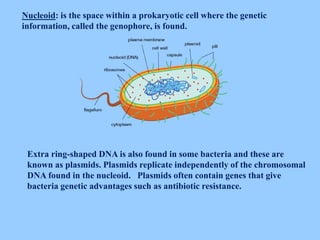
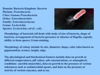
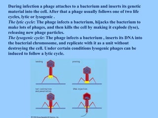
Microbiology
Specialty 241 “Hotel and Restaurant Business”
Topic 1. GENERAL MICROBIOLOGY
Theme 1 “Basic of morphology and systematic of
microorganisms. Bacteria”
Stabnikov Viktor, PhD
NATIONAL UNIVERSITY OF FOOD
TECHNOLOGIES
1. Place of microorganisms among living organisms
Microorganisms are very small, their size is measured in micrometers
(microns) : 1 millimeter (mm) = 1000 microns (мкм). 1 micron = 1000
nanometers (nm). Sizes of most bacteria are within the limits of 0,5-5 microns.
Microorganisms are visible only under a microscope.
Before microorganisms have been opened by Antoni van Leeuwenhoek (XVII
of century) and 100 after it, biologists placed all the living organisms into two
kingdoms: plants and animals.
When the first unicellular organisms were discovered by Antoine van
Leeuwenhoek in 1674, they were placed in one of the two kingdoms of living
beings, according to their characteristics. It remained so until 1866, when the
German biologist Ernst Haeckel proposed a third kingdom of life, which he
called Protista, and included all unicellular organisms in it.
Later, it was shown important differences in cells, mainly according to the
presence or absence of distinct nucleus, so, microorganisms were divided in
prokaryotes (without a distinct nucleus) and eukaryotes (with a distinct
nucleus) in 1925.
2. Structure and classification of microorganisms
Bacteria (prokaryotes) include the large group of microorganisms. More than
2400 types of bacteria are presently known.
They occur in a wide variety of forms but the three main basic shapes of
bacteria are spheres (also known as cocci), rods (also known as bacilli), and
spirals.
A spherical shaped cells are quite small, being only 0,5 µm to 1,0 µm in
diameter.
Coccus (cocci plural) bacteria are round, oval, or spherical in shape.
These cells can exist in several different arrangements that include:
Cocci that remain in a chain are called a streptococcus (2).
Cocci may divide randomly and form grape-like cluster of cell called a
staphylococcus (2).
Cocci remaining in a pair after reproduction represent a diplococcoccus (4).
Arrangement from four spheres that form a square called tetrad (5).
A cube-like packet of eight cocci is called a sarcina (6).
1 2 3 4 5 6
The size of rod shape bacteria can varies from 0,5 µm to 20 µm. Most rods
occur single, in pairs or arranged into a long chain.
Rod shape bacteria are subdivided into a bacterium, that does not
form spores, and bacillus which can form spore.
Bacteria of spiral form are characterized the different amount of coils : (1)
rods shaped of a comma, named vibrio (1). They also have a flagellum, which is
used for movement; rods with one or a few curls – spirillum (2) ; long rods with
by a great number of curls – spirochetes (3) .
1
2
3
.
Sizes of cocci, rods, and spirillum.
There are also another exotic shapes of bacteria. Some species have square, star
shape, multilobed or segmented.
STRUCTURE OF BACTERIAL CELL
Structures of Bacteria :
Cell Wall: Protects inside contents of cells: contains peptidoglycan and some
lipids (fats).
Cell Membrane: Contains enzymes for cellular respiration, murein and
peptidoglycan synthesis, oxidative phosphorylation as well as transport of
substances in and out of the cell.
Cytoplasm: It is an amorphous gel containing enzymes, ions and various small
structures similar to organelles that contain structures like cytoplasmic
inclusions.
Ribosomes: Proteins are synthesized on them
Mesosomes: Areas in the cell membrane of prokaryotic (bacterial) cells that
fold inward. Generates energy as ATP .
Nucleoid: is the space within a prokaryotic cell where the genetic
information, called the genophore, is found.
Extra ring-shaped DNA is also found in some bacteria and these are
known as plasmids. Plasmids replicate independently of the chromosomal
DNA found in the nucleoid. Plasmids often contain genes that give
bacteria genetic advantages such as antibiotic resistance.
Pigments: may be different colors such as
reds, blues or purple
Appendages
Capsule: Polysaccharide coat: a sugar-like
coat or covering that helps protect
microorganisms.
Flagella: Long whip like appendages used to
propel or for the microorganism to swim.
Bacterial flagella are long, thin (about 20 nm),
whip like appendages that move the
bacteria towards nutrients and other
attractants. Flagella are free at one end and
attached to the cell at the other end.
Arrangement and Types of Bacterial Flagella.
The number and location of flagella are distinctive for each genus. There
are four types of flagellar arrangement:
Monotrichous (Mono means one): Single polar flagellum.
Amphitrichous: Single flagellum at both ends.
Lophotrichous: A cluster of flagella on one or both ends.
Peritrihous: flagella are spread over the whole surface
Fimbria can be found on many Gram-
negative and some Gram-positive
bacteria that is thinner and shorter than
a flagellum. This appendage ranges
from 3-10 nanometers in diameter and
can be up to several micrometers long.
Fimbriae are used by bacteria to adhere
to one another and to adhere to animal
cells. A bacterium can have as many as
1,000 fimbriae.
The terms pilus and fimbria can be used interchangeably
Staining methods
Gram staining is a method for staining samples of bacteria that
differentiates between the two main types of bacterial cell wall. It is
named after the inventor, the Danish scientist Hans Christian
Gram (1853-1928), who developed the technique in 1884.
The Gram stain is a very important preliminary step in the
characterization and classification of bacteria.
Gram-positive bacteria have a thick cell
wall made of peptidoglycan which is
capable of retaining the violet dye.
Gram negative bacteria have a thin cell
wall made of a layer of peptidoglycan
surrounded by an outer membrane
containing lipids.
The four basic steps of the Gram Stain are:
1) Application of the stain Crystal Violet (CV) to a heat-fixed smear of bacterial
culture.
2) Addition of Gram’s Iodine.
The crystal violet and iodine form an insoluble complex (CV-I complex) which
serves to turn the smear a dark purple color. At this stage, all cells will turn
purple.
3) Decolorization with 95% ethyl alcohol.
Alcohol dissolves the lipid outer membrane of Gram negative bacteria, thus
leaving the peptidoglycan layer exposed and increases the porosity of the cell
wall. The CV-I complex is then washed away from the thin peptidoglycan layer,
leaving Gram negative bacteria colorless.
The decolorization step must be performed carefully, otherwise over-
decolorization may occur.
4) Counterstain with Safranin.
The decolorized Gram negative cells can be rendered visible with a suitable
counterstain. Pink colour which adheres to the Gram positive bacteria is
masked by the purple of the crystal violet.
Staining by Gram:
Staphylococcus (Gram-positive
cocci), and Escherichia coli
(Gram-negative rods).
Bacterial reproduction
Asexual reproduction in bacteria occurs by binary fission. This type of asexual
reproduction produces identical cells. It occurs during favourable conditions.
The size of bacteria becomes double. Bacteria absorb nutrients. It synthesizes
its DNA. Bacteria also develop other material for cell division. This material
includes enzymes, cell wall material and cell membrane materials. The DNA
replicates and new DNAs move to the opposite poles. The membrane grows
inward at the middle of the cell. It forms a mesosome. Mesosome form
materipl for cell membrane. The inward growth of membrane continues.
Finally both part of membrane joins in the middle. It divides the bacteria in
two. Asexual reproduction is completed in a very short period of time around
30 minutes.
Bacterial spores
Some bacteria able to form spores under unfavorable conditions. Spores
help in the survival of the organisms during adverse environmental
conditions Spore formation (sporulation) occurs when nutrients, such as
sources of carbon and nitrogen are depleted. Bacterial spores are highly
resistant to heat, dehydration, radiation and chemicals.
Spores are highly resistant to heating; spores are not killed by boiling
(100oC) but are killed at 121oC.
Spores are highly resistant to many chemicals, including most disinfectants.
Spores can survive for many years in soil and other inanimate objects.
Spores formed by only two genera of Gram positive rods Bacillus and
Clostridium.
Bacillus
Clostridium
The shape and the position of spores vary in different species and can be
useful for classification and identification purposes. Endospores may be
located in the middle of the bacterium (central), at the end of the
bacterium (terminal) and near the end of the bacteria (subterminal) and
may be spherical or elliptical.
Among spore forming bacteria there are many species that
can cause a number of diseases, including botulism, anthrax, tetanus and
acute food poisoning.
Classification of bacteria
Classification is the arrangement of organisms into groups (taxa) on the basis
of similarities or relationships.
Classification and adequate description of bacteria require knowledge of their
morphologic, biochemical, physiological, and genetic characteristics.
Several levels or ranks are used in bacterial classification.
For classification purposes, organisms are usually organized into
subspecies, species, genera, families, and higher orders.
The most important level of this type of classification is the species level.
Species, groups of similar organisms within a genus.
The name of a species is a binary combination consisting of the name of the
genus and a species epithet, for example, Escherichia coli. E. coli is the
type species of the genus (Escherichia). Escherichia is the type genus of the
family Enterobacteriaceae. The first letter of the genus is capitalized,
remainder is written in lower case. Both genus and species should be printed in
italic. For any subsequent citation of the same microorganisms, genus is
abbreviated e.g. E. coli.
Domain: Bacteria Kingdom: Bacteria
Phylum: Proteobacteria
Class: Gamma Proteobacteria
Order: Enterobacteriales
Family: Enterobacteriaceae
Genus: Escherichia
Species: Escherichia coli (E. coli) .
Morphology of bacterial cell deals with study of size of bacteria, shape of
bacteria, arrangement of bacteria (presence or absence of flagella, capsule,
ability to form spores, Gram-staining.
Morphology of colony include its size, diameter, shape, color (also known as
pigmentation, texture, height, edge.
The physiological and biochemical features include data on growth at
different temperatures, pH values, salt concentrations, or atmospheric
conditions (aerobic/anaerobic), data on growth in the presence of various
substances such as antimicrobial agents, and data on the presence or
activity of various enzymes, and so on.
Actinomycetes
Actinomycetes are gram-positive, generally
anaerobic bacteria. These are the organisms with
characteristics common to both bacteria and fungi.
They are unicellular like bacteria, but produce a
mycelium which is non-septate and more slender.
Actinomycetes are numerous and widely
distributed in soil, compost, etc. Plate count
estimates give values ranging from 104 to 108 per
gram of soil. Among them there are producers of
antibiotics, such as Streptomycin, Terramycin,
Aureomycin. Some of them are pathogenic.
Viruses
A virus is a small infectious agent that cannot reproduce by itself and can only
replicate inside the cells of another organism. The word is from the Latin
''virus'' referring to poison. Most viruses have either DNA or RNA
(deoxyribonucleic acid and ribonucleic acid respectively) as their genetic
material and it contains the instructions for making new viruses. The nucleic
acid may be single- or double-stranded.
A virion (virus particle) has three main parts: Nucleic acid; Protein Coat
(capsid) – This is covering over the nucleic acid that protects it; Lipid
membrane (envelope) – this covers the capsid.
Many viruses also
develop spikes made of glycoprotein
on their envelopes that help them to
attach to specific cell surfaces.
Many viruses do not have this
envelope and are called naked
viruses.
Size of virus varies from 10 to 300
nm.
Phages
A bacteriophage, also known as a phage, is a virus that infects and replicates
within a bacterium. Like all viruses, phages are simple organisms that consist
of a core of genetic material (nucleic acid) surrounded by a protein capsid.
All phages contain a head
structure which can vary in
size and shape. The head or
capsid is composed of many
copies of one or more different
proteins. Inside the head is
found the nucleic acid. The
head acts as the protective
covering for the nucleic acid.
Base plate: is the hexagonal plate with tail fibers, usually 6 in number and used for
attachment to the host cell wall during infection. Tail fibers can recognize specific
receptor sites in the host cell wall.
During infection a phage attaches to a bacterium and inserts its genetic
material into the cell. After that a phage usually follows one of two life
cycles, lytic or lysogenic .
The lytic cycle: The phage infects a bacterium, hijacks the bacterium to
make lots of phages, and then kills the cell by making it explode (lyse),
releasing new phage particles.
The lysogenic cycle: The phage infects a bacterium , inserts its DNA into
the bacterial chromosome, and replicate with it as a unit without
destroying the cell. Under certain conditions lysogenic phages can be
induced to follow a lytic cycle.
The stages of the lytic cycle are:
1. Attachment: Proteins in the "tail" of the phage bind to a specific receptor
(in this case, a sugar transporter) on the surface of the bacterial cell.
2. Entry: The phage injects its double-stranded DNA genome into the cytoplasm
of the bacterium.
3. DNA copying and protein synthesis: Phage DNA is copied, and phage genes
are expressed to make proteins, such as capsid proteins.
4. Assembly of new phage: Capsids assemble from the capsid proteins and are
stuffed with DNA to make lots of new phage particles.
5. Lysis: Late in the lytic cycle, the phage expresses genes for proteins that poke
holes in the plasma membrane and cell wall. Cell bursting, or lysis, releases
hundreds of new phages, which can find and infect other host cells nearby.

Topic 2 bacteria

  • 1.
    Microbiology Specialty 241 “Hoteland Restaurant Business” Topic 1. GENERAL MICROBIOLOGY Theme 1 “Basic of morphology and systematic of microorganisms. Bacteria” Stabnikov Viktor, PhD NATIONAL UNIVERSITY OF FOOD TECHNOLOGIES
  • 2.
    1. Place ofmicroorganisms among living organisms Microorganisms are very small, their size is measured in micrometers (microns) : 1 millimeter (mm) = 1000 microns (мкм). 1 micron = 1000 nanometers (nm). Sizes of most bacteria are within the limits of 0,5-5 microns. Microorganisms are visible only under a microscope. Before microorganisms have been opened by Antoni van Leeuwenhoek (XVII of century) and 100 after it, biologists placed all the living organisms into two kingdoms: plants and animals. When the first unicellular organisms were discovered by Antoine van Leeuwenhoek in 1674, they were placed in one of the two kingdoms of living beings, according to their characteristics. It remained so until 1866, when the German biologist Ernst Haeckel proposed a third kingdom of life, which he called Protista, and included all unicellular organisms in it. Later, it was shown important differences in cells, mainly according to the presence or absence of distinct nucleus, so, microorganisms were divided in prokaryotes (without a distinct nucleus) and eukaryotes (with a distinct nucleus) in 1925.
  • 3.
    2. Structure andclassification of microorganisms Bacteria (prokaryotes) include the large group of microorganisms. More than 2400 types of bacteria are presently known. They occur in a wide variety of forms but the three main basic shapes of bacteria are spheres (also known as cocci), rods (also known as bacilli), and spirals. A spherical shaped cells are quite small, being only 0,5 µm to 1,0 µm in diameter.
  • 4.
    Coccus (cocci plural)bacteria are round, oval, or spherical in shape. These cells can exist in several different arrangements that include: Cocci that remain in a chain are called a streptococcus (2). Cocci may divide randomly and form grape-like cluster of cell called a staphylococcus (2). Cocci remaining in a pair after reproduction represent a diplococcoccus (4). Arrangement from four spheres that form a square called tetrad (5). A cube-like packet of eight cocci is called a sarcina (6). 1 2 3 4 5 6
  • 5.
    The size ofrod shape bacteria can varies from 0,5 µm to 20 µm. Most rods occur single, in pairs or arranged into a long chain. Rod shape bacteria are subdivided into a bacterium, that does not form spores, and bacillus which can form spore.
  • 6.
    Bacteria of spiralform are characterized the different amount of coils : (1) rods shaped of a comma, named vibrio (1). They also have a flagellum, which is used for movement; rods with one or a few curls – spirillum (2) ; long rods with by a great number of curls – spirochetes (3) . 1 2 3
  • 7.
    . Sizes of cocci,rods, and spirillum. There are also another exotic shapes of bacteria. Some species have square, star shape, multilobed or segmented.
  • 8.
  • 9.
    Structures of Bacteria: Cell Wall: Protects inside contents of cells: contains peptidoglycan and some lipids (fats). Cell Membrane: Contains enzymes for cellular respiration, murein and peptidoglycan synthesis, oxidative phosphorylation as well as transport of substances in and out of the cell. Cytoplasm: It is an amorphous gel containing enzymes, ions and various small structures similar to organelles that contain structures like cytoplasmic inclusions. Ribosomes: Proteins are synthesized on them Mesosomes: Areas in the cell membrane of prokaryotic (bacterial) cells that fold inward. Generates energy as ATP .
  • 10.
    Nucleoid: is thespace within a prokaryotic cell where the genetic information, called the genophore, is found. Extra ring-shaped DNA is also found in some bacteria and these are known as plasmids. Plasmids replicate independently of the chromosomal DNA found in the nucleoid. Plasmids often contain genes that give bacteria genetic advantages such as antibiotic resistance.
  • 11.
    Pigments: may bedifferent colors such as reds, blues or purple Appendages Capsule: Polysaccharide coat: a sugar-like coat or covering that helps protect microorganisms.
  • 12.
    Flagella: Long whiplike appendages used to propel or for the microorganism to swim. Bacterial flagella are long, thin (about 20 nm), whip like appendages that move the bacteria towards nutrients and other attractants. Flagella are free at one end and attached to the cell at the other end.
  • 13.
    Arrangement and Typesof Bacterial Flagella. The number and location of flagella are distinctive for each genus. There are four types of flagellar arrangement: Monotrichous (Mono means one): Single polar flagellum. Amphitrichous: Single flagellum at both ends. Lophotrichous: A cluster of flagella on one or both ends. Peritrihous: flagella are spread over the whole surface
  • 14.
    Fimbria can befound on many Gram- negative and some Gram-positive bacteria that is thinner and shorter than a flagellum. This appendage ranges from 3-10 nanometers in diameter and can be up to several micrometers long. Fimbriae are used by bacteria to adhere to one another and to adhere to animal cells. A bacterium can have as many as 1,000 fimbriae. The terms pilus and fimbria can be used interchangeably
  • 15.
    Staining methods Gram stainingis a method for staining samples of bacteria that differentiates between the two main types of bacterial cell wall. It is named after the inventor, the Danish scientist Hans Christian Gram (1853-1928), who developed the technique in 1884. The Gram stain is a very important preliminary step in the characterization and classification of bacteria. Gram-positive bacteria have a thick cell wall made of peptidoglycan which is capable of retaining the violet dye. Gram negative bacteria have a thin cell wall made of a layer of peptidoglycan surrounded by an outer membrane containing lipids.
  • 16.
    The four basicsteps of the Gram Stain are: 1) Application of the stain Crystal Violet (CV) to a heat-fixed smear of bacterial culture. 2) Addition of Gram’s Iodine. The crystal violet and iodine form an insoluble complex (CV-I complex) which serves to turn the smear a dark purple color. At this stage, all cells will turn purple. 3) Decolorization with 95% ethyl alcohol. Alcohol dissolves the lipid outer membrane of Gram negative bacteria, thus leaving the peptidoglycan layer exposed and increases the porosity of the cell wall. The CV-I complex is then washed away from the thin peptidoglycan layer, leaving Gram negative bacteria colorless. The decolorization step must be performed carefully, otherwise over- decolorization may occur. 4) Counterstain with Safranin. The decolorized Gram negative cells can be rendered visible with a suitable counterstain. Pink colour which adheres to the Gram positive bacteria is masked by the purple of the crystal violet.
  • 17.
    Staining by Gram: Staphylococcus(Gram-positive cocci), and Escherichia coli (Gram-negative rods).
  • 18.
    Bacterial reproduction Asexual reproductionin bacteria occurs by binary fission. This type of asexual reproduction produces identical cells. It occurs during favourable conditions. The size of bacteria becomes double. Bacteria absorb nutrients. It synthesizes its DNA. Bacteria also develop other material for cell division. This material includes enzymes, cell wall material and cell membrane materials. The DNA replicates and new DNAs move to the opposite poles. The membrane grows inward at the middle of the cell. It forms a mesosome. Mesosome form materipl for cell membrane. The inward growth of membrane continues. Finally both part of membrane joins in the middle. It divides the bacteria in two. Asexual reproduction is completed in a very short period of time around 30 minutes.
  • 19.
    Bacterial spores Some bacteriaable to form spores under unfavorable conditions. Spores help in the survival of the organisms during adverse environmental conditions Spore formation (sporulation) occurs when nutrients, such as sources of carbon and nitrogen are depleted. Bacterial spores are highly resistant to heat, dehydration, radiation and chemicals. Spores are highly resistant to heating; spores are not killed by boiling (100oC) but are killed at 121oC. Spores are highly resistant to many chemicals, including most disinfectants. Spores can survive for many years in soil and other inanimate objects. Spores formed by only two genera of Gram positive rods Bacillus and Clostridium. Bacillus Clostridium
  • 20.
    The shape andthe position of spores vary in different species and can be useful for classification and identification purposes. Endospores may be located in the middle of the bacterium (central), at the end of the bacterium (terminal) and near the end of the bacteria (subterminal) and may be spherical or elliptical. Among spore forming bacteria there are many species that can cause a number of diseases, including botulism, anthrax, tetanus and acute food poisoning.
  • 21.
    Classification of bacteria Classificationis the arrangement of organisms into groups (taxa) on the basis of similarities or relationships. Classification and adequate description of bacteria require knowledge of their morphologic, biochemical, physiological, and genetic characteristics. Several levels or ranks are used in bacterial classification. For classification purposes, organisms are usually organized into subspecies, species, genera, families, and higher orders. The most important level of this type of classification is the species level. Species, groups of similar organisms within a genus. The name of a species is a binary combination consisting of the name of the genus and a species epithet, for example, Escherichia coli. E. coli is the type species of the genus (Escherichia). Escherichia is the type genus of the family Enterobacteriaceae. The first letter of the genus is capitalized, remainder is written in lower case. Both genus and species should be printed in italic. For any subsequent citation of the same microorganisms, genus is abbreviated e.g. E. coli.
  • 22.
    Domain: Bacteria Kingdom:Bacteria Phylum: Proteobacteria Class: Gamma Proteobacteria Order: Enterobacteriales Family: Enterobacteriaceae Genus: Escherichia Species: Escherichia coli (E. coli) . Morphology of bacterial cell deals with study of size of bacteria, shape of bacteria, arrangement of bacteria (presence or absence of flagella, capsule, ability to form spores, Gram-staining. Morphology of colony include its size, diameter, shape, color (also known as pigmentation, texture, height, edge. The physiological and biochemical features include data on growth at different temperatures, pH values, salt concentrations, or atmospheric conditions (aerobic/anaerobic), data on growth in the presence of various substances such as antimicrobial agents, and data on the presence or activity of various enzymes, and so on.
  • 23.
    Actinomycetes Actinomycetes are gram-positive,generally anaerobic bacteria. These are the organisms with characteristics common to both bacteria and fungi. They are unicellular like bacteria, but produce a mycelium which is non-septate and more slender. Actinomycetes are numerous and widely distributed in soil, compost, etc. Plate count estimates give values ranging from 104 to 108 per gram of soil. Among them there are producers of antibiotics, such as Streptomycin, Terramycin, Aureomycin. Some of them are pathogenic.
  • 24.
    Viruses A virus isa small infectious agent that cannot reproduce by itself and can only replicate inside the cells of another organism. The word is from the Latin ''virus'' referring to poison. Most viruses have either DNA or RNA (deoxyribonucleic acid and ribonucleic acid respectively) as their genetic material and it contains the instructions for making new viruses. The nucleic acid may be single- or double-stranded. A virion (virus particle) has three main parts: Nucleic acid; Protein Coat (capsid) – This is covering over the nucleic acid that protects it; Lipid membrane (envelope) – this covers the capsid. Many viruses also develop spikes made of glycoprotein on their envelopes that help them to attach to specific cell surfaces. Many viruses do not have this envelope and are called naked viruses. Size of virus varies from 10 to 300 nm.
  • 25.
    Phages A bacteriophage, alsoknown as a phage, is a virus that infects and replicates within a bacterium. Like all viruses, phages are simple organisms that consist of a core of genetic material (nucleic acid) surrounded by a protein capsid. All phages contain a head structure which can vary in size and shape. The head or capsid is composed of many copies of one or more different proteins. Inside the head is found the nucleic acid. The head acts as the protective covering for the nucleic acid. Base plate: is the hexagonal plate with tail fibers, usually 6 in number and used for attachment to the host cell wall during infection. Tail fibers can recognize specific receptor sites in the host cell wall.
  • 26.
    During infection aphage attaches to a bacterium and inserts its genetic material into the cell. After that a phage usually follows one of two life cycles, lytic or lysogenic . The lytic cycle: The phage infects a bacterium, hijacks the bacterium to make lots of phages, and then kills the cell by making it explode (lyse), releasing new phage particles. The lysogenic cycle: The phage infects a bacterium , inserts its DNA into the bacterial chromosome, and replicate with it as a unit without destroying the cell. Under certain conditions lysogenic phages can be induced to follow a lytic cycle.
  • 27.
    The stages ofthe lytic cycle are: 1. Attachment: Proteins in the "tail" of the phage bind to a specific receptor (in this case, a sugar transporter) on the surface of the bacterial cell. 2. Entry: The phage injects its double-stranded DNA genome into the cytoplasm of the bacterium. 3. DNA copying and protein synthesis: Phage DNA is copied, and phage genes are expressed to make proteins, such as capsid proteins. 4. Assembly of new phage: Capsids assemble from the capsid proteins and are stuffed with DNA to make lots of new phage particles. 5. Lysis: Late in the lytic cycle, the phage expresses genes for proteins that poke holes in the plasma membrane and cell wall. Cell bursting, or lysis, releases hundreds of new phages, which can find and infect other host cells nearby.