Download to read offline
![MICI 1100 Health Sciences Microbiology Course Coordinator: Dr David Haldane Rm 326 Mackenzie Building, QE II HSC [email_address] Welcome to](https://image.slidesharecdn.com/mici1100sept08lectures1-5-101214075930-phpapp01/85/Mici-1100-sept_08_lectures_1-5-1-320.jpg)

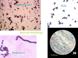
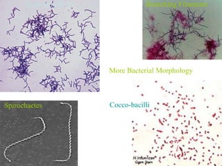
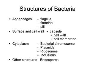
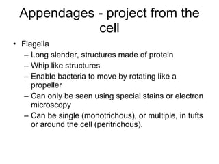
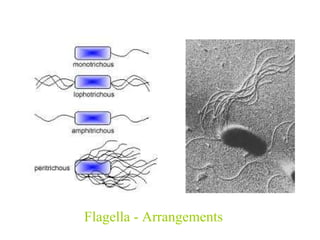

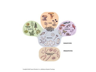
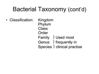
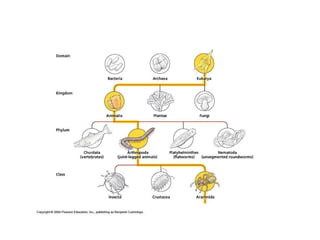
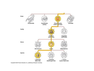
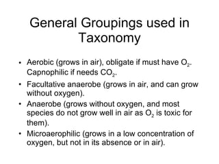
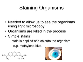
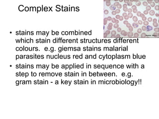
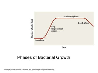
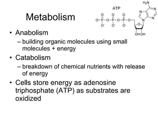

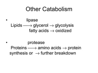
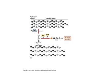
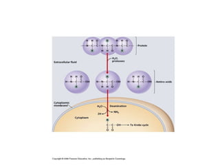
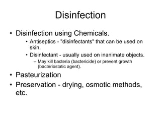
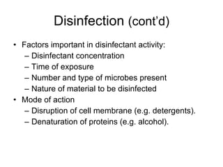
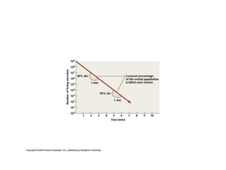
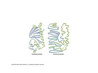
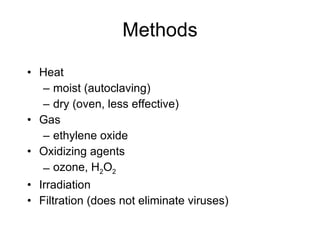
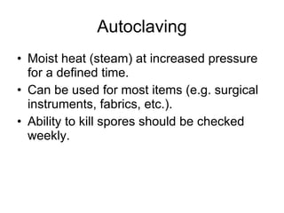
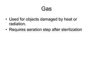
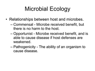
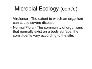

This document provides an overview of the objectives and content covered in the MICI 1100 Health Sciences Microbiology course at QE II HSC, including introductions to microbiology, bacterial structure and classification, growth and metabolism, pathogenicity, and control of microbial growth. Key topics covered include bacterial morphology, staining techniques, taxonomy, requirements for growth, phases of growth, and methods of sterilization and disinfection.