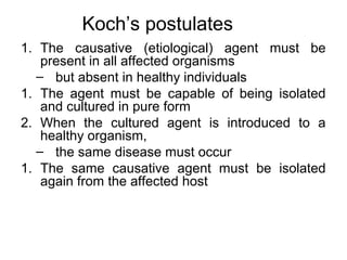
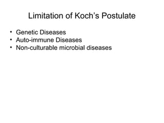
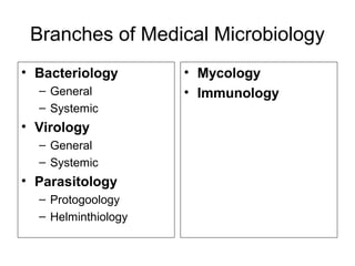
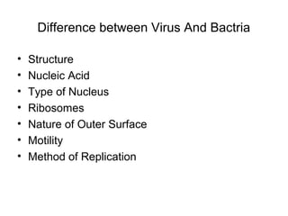
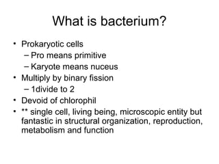
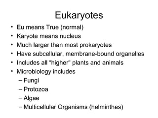
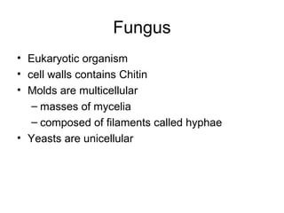
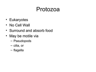
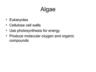
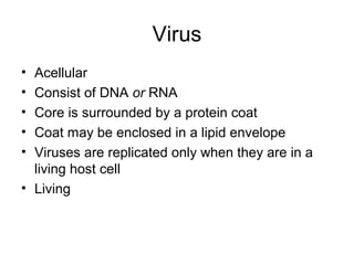
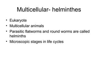
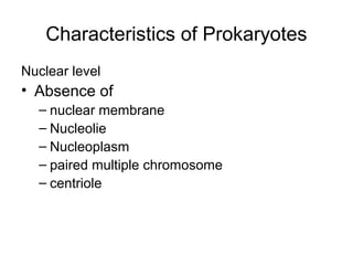
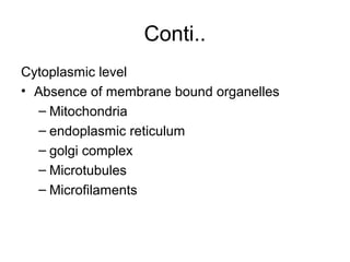
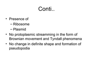
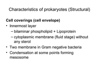
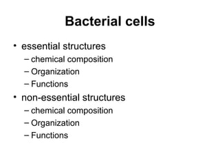
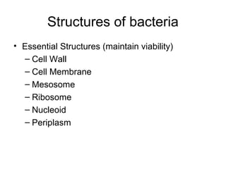
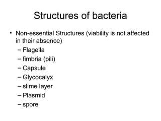
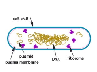
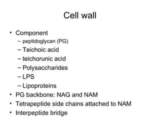
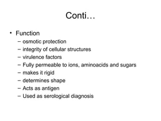
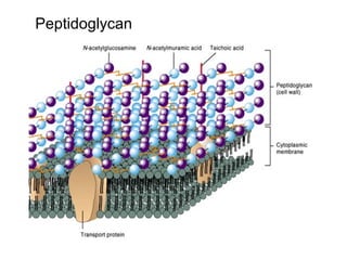
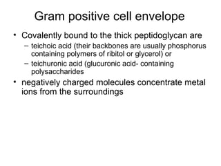
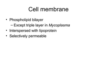
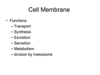
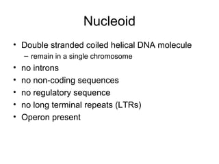
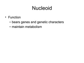
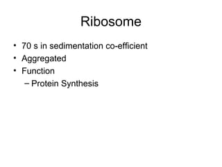
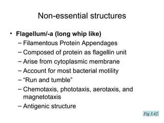
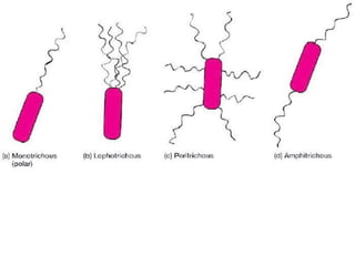
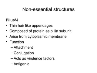
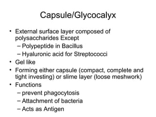
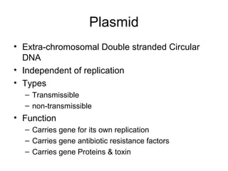
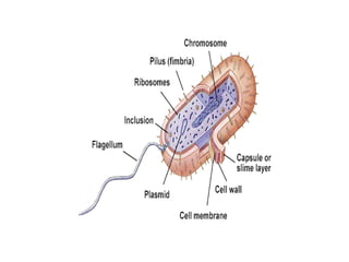
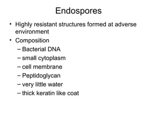
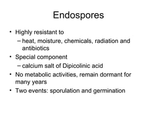
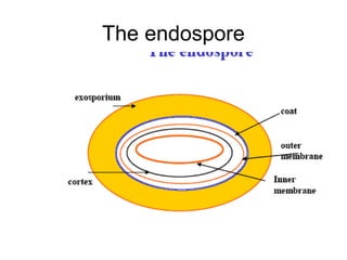
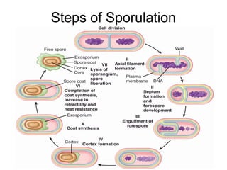
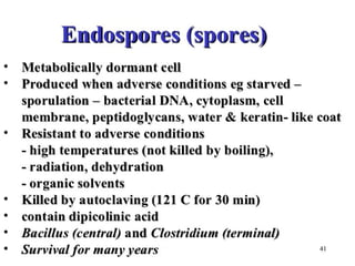
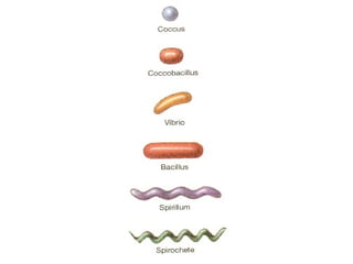
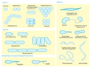
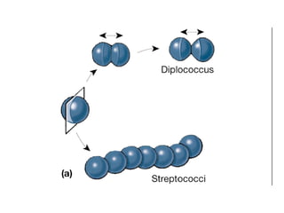
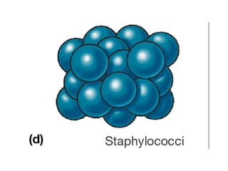
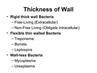
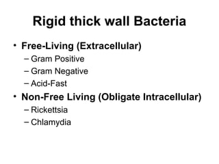
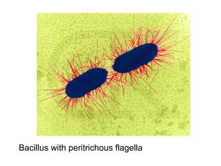
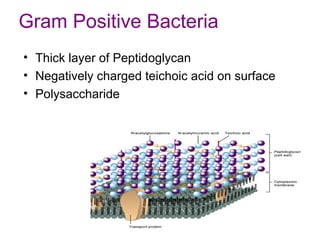
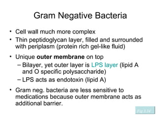
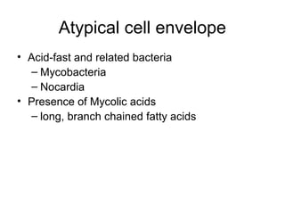
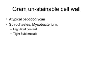
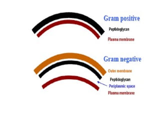
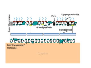
VIP Call Girls Tirunelveli Aaradhya 8250192130 Independent Escort Service Tir...
Introduction of Bacterial cells 2017
1. Medical Microbiology:
Introduction
Dr. Md. Abdullah Yusuf
Assistant Professor, Dept. of Microbiology
National Institute of Neurosciences & Hospital
Dhaka, Bangladesh
Email: ayusuf75@yahoo.com
2. Learning Objectives
Introduction and history of Microbiology
• Important events of history
• Koch’s postulates
• Branches of Medical Microbiology
• Role of Microbes in environment & medical
science
3. Important events of history
• Dutch Biologist “Anton Van Leeuwenhock”
discovered “animicules” in a drop of water by his
simple microscope in 1674
• Otto Muller (Danish biologist) organized
bacteria into genus and species in1774
• In 1840, Friedrich Henle (German pathologist)
proposed germ theory of disease
4. Conti…
• Robert Koch
– Father of medical Microbiology
• Luis Pasteur (Father of Modern Microbiology)
– identified causative agent of anthrax, rabies,
plague, cholera and tuberculosis
• Discovery of penicillin
– 1928 (Alexander Flemming)
• Discovery of sulphanilamide
– 1935 (Gerhard Domagk’s)
• Discovery of streptomycin
– 1928 (Selman Waksman)
5. Koch’s postulates
1. The causative (etiological) agent must be
present in all affected organisms
– but absent in healthy individuals
1. The agent must be capable of being isolated
and cultured in pure form
2. When the cultured agent is introduced to a
healthy organism,
– the same disease must occur
1. The same causative agent must be isolated
again from the affected host
7. Branches of Medical Microbiology
• Bacteriology
– General
– Systemic
• Virology
– General
– Systemic
• Parasitology
– Protogoology
– Helminthiology
• Mycology
• Immunology
8. Difference between Virus And Bactria
• Structure
• Nucleic Acid
• Type of Nucleus
• Ribosomes
• Nature of Outer Surface
• Motility
• Method of Replication
10. What is bacterium?
• Prokaryotic cells
– Pro means primitive
– Karyote means nuceus
• Multiply by binary fission
– 1divide to 2
• Devoid of chlorophil
• ** single cell, living being, microscopic entity but
fantastic in structural organization, reproduction,
metabolism and function
11. Eukaryotes
• Eu means True (normal)
• Karyote means nucleus
• Much larger than most prokaryotes
• Have subcellular, membrane-bound organelles
• Includes all “higher" plants and animals
• Microbiology includes
– Fungi
– Protozoa
– Algae
– Multicellular Organisms (helminthes)
12. Fungus
• Eukaryotic organism
• cell walls contains Chitin
• Molds are multicellular
– masses of mycelia
– composed of filaments called hyphae
• Yeasts are unicellular
13. Protozoa
• Eukaryotes
• No Cell Wall
• Surround and absorb food
• May be motile via
– Pseudopods
– cilia, or
– flagella
14. Algae
• Eukaryotes
• Cellulose cell walls
• Use photosynthesis for energy
• Produce molecular oxygen and organic
compounds
15. Virus
• Acellular
• Consist of DNA or RNA
• Core is surrounded by a protein coat
• Coat may be enclosed in a lipid envelope
• Viruses are replicated only when they are in a
living host cell
• Living
16. Multicellular- helminthes
• Eukaryote
• Multicellular animals
• Parasitic flatworms and round worms are called
helminths
• Microscopic stages in life cycles
19. Conti..
• Presence of
– Ribosome
– Plasmid
• No protoplasmic streamming in the form of
Brownian movement and Tyndall phenomena
• No change in definite shape and formation of
pseudopodia
20. Characteristics of prokaryotes (Structural)
Cell coverings (cell envelope)
• Innermost layer
– bilaminar phospholipid + Lipoprotein
– cytoplasmic membrane (fluid stage) without
any sterol
• Two membrane in Gram negative bacteria
• Condensation at some points forming
mesosome
21. Conti…
2) Outer to Cell Membrane
– cell wall containing specialized unique
chemicals called peptidoglycan
3) Outer to Cell Wall
– investing layer either capsule/glycocalyx/slime
layer
4) S layer
– Single type protein lattice outer to CW
22. Characteristics of Prokaryotes
• No genetic exchange during chromosome
replication
• Show secretory and excretory activities
• Show chemical selectivity, motility and alteration
of life stage (spore)
• Wide range of temperature and pH tolerance
• Heterogenous nutritional requirements and host
specificity
• Can produce self protective molecule bacteriocin
• Become infected by particular virus
• Can acquire extrachromosomal DNA (plasmid)
23. Difference Between Eukaryotes and
Prokaryotes
• Nature of Nucleus
• Mitotic Division
• Presence of histones in DNA
• Membrane bound Organelles
• Size of Ribosomes
• Peptidoglycan in Cell Wall
27. Structures of bacteria
• Non-essential Structures (viability is not affected
in their absence)
– Flagella
– fimbria (pili)
– Capsule
– Glycocalyx
– slime layer
– Plasmid
– spore
28.
29. Cell wall
• Component
– peptidoglycan (PG)
– Teichoic acid
– teichorunic acid
– Polysaccharides
– LPS
– Lipoproteins
• PG backbone: NAG and NAM
• Tetrapeptide side chains attached to NAM
• Interpeptide bridge
30. Conti…
• Function
– osmotic protection
– integrity of cellular structures
– virulence factors
– Fully permeable to ions, aminoacids and sugars
– makes it rigid
– determines shape
– Acts as antigen
– Used as serological diagnosis
32. Gram positive cell envelope
• Covalently bound to the thick peptidoglycan are
– teichoic acid (their backbones are usually phosphorus
containing polymers of ribitol or glycerol) or
– teichuronic acid (glucuronic acid- containing
polysaccharides
• negatively charged molecules concentrate metal
ions from the surroundings
33. Conti…
• Teichoic acids can also direct autolytic enzymes
to sites of peptidoglycan digestion (autolysis).
• This is needed to insert sections of cell wall for
growth and division.
• Lipoteichoic acid is primarily associated with the
cell membrane.
34. The Gram negative cell envelope
• Covalently linked to the thin peptidoglycan is the
Braun lipoprotein which binds the outer
membrane to the cell wall.
• Like other membranes it contains proteins and
phospholipids.
• Unlike other membranes it contains
lipopolysaccharide
35. Conti…
• Lipopolysaccharide
– helps to provide a permeability barrier
• LPS consists of three regions
– an outer O antigen
– a middle core
– an inner lipid A region
• core contains several sugars
– lipid A contains β hydroxyfatty acids
(uncommon in nature)
– The molecule displays endotoxin activity
36. Conti…
• Porins in the outer membrane form channels to
allow passage of small hydrophilic nutrients
(such as sugars) through the outer membrane.
37. Cell membrane
• Phospholipid bilayer
– Except triple layer in Mycoplasma
• Interspersed with lipoprotein
• Selectively permeable
38. Cell Membrane
• Functions
– Transport
– Synthesis
– Excretion
– Secretion
– Metabolism
– division by mesosome
39. Nucleoid
• Double stranded coiled helical DNA molecule
– remain in a single chromosome
• no introns
• no non-coding sequences
• no regulatory sequence
• no long terminal repeats (LTRs)
• Operon present
41. Ribosome
• 70 s in sedimentation co-efficient
• Aggregated
• Function
– Protein Synthesis
42. Non-essential structures
• Flagellum/-a (long whip like)
– Filamentous Protein Appendages
– Composed of protein as flagellin unit
– Arise from cytoplasmic membrane
– Account for most bacterial motility
– “Run and tumble”
– Chemotaxis, phototaxis, aerotaxis, and
magnetotaxis
– Antigenic structure
Fig 3.42
43.
44. Non-essential structures
Pilus/-i
• Thin hair like appendages
• Composed of protein as pillin subunit
• Arise from cytoplasmic membrane
• Function
– Attachment
– Conjugation
– Acts as virulence factors
– Antigenic
45. Capsule/Glycocalyx
• External surface layer composed of
polysaccharides Except
– Polypeptide in Bacillus
– Hyaluronic acid for Streptococci
• Gel like
• Forming either capsule (compact, complete and
tight investing) or slime layer (loose meshwork)
• Functions
– prevent phagocytosis
– Attachment of bacteria
– Acts as Antigen
46. Plasmid
• Extra-chromosomal Double stranded Circular
DNA
• Independent of replication
• Types
– Transmissible
– non-transmissible
• Function
– Carries gene for its own replication
– Carries gene antibiotic resistance factors
– Carries gene Proteins & toxin
47.
48. Endospores
• Highly resistant structures formed at adverse
environment
• Composition
– Bacterial DNA
– small cytoplasm
– cell membrane
– Peptidoglycan
– very little water
– thick keratin like coat
49. Endospores
• Highly resistant to
– heat, moisture, chemicals, radiation and
antibiotics
• Special component
– calcium salt of Dipicolinic acid
• No metabolic activities, remain dormant for
many years
• Two events: sporulation and germination
53. Classification of bacteria
• Basis of Classification
• differences between Gram positive and Gram
negative bacteria
• atypical characters of bacteria in relation to
staining
54. Basis of Classification
• Morphological Classification
• Staining Properties
• Thickness of Wall
• According to Motility
55. Morphological Classification
Morphology of Bacteria
• Cocci
– rounded or oval (Staphylococcus)
• Bacilli
– elongated rod like (Esch.coli)
• Vibrio (from vibration)
– coma shaped (Vibrio cholerae)
• Spiral bacteria
– long slender curved body with wave like spiral
(Spirillum minus, Helicobacter pylori)
56. Others Shapes
• Cocco-bacillus
– longer than coccus, shorter than bacillus
(Bordetella, Brucella)
• Filamentous
– long curved body like ribbon with branching
(Actinomycetes, Nocardia)
74. Types of Staining
Differential Staining
• using primary and counter dye
• performing a step of decolourisation
• to see differential characters of bacteria
– Gram’s stain
– Ziehl-Neelsen stain
– Albert stain
75. Types of Staining
Special Staining
• to see unique property of particular
bacteria
– Fluorescense stain
76. Gram’s staining
• Named according to inventor Christian Gram
• Based on the property of bacterial cell envelope
• stained at first by a primary dye & mordanted
• Decolorized by acetone or alcohol
• Counter stain is added
• Gram positive bacteria
– can resist decolourisation and retain the primary dye
• Gram negative bacteria
– can not resist decolourisation and take the counter
dye
77. Gram Positive Bacteria
• Thick layer of Peptidoglycan
• Negatively charged teichoic acid on surface
• Polysaccharide
78. Gram Negative Bacteria
• Cell wall much more complex
• Thin peptidoglycan layer, filled and surrounded
with periplasm (protein rich gel-like fluid)
• Unique outer membrane on top
– Bilayer, yet outer layer is LPS layer (lipid A
and O specific polysaccharide)
– LPS acts as endotoxin (lipid A)
• Gram neg. bacteria are less sensitive to
medications because outer membrane acts as
additional barrier.
Fig 3.34
79. Atypical cell envelope
• Acid-fast and related bacteria
– Mycobacteria
– Nocardia
• Presence of Mycolic acids
– long, branch chained fatty acids