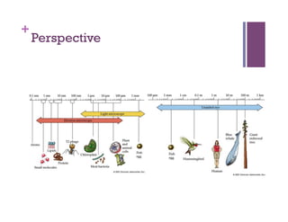
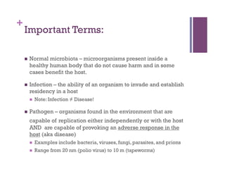
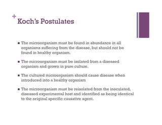
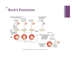
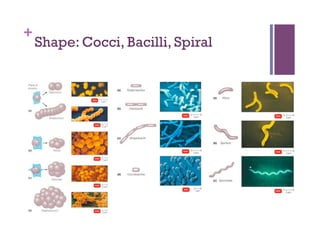
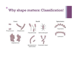
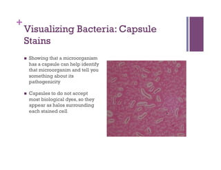
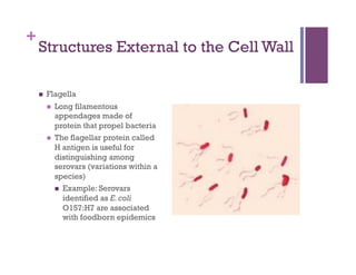
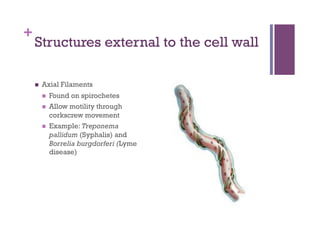
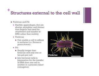
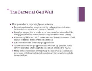
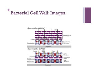
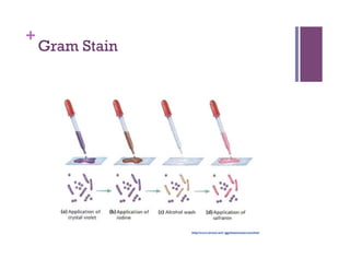
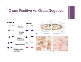
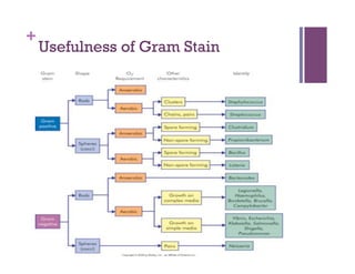
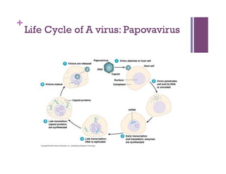
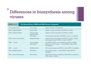
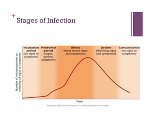
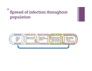
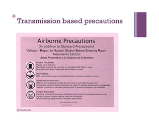
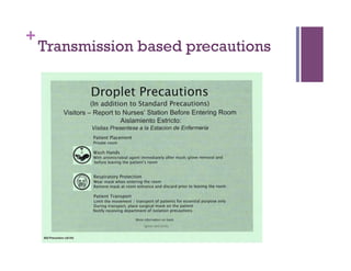
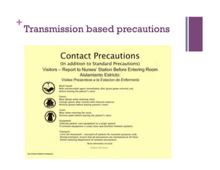
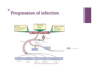
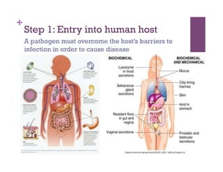
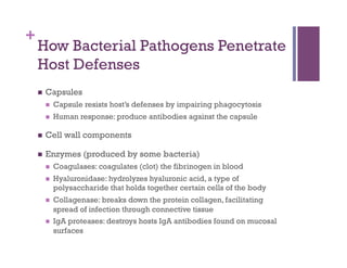
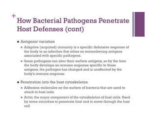
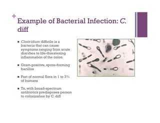
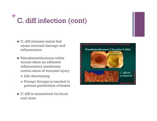
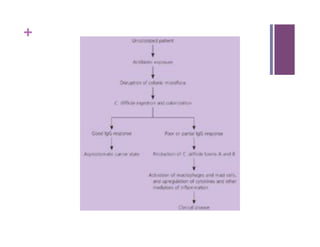
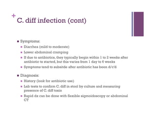
2. +
Objectives
1. Discuss basic characteristics of bacteria, including
structures that lead to pathogenicity, drug susceptibility,
and accurate identification
2. Discuss basic characteristics of fungi, helminths, and
viruses
3. Describe three main modes of disease transmission
4. Describe how bacteria penetrate host defenses and cause
damage
3. +
Objectives
6. Describe how bacterial infections are diagnosed and
treated
7. Describe how viruses enter the host can cause illness, and
how do we diagnose and treat viral infections
8. Describe how fungi invade and damage host cells, and how
fungal infections are diagnosed and treated
9. Discuss methods of microbial control
4. +
Overview of basic
principles
A quick reference
5. +
The Basics:
Cell Theory: All living things are composed of cells (circa
1665)
Biogenesis: All living cells can arise only from preexisting
cells
Formed basis of aseptic techniques: techniques that prevent
contamination by microorganisms, because we know that
microorganisms can only come from living microorganisms.
Heritable material (DNA) is passed from parent to offspring
and dictates specific traits expressed in the offspring.
Germ Theory: Germs can cause disease
7. +
Important Terms:
Normal microbiota – microorganisms present inside a
healthy human body that do not cause harm and in some
cases benefit the host.
Infection – the ability of an organism to invade and establish
residency in a host
Note: Infection ≠ Disease!
Pathogen – organisms found in the environment that are
capable of replication either independently or with the host
AND are capable of provoking an adverse response in the
host (aka disease)
Examples include bacteria, viruses, fungi, parasites, and prions
Range from 20 nm (polio virus) to 10 m (tapeworms)
8. +
Koch’s Postulates
The microorganism must be found in abundance in all
organisms suffering from the disease, but should not be
found in healthy organism.
The microorganism must be isolated from a diseased
organism and grown in pure culture.
The cultured microorganism should cause disease when
introduced into a healthy organism
The microorganism must be reisolated from the inoculated,
diseased experimental host and identified as being identical
to the original specific causative agent.
10. +
Tools in Microbiology: Microscopy
Most initial observations of microorganisms are made with
stained preparations under the microscope
Stains:
Stains are used to color the microorganisms in a way that
emphasizes what you want to look at.
Microscopy can aid in classification of microorganism
Determine overall cell shape, size, and capsule
Using microscopy, you can tell if the microorganism is unicellular
(like a bacteria) or multicellular (like a parasite)
Based on the organisms response to dyes, you can also note
characteristics of the organisms cell wall (see gram stain and acid
fast stain)
12. +
Anatomy of a Bacterial Cell
Bacteria are Prokaryotic, Unicellular
Organisms
No nucleus – Their DNA is usually a
singular circularly arranged
chromosome
Their DNA is not associated with
histones
They lack membrane-enclosed
organelles (i.e. mitochondria)
Their cell walls almost always
contain the complex polysaccharide
peptidoglycan
They usually divide by binary
fission. During this process, the DNA
is copied and the cell splits into two
cells.
15. +
Structures External to the Cell Wall
Glycocalyx
A viscous, gelatinous polymer that is composed of
polysaccharides, polypeptides, or both
If it is organized and firmly attached to the cell wall, it is called a
capsule
If it is unorganized and loosely attached, it is described as a slime
layer
Contributes to virulence:
Protects pathogenic bacteria from phagocytosis (i.e. Bacillus
anthracis and Streptococcus pneumoniae)
Enables bacterium to survive by attaching to various surfaces
(Streptococcus mutans)
Protects bacterium against dehydration and can inhibit
movement of nutrients out of the cell
16. +
Visualizing Bacteria: Capsule
Stains
Showing that a microorganism
has a capsule can help identify
that microorganim and tell you
something about its
pathogenicity
Capsules to do not accept
most biological dyes, so they
appear as halos surrounding
each stained cell
17. +
Structures External to the Cell Wall
Flagella
Long filamentous
appendages made of
protein that propel bacteria
The flagellar protein called
H antigen is useful for
distinguishing among
serovars (variations within a
species)
Example: Serovars
identified as E. coli
O157:H7 are associated
with foodborn epidemics
18. +
Structures external to the cell wall
Axial Filaments
Found on spirochetes
Allow motility through
corkscrew movement
Example: Treponema
pallidum (Syphalis) and
Borrelia burgdorferi (Lyme
disease)
19. +
Structures external to the cell wall
Fimbriae and Pili
Hairlike appendages, that are
shorter, straighter, and thinner
than flagella and used for
attachment and transfer of
DNA rather than motility
Fimbriae
Can enable a cell to adhere
to surfaces (i.e. Neisseria
gonorrhoeae)
Pili
Usually longer than
fimbriae and only one or
two pili per cell
Join bacterial cells in
preparation for the transfer
of DNA from one cell to
another in a process called
conjugation
20. +
The Bacterial Cell Wall
Composed of a peptidoglycan network
Repeating disaccharide attached by polypeptides to form a
lattice that surrounds and protects the cell
Disacharide portion is made up of monosaccharides called N-
acetylglucosamine (NAG) and N-acetylmuramic acid (NAM)
Alternating NAM and NAG molecules are linked in rows of 10-65
sugars to form a carbohydrate backbone
Adjacent rows are linked by polypeptides
The structure of the polypeptide link varies by species, but it
always includes a tetrapeptide side chain attached to NAMs.
Many antibiotics work by targeting the cell wall (i.e. penicillin
interferes with final linking of pepdidoglycan rows by peptide
cross-bridges
26. +
Bacterial Metabolism: Generating
Energy
Energy Growth End Product ATP molecules
Process Conditions produced
Aerobic Aerobic Water 38/glucose
Respiration
Anaerobic Anaerobic Us. an inorganic Between 2 and 38
Respiration substance (i.e.
methane, hydrogen
sulfide)
Fermentation Aerobic or An organic molecule 2
anaerobic (i.e. lactic acid)
27. +
Objective 1
Discuss basic characteristics of bacteria, including structures
that lead to pathogenicity, drug susceptibility, and accurate
identification
Glycocalyx - Protects pathogenic bacteria from phagocytosis,
Enables bacterium to survive by attaching to various surfaces
(Streptococcus mutans), Protects bacterium against dehydration
and can inhibit movement of nutrients out of the cell
Flagella – H-antigen is useful for distinguishing among serovars
Axial filaments – allows motility so bacterium can travel to site
of infection
Fimbriae – allows bacterium to adhere to surfaces
Components of the cell wall allow bacteria to be identified
through the gram stain and acid fast stain
29. +
Fungi: the basics
Easily visible by Light Microscopy (1-200 u)
Plant-like
organisms which grow by
continuous extension and branching
(hyphae)
Growth
Some grow only as yeasts (ie. Candida
Albicans, Cryptococcus )
Some grow only as molds (ie. Aspergillus)
31. +
Fungi: The basics (continued)
All fungi require some organic compounds for energy (not
phototrophs)
Most fungi are aerobic
Fungi can reproduce via a few routes:
Asexually by fragmentatino of their hyphae
Suxually or asexually by formation of spores
Fungi usually grow better in an environment with a pH of
about 5
Fungi can also grow on substances with very low moisture
content and are relatively resistant to osmotic pressure
33. +
Helminths: the basics
Multicellular
Eukaryotic
Most possess digestive, circulatory, nervous, excretory, and
reproductive systems
Most Helmonths that cause disease are parasitic, there are
some distinguishing features of parasitic helminths:
They may lack a digestive system
Their nervous system is reduced
Their means of locomotion is occasionaly reduced or completely
lacking
Their reproductive system is often complex. An individual
produces a large number of eggs, by which a host is infected
37. +
Viruses: The basics
Viruses are considered to be obligatory intracellular
parasites
They require living host cells in order to multiply
They use host machinery to synthesize their own proteins and
replicate their own DNA
Viruses have few or no enzymes of their own for metabolism
They lack enzymes for ATP generation and protein synthesis
This makes it difficult when designing antivirals, because most
antimicrobial drug targets are enzymes unique the to pathogen,
when an organism has no unique enzymes, you have no potential
drug target
38. +
Viral Anatomy
Nucleic Acid
DNA or RNA (But never both)
ssDNA
ds DNA
ss RNA
ds RNA
Capsid (Coat Protein)
protects viral genome from host endonucleases
capsomeres
Binding Sites
Envelope (not present on all viruses)
Derived from the host cell
Made up of lipids, proteins and carobhydrates
Binding Sites
43. +
Objective 2
Discuss basic characteristics of fungi, helminths, and viruses
Fungi
multicellular or unicellular (yeasts)
Prefer acidic (pH ~5) environments
Can thrive in dry environments and are relatively resistant to
osmotic pressure
Helminths
Multicellular
Most that cause disease are parasitic and have the following
features:
They may lack a digestive system
Their nervous system is reduced
Their means of locomotion is occasionally reduces or completely
lacking
Their reproductive system is often complex
44. +
Objective 2 (cont)
Viruses
Require a host to multiply
Use hosts machinery to multiply
Made up to 2-3 main components:
Nucleic Acid (DNA or RNA)
Capsid
Envelope (mot present on all viruses)
48. +
Contact Transmission
Spread of agent of disease by direct contact, indirect contact,
or droplet transmission
Direct contact (aka person to person) occurs when agent is
transferred by physical contact between its source and a
susceptible host, no intermediate object is involved
Indirect contact occurs when pathogen is transmitted from its
reservoir to susceptible host by means of a nonliving object
(fomite).
Droplet transmission occurs when microbes are spread in
droplet nuclei that travel short distances
49. +
Vehicle Transmission
Transmission of disease agents by a medium, such as water,
food, or air
Note: The difference between airborne and droplet
transmission
Airborne transmission refers to the spread of agents of infection
by droplet nuclei in dust that travel more than 1 meter from the
reservoir to the host (in droplet transmission the droplet travels
less than 1 meter from the reservoir).
Example: Measles, Tuberculosis can also be spread by airborne
transmission, even though it is also considered to be spread by
droplet transmission
50. +
Vector-based Transmission
Vectors – animals that carry pathogens from one host to another
Arthropods are an important group of vectors. They transmit
disease by 2 general methods:
Mechanical transmission:
Passive transport of pathogens on the insect’s feet or other body
parts
Example: houseflies can transfer pathogens of typhoid fever and
bacillary dysentery
Biological transmission
Arthropod bites an infected person or animal and ingests some of
the infected blood
Pathogen then reproduce in the vector
Pathogen either migrates to salivary gland of vector or gut
Vector spreads pathogen to susceptible host by defecating,
vomiting, or biting the host
Example: malaria
51. +
Objective 3
Describe three modes of disease transmission
Contact transmission: Spread of agent of disease by direct
contact, indirect contact, or droplet transmission
Vehicle based transmission: Transmission of disease agents by a
medium, such as water, food, or air
Vector based transmission: transmission of disease through an
animal vector (i.e. arthropod)
52. +
Nosocomial (hospital acquired)
infections
In the US, the CDC estimates that 5 to 15% of all hospital
patients acquire some type of nosocomial infection
The rate of nosocomial infections has increased 36% during
the last 20 years.
2 million people per year contract nosocomial infections, and
nearly 20,000 die as a result.
This means, nosocomial infections are the 8th leading cause
of death in the US.
58. +
Step 1: Entry into human host
A pathogen must overcome the host’s barriers to
infection in order to cause disease
59. +
Portal of Entry: Mucous
Membranes
Lining of respiratory tract, GI tract, GU tract, and conjunctiva
(lining of the eye)
Respiratory tract – easiest and most frequently traveled.
Examples: cold, flu, tuberculosis, pertussis, measles, strep throat,
diptheria, pneumonia
GI – via contaminated food, water, and fingers
Examples: Hepatitis A, shigellosis, cholera
GU – some microbes can penetrate and unbroken mucous
membrane, others require a cut or abrasion
Example: chlamydia, syphilis, herpes, gonorrhea
60. +
Other portals of entry
Skin Parenteral
Skin is the largest organ of Microorganisms are
the body. When unbroken is deposited into the tissues
an effective barrier for most below the skin or mucus
microorganisms. membranes
Some microbes can gain Punctures, injections, bites,
entrance thru openings in scratches, surgery, splitting
the skin: hair follicles and of skin due to swelling or
sweat glands dryness
Example: Nectoriasis
Caused by hookworm
Example: Athletes Foot
Caused by Fungi: Tinea
pedis
61. +
Step 2: Travel to site of infection
The blood stream, lymphatic system, and interstitial fluids
provide mediums for pathogens to travel throughout the
body, once inside
62. +
Step 3: Cause damage
Pathogens can cause damage via three main pathways:
Direct damage
Release of Exotoxins, Endotoxins and/or irritant enzymes/
proteins
Induction of host immune response
63. +
How Bacterial Pathogens Penetrate
Host Defenses
Capsules
Capsule resists host’s defenses by impairing phagocytosis
Human response: produce antibodies against the capsule
Cell wall components
Enzymes (produced by some bacteria)
Coagulases: coagulates (clot) the fibrinogen in blood
Hyaluronidase: hydrolyzes hyaluronic acid, a type of
polysaccharide that holds together certain cells of the body
Collagenase: breaks down the protein collagen, facilitating
spread of infection through connective tissue
IgA proteases: destroys hosts IgA antibodies found on mucosal
surfaces
64. +
How Bacterial Pathogens Penetrate
Host Defenses (cont)
Antigenic variation
Adaptive (acquired) immunity is a specific defensive response of
the body to an infection that relies on remembering antigens
associated with specific pathogens.
Some pathogens can alter their surface antigens, so by the time
the body develops an immune response specific to those
antigens, the pathogen has changed and is unaffected by the
body’s immune response
Penetration into the host cytoskeleton
Adhesins: molecules on the surface of bacteria that are used to
attach to host cells
Actin: the major component of the cytoskeleton of host cells. Used
by some microbes to penetrate host and to move through the host
cell
65. +
How Bacterial Pathogens Cause
Damage
Using host’s nutrients: Siderophores
Direct Damage
Production of Toxins
Exotoxins
Endotoxins
Inducing a hypersensitivity reaction
66. +
Siderophores
Proteins secreted by some pathogens that allow the pathogen
to obtain free iron from the host
Iron is required for the growth of most pathogenic bacteria,
but the concentration of free iron is low because most iron is
tightly bound to proteins
When iron is needed by a pathogen, siderophores are
released into the medium where they take the iron away from
iron transport proteins by binding the iron more tightly.
Once the iron-siderophore complex is formed, it binds to the
bacterial surface and is used by the bacteria
67. +
Direct damage
Bacteria can cause direct damage as they use the host cells
for nutrients and produce waste products
As bacteria metabolize and multiply in cells, the cells usually
rupture
68. +
Toxins
Exotoxins:
produced inside some bacteria as a part of their growth and
metabolism and secreted by the bacterium into the surrounding
medium or released following lysis
Many exotoxins are encoded by genes on the bacterial plasmid
They destroy particular parts of the host cell or inhibit certain
metabolic functions
The body produces antitoxins, which are antibodies that provide
immunity to exotoxins
69. +
Toxins (Cont)
Endotoxins
Are part of the outer portion of the cell wall of gram-negative
bacteria
Are lipopolysaccharides
Are released when gram negative bacteria die and their cell walls
undergo lysis
Antibiotics used to treat diseases caused by gram-negative
bacteria can lead to release of endotoxin and thus may lead to
immediate worsening of the symptoms, but the condition usually
improves as the endotoxin breaks down
Stimulates macrophages to release cytokines in very high (toxic)
concentrations. Symptoms include: chills, fever, weakness,
generalized aches. Can lead to miscarriage, shock, and death
70. +
Objective 4
Describe how bacteria penetrate host defenses and cause
damage
Penetrate host defenses:
Capsules
Cell wall components
Enzymes
Antigenic variation
Penetration into cytoskelton
Cause damage
Using host’s nutrients
Direct damage
Toxins
Hypersensitivity reaction
71. +
Diagnosing and treating a
bacterial infection
Culture
Gram stain
Set up culture with best specimen for symptoms
Identification based on biochemical reactions
Sensitivities to determine best antibiotic therapy
All usually completed in 24-72 hours
Special consideration for Mycobacteria
distinctive waxy cell coat requiring Acid Fast Stains
virulence is related to cell wall “cord factor” which inhibits
phagosome-lysosome fusion
Special culture methods used; may take up to 6 weeks
TX: antibiotics
72. +
Objective 5
Describe how bacterial infections are diagnosed and treated
Diagnosis
Culture
Drug sensitivity tests
Gram stain/ acid fast stain
Treatment
antibiotics
73. +
Example of Bacterial Infection: C.
diff
Clostridium difficile is a
bacteria that can cause
symptoms ranging from acute
diarrhea to life-threatening
inflammation of the colon
Gram-positive, spore-forming
bacillus
Part of normal flora in 1 to 3%
of humans
Tx. with broad-spectrum
antibiotics predisposes person
to colonization by C. diff
74. +
C. diff infection (cont)
C. diff releases toxins that
cause mucosal damage and
inflammation
Pseudomembranous colitis
occurs when an adherent
inflammatory membrane
covers areas of mucosal injury
Life threatening
Prompt therapy is needed to
prevent perforation of bowel
C. diff is transmitted via fecal-
oral route
76. +
C. diff infection (cont)
Symptoms:
Diarrhea (mild to moderate)
Lower abdominal cramping
If due to antibiotics, they typically begin within 1 to 2 weeks after
antibiotic tx started, but this varies from 1 day to 6 weeks
Symptoms tend to subsitde after antibiotic has been d/c’d
Diagnosis:
History (look for antibiotic use)
Lab tests to confirm C. diff in stool by culture and measuring
presence of C. diff toxin
Rapid dx can be done with flexible sigmoidoscopy or abdominal
CT
77. +
C. Diff (cont)
Treatment
Immediate d/c of antibiotics
Specific tx aimed at eradicate C. diff if symptoms are severe or
persistent
Metronidazole (PO) is first choice, but vancomycin (PO) is used
in people who can’t tolerate metronidazole or don’t respond
Nursing concerns
Maintaining and monitoring hydration
Maintaining and monitoring skin integrity
Monitoring stool output and response to tx
Contact precautions to prevent spread
78. +
Viral Mechanisms for Invading
Host
Viruses need a host for replication
Viruses gain access to cells because they have attachment
sites for receptors on their target cells.
When an attachment site is brought together with an
appropriate receptor, the virus can bind to and penetrate
those cells
79. +
Common terms in viral infection
Active Infection: After recent entry, the virus replicates
with the production of antigens. Antibodies and/or a cell
mediated response is usually absent in early acute
infection.
Past Infection: The virus entered long ago, an immune
response occurred and the virus has been cleared. This is
characterized by the absence of antigen and the presence
of antibodies and/or a cell-mediated response.
Latent Infection: The virus entered in the past and remains
there, BUT is inactive. Antigens are absent, but antibodies
and/or a cell mediated response is present. The virus can
reactivate.
Recurrent Infection: The latent virus has become active.
Antigen and antibodies are present.
80. +
Viral induced injury
Infection of a host cell by a virus usually leads to the death of
the host cell. This can occur one of a few ways:
Accumulation of large numbers of multiplying viruses
Effects of viral proteins on the permeability of the host cell’s
plasma membrane
Inhibition of host DNA, RNA, or protein synthesis
Some viruses cause lysosomes to release their enzymes
Some viruses cause adjacent infected cells to fuse to form a vary
large multinucleate call called a syncytium
Some viruses cause antigenic changes on the surfface of infected
cells. This elicits a host antibody response against the infected
cell, and thus targets the cell for destruction by the host’s immune
system
Some viruses cause chromosomal damage to the host cell
81. +
Diagnosis: methods of identifying a
virus
Detection of virus by Culture
Detection of Antigens-- ELISA
Detection of specific Antibodies
82. +
Prevention and treatment of viral
diseases
Let them run their course and treat symptoms
Antivirals (there are not as many antiviral medications
available)
Vaccines
83. +
Objective 6
How do viruses enter the host can cause illness, and how do we
diagnose and treat viral infections?
Viruses gain access to cells because they have attachment sites for receptors
on their target cells.
Viruses can cause damage in the following ways:
Accumulation of large numbers of multiplying viruses
Viral proteins effect the permeability of the host cell’s plasma membrane
Inhibition of host DNA, RNA, or protein synthesis
Cause lysosomes to release their enzymes
Cause adjacent infected cells to fuse to form a vary large multinucleate call
Cause antigenic changes on the surface of infected cells, targeting them for
destruction by the host’s immune system
Cause chromosomal damage to the host cell
Diagnosis through culture and nucleic acid matching, ELISA, Western Blot
Treatment: treat symptoms, give antivirals (rare), prevent spread with vaccines
84. +
Example of a viral infection:
Influenza (flu)
Influenza is caused by RNA viruses in the family
Orthomyxoviridae
Can affect birds and mammals (which can serve as reservoirs)
There are 3 types that cause epidemics in humans: A, B, and C
Airborne transmission and direct contact transmission
Coughs and sneezes (air)
Contact with bird droppings or nasal secretions (direct-contact)
A person is contagious beginning about the day before
symptoms appear to 5 to 7 days later
85. +
Influenza (Cont)
Influenza binds to epithelial
cells in nose, throat and lungs
(See diagram)
Viral proteins can degrade
host cell mRNA, inhibit
translation of host cell mRNA
New viruses buds off from the
host cell to spread infection
and the host cell dies
Influenza evades immune
response through antigenic
variation
86. +
Influenza
Most Common Symptoms:
Chills
Fever
Sore throat
Muscle pains
Severe headache
Coughing
Weakness/fatigue
General discomfort
Nausea and vomiting (esp
children)
87. +
Influenza diagnosis
Symptom assessment
Tests (rare)
Rapid flu test
Done in a doctor’s office
Direct fluorescent antibody stain (DFA)
Done in a lab
Viral culture
Done in a lab
Takes 3-10 days
Nucleic acid amplification
Influenza A or B antibody tests
88. +
Influenza treatment
Treatment:
Fluids, rest
Treat symptoms (acetaminophen for fever, etc)
Neuraminidase inhibitors (antiviral)
Prevent the release of new infectious viruses and halt viral
replication
M2 inhibitors (antiviral)
Prevent uncoating of virus once in cell, thus preventing
replication
Preventing spread:
Handwashing
Seasonal Vaccines (esp. health care workers, over 50, etc)
89. +
Influenza: Nursing concerns
Encourage rest
Encourage fluids
Monitor lung sounds
Provide supportive therapy as indicated (antipyretics, or
antitussives)
90. +
Fungal Infection
Infection is called mycoses or mycotic infection.
Fungal infections tent to be chronic because fungi grow
slowly
Do not have well defined set of virulence factors
Classified into one of five groups according to extent of
tissue involvement and mode of entry into the host
Systemic
Subcutaneous
Cutaneous
Superficial
Opportunistic
91. +
Systemic Mycoses
Fungal infections deep within the body
Not restricted to any particular region of the body, but can
affect a number of tissues and organs
Usually caused by fungi that live in the soil
Inhalation of spores is the route of transmission; infection
typically begins in the lungs and then spreads to other
tissues.
Do not tend to be contagious from animal to human or from
human to human
Example: Histoplasmosis and coccidioidomycosis
92. +
Subcutaneous mycoses
Fungal infections beneath the skin
Caused by fungi that live in soil and on vegetation
Infection often begins by direct implantation of spores or
mycelial fragments into a puncture wound on the skin
Example: Sporotrichosis (acquired by gardeners and
farmers)
93. +
Cutaneous mycoses
Dermatophytes: fungi that infect only the epidermis, hair, and
nails
Cutaneous mycoses (aka dermatomycoses) are infections
caused by dermatophytes.
Dermatophytes secrete karatinase, an enzyme that degrades
karatin, a protein in hair, skin, and nails
Infection is transmitted from human to human or animal to
human by direct contact or by contact with infected hairs and
epidermal cells
Example: Epidermophyton floccosum
94. +
Superficial Mycoses
Caused by fugni localized
along hair shafts and in
superficial (surface)
epidermal cells
Prevalent in tropical climates
Example: Pitvriases versicolor
(shown on the right, above),
Tinea Capitus (shown on right,
below)
95. +
Opportunistic Mycoses
Opportunistic pathogen: Example: Pneumocystis and
generally harmless in normal Candidiasis (shown below)
habitat, but can become
pathogenic in a host who is:
seriously debilitated or
traumatized
under treatment with broad-
spectrum antibiotics
Immunocompromised due
to drugs or an immune
disorder
diagnosed with a lung
disease
96. +
How do fungi invade host cells?
Candida albicans and Trichophyton secrete proteases that
modify host cell membranes to allow attachment of the fungi
Cryptococcus neoformans produces a capsule that helps it
resist phagocytosis
97. +
How do Fungi cause damage?
Some produce metabolic products that are toxic to humans
i.e. Trichothecenes: fungal toxins that inhibit protein synthesis in
host cells
i.e. Alfatoxin is produced by a mold that can be found in peanut
butter. Excessive amounts of this toxin is known to have
carcinogenic properties.
Some provoke allergic reactions
98. +
Diagnosis and Treatment of Fungal
Infection
Cultures may be done.
Many
fungi can be identified by light
microscopy.
Specimens and biopsies: Special stains
Medications are available for treatment.
Topicals: end in –azole
Systemic: ketoconazole, amphotericin B, Nystatin
Note: Some fungi have developed resistance to
antifungals
99. +
Objective 7
Describe how fungi invade and damage host cells, and how
fungal infections are diagnosed and treated
Invade host:
Secrete proteases that allow attachment
Produce a capsule that resists phagocytosis
Cause damage:
Producing toxins
Elicit allergic response
Diagnosis: culture and light microscopy, gross assessment
Treatment: antifungals
100. +
Helminth induced injury
Use host tissues for their own growth or produce large
parasitic masses; the resulting cellular damage evokes
symptoms
Waste products of these parasites can also contribute to
disease
101. +
Protozoa induced injury
Reproduce within host cell, causing it to rupture
Example: Plasmodium
Prevent normal cell function
Example: Toxoplasma
Digest cells and tissue fluids
Example: Giardia lamblia
Note: some protozoa can avoid host defenses by varying their
antigens like viruses. For example Trypanosoma (causes
sleeping sickness) can produce up to 1000 different antigens
103. +
Requirements for Growth
Temperature
Every bacterium has an ideal
temperature range and
temperatures outside that
range prohibit growth and life
Psychrophiles (cold-loving)
Mesophiles (moderate temp-
loving)
Thermophiles (heat loving)
104. +
Requirements for Growth
pH Trace Elements (i.e. zinc, iron,
Most bacteria prefer a pH copper, molybdenum)
between 6.5 and 7.5
Oxygen (in some bacteria)
Human immune response
often uses pH to destroy Organic Growth Factors
bacteria
Organic compounds that an
Osmotic Pressure organism is unable to
synthesize and must be
Carbon directly obtained from the
environment (i.e. vitamins,
Nitrogen, Sulfur and amino acids, purines,
Phosphorus pyrimidines)
105. +
Terms used in microbial control
Sterilization: destroying all forms of life
Disinfection: destroying pathogens or unwanted organisms
Disinfectant: antimicrobial agent used on inanimate objects
Antiseptic: antimicrobial agent used on living tissue
-cidal: kills (i.e. bactericidal = kills bacteria)
Static: inhibits growth (i.e. bacteristatic = inhibits growth of
bacteria)
106. +
Actions of microbial control agents
Alteration of membrane Damage to proteins and
permeability nucleic acids
Plasma membrane is located Many bacterial proteins are
just inside the cell wall enzymes that are vital to all
cellular activities
Membrane actively
Destroying the shape of
regulates passage of proteins destroys the
nutrients into the cell and function of proteins
the elimination of wastes
H-bonds and disulfide
from the cell bridges are critical to
Damage to the membrane protein structure but can
causes cellular contents to be broken by heat and
leak into the surrounding chemicals
medium and interferes with Heat, radiation, or chemicals
growth can destroy bacterial DNA
and RNA
108. +
Objective 9
Discuss methods of microbial control
Physical: Heat, Filtration, Cold, High Pressure, Dessication,
Osmotic Pressure, Radiation
Chemical: Phenols, Phenolics, Bisphenols, Biguanides, Halogens,
Alcohols, Heavy metals, Surface-Active Agents, Chemical Food
preservatives, Aldehydes, Gaseous Chemosterilizers, Peroxygens
The above act either by altering the membrane permeability of
the microorganism or damaging proteins and nucleic acids of the
microorganism