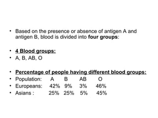
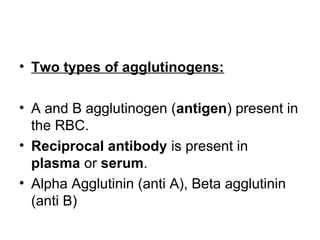
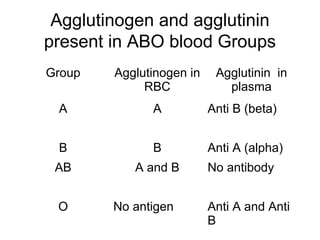
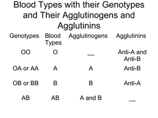
BLOOD GROUPS
ABO Grouping
• The red cell surface contains genetically
determined antigens called agglutinogens of a
glycoprotein nature (the carbohydrate of which
determines the blood group specificity of the
RBCs).
• Human beings are divided into four main groups
according to the presence (or absence) in their
red cells of agglutinogens.
ABO Grouping
• There are four possibilities:
• A) Persons whose red cells contain agglutinogen A, belong to
• group A.
• B) Persons whose red cells contain agglutinogen B, belong to
• group B.
• C) Persons whose red cells contain both types of agglutinogens A
• and B, belong to group AB.
• D) Persons whose red cells contain ‘no’ agglutinogen, belong to
• group O.
• We classify persons as belonging to one group or the other on the
basis of presence or absence of two agglutinogens.
ABO BLOOD GROUPS
• According to Landsteiner’s Law:
• If an agglutinogen is present in the red cells of a blood
the corresponding or specific agglutinin must be
absent from the plasma of the person,
• otherwise agglutinogen-agglutinin reaction will occur and
the red cells will be agglutinated or clumped.
• If an agglutinogen is absent in the RBC, the
corresponding agglutinin must be present in the
plasma.
• Based on the presence or absence of antigen A and
antigen B, blood is divided into four groups:
• 4 Blood groups:
• A, B, AB, O
• Percentage of people having different blood groups:
• Population: A B AB O
• Europeans: 42% 9% 3% 46%
• Asians : 25% 25% 5% 45%
• Two types of agglutinogens:
• A and B agglutinogen (antigen) present in
the RBC.
• Reciprocal antibody is present in
plasma or serum.
• Alpha Agglutinin (anti A), Beta agglutinin
(anti B)
Agglutinogen and agglutinin
present in ABO blood Groups
Group Agglutinogen in
RBC
Agglutinin in
plasma
A A Anti B (beta)
B B Anti A (alpha)
AB A and B No antibody
O No antigen Anti A and Anti
B
Determination of ABO Group
• Blood typing is done on the basis of agglutination.
• Agglutination occurs if agglutinogen is mixed with its
corresponding antibody, which is called isoagglutinin.
• Agglutination occurs when A agglutinogen is mixed with alpha
agglutinin (anti-A) or when B agglutinogen is mixed with beta
agglutinin (anti-B).
• For blood typing, RBC of the individual (recipient) and test sera are
used.
• Materials required for Blood Typing:
• Antiserum A (Alpha agglutinin)
• Antiserum B (Beta agglutinin)
BLOOD TYPING
• Take a slide, Put one drop of Antiserum A on one side
and one drop of Antiserum B on the other side.
• One drop of RBC suspension is mixed with each
serum. The presence or absence of agglutination is
observed.
• If agglutination occurs with antiserum A:
• A Blood Group.
• If Agglutination occurs with antiserum B:
• B Blood Group
BLOOD TYPING
• If Agglutination occurs with antiserum A&B:
• AB Blood Group
• If Agglutination does not occur with antiserum A&B:
• O Blood Group
• While transfusing blood, agglutinogen of the donor and the
agglutinin of the recipient are given importance.
• Cross matching is done by mixing the serum of the recipient and
the RBCs of donor.
• Cross matching is always done before blood transfusion.
• If agglutination of RBCs from a donor occurs during cross
matching, the blood from that person is not used for transfusion.
Inheritance of ABO Agglutinogens
and Agglutinins
• The A and B antigens appear during the 6th
month of fetal
life.
• The conc. at birth is 1/5 of the adult concentration. It
rises to the adult level at puberty.
• Antigens are present not only on RBCs but also present
in many organs like salivary glands, pancreas, kidney,
liver, lungs, etc.
• The A and B antigens are inherited from the parents as
Mendelian phenotypes.
Inheritance of ABO Agglutinogens
and Agglutinins
• The blood group of a person depends upon the two
genes received from each parent.
• Two genes, one on each of two paired chromosomes,
determine the O-A-B blood type.
• These genes can be any one of three types but only one
type on each of the two chromosomes: type O, type A,
or type B.
• Gene A and Gene B are dominant by themselves and
gene O is recessive (functionless).
Inheritance of ABO Agglutinogens
and Agglutinins
Gene
received
from
parents
Group of the
offspring
Genotype
A+A
A+O
A AA
AO
B+B
B+O
B BB or
BO
A+B
O+O
AB
O
AB
OO
Blood Types with their Genotypes
and Their Agglutinogens and
Agglutinins
Genotypes Blood
Types
Agglutinogens Agglutinins
OO O __ Anti-A and
Anti-B
OA or AA A A Anti-B
OB or BB B B Anti-A
AB AB A and B __
Rh Factor
• Present in RBC, many Rh antigens are present
but only the D is more antigenic.
• There are six common types of Rh antigens,
each of which is called an Rh factor.
• These types are designated C, D, E, c,d, and e.
• Because of the manner of inheritance of these
factors, each person has one of each of the
three pairs of antigens
• Persons with Rh D antigen are called Rh positive (85% of all white
people).
• Those without D antigen are called Rh negative (15%).
• In American blacks, Rh positive (95%), whereas in African blacks, it is
100%
• There is no natural corresponding antibody (anti-D) present for the D
antigen.
• However, if Rh negative person is exposed for Rh positive blood for the
first time, then anti-D is formed in that person.
• Rh positive person can receive Rh negative blood without the risk of
developing complications.
Inheritance of Rh Antigen
• Father DD(Rh +ive) Mother dd (Rh –ive)
• Offsprings: Dd Dd Dd Dd
• All are Rh positive (Heterozygous)
• Father dd (Rh -ive) Mother dd (Rh –ive)
• Offsprings: dd dd dd dd
• All are Rh negative
• Father Dd(Rh +ive) Mother dd (Rh –ive)
• Offsprings: Dd dd Dd dd
• 50% are Rh positive (Heterozygous)
• Rhesus factor is inherited dominant factor. It can be homozygous Rhesus
positive with DD or heterozygous Rhesus positive with Dd.
• Rhesus –ive can only with complete absence of D (homozygous dd).
Other blood Groups Systems
• Many other blood group systems have been
discovered.
• Different factors have been discovered and
include Kell, Diego, Duffy, Luther, Leavy and
Ceilano factors, all named after women who
first showed antibody production in response
to antigens in their husbands’ red blood cells
which were transmitted to their children.
MNSs Syetem
• This system is the result of closely linked genes on chromosomes 4
which determine the MN and Ss antigens.
• Inherited haplotypes:
• MS, Ms, NS, Ns
• Anti- M and Anti-N are naturally occurring IgM agglutinins.
• The production of Anti-S or Anti-s usually requires the stimulus of
pregnancy or transfusion and these IgG class antibodies may
destroy RBCs.
Transfusion reactions due to ABO
Incompatibility
• 1) Immediate effects:
• A febrile reaction (may be due to leukocyte antibodies).
• 2) Allergic reactions like urticaria, due to sensitizing
antibodies usually in asthmatic blood recipients reacting
with exogenous antigens in the donor blood.
• 3) Haemolytic transfusion reactions:
• Due to mismatched transfusion (e.g, ABO
incompatibility) results in heated sensation or pain in the
transfused vein, followed by flushing of the back and a
feeling of constriction in the chest.
• 4) Hypotension and shock may follow.
• 5) Circulatory overload (in cardiac pts. or aged
persons) may precipitate it. ventricular failure resulting in
pulmonary edema.
• 6) With massive transfusions of stored blood there is
increased amount of K in the plasma and decreased
amount in RBC’s.
• Hyperkalaemia may produce ECG changes and sudden
death especially in infants on exchange transfusions.
• 7) Generalized bleeding tendency may occur
due to thrombocytopenia (repeated transfusions
containing non-viable platelets.
• 8) Jaundice may occur due to hepatitis virus.
• 9) Infections like malaria, brucellosis, filariasis
and kala-azar may also be introduced by
transfusion.
• 10) Cardiac shock:
• Hb released into the plasma increase the viscosity of blood.
• This increases workload on the heart leading to heart failure.
• Toxic substances released from hemolyzed cells also reduce
arterial B.P and develop circulatory shock.
• 11) Renal shut down:
• Toxic substances cause constriction of B.V’s in kidney. When enters
the renal tubule, they precipitate and obstruct the renal tubule.
• This stops urine formation and renal shut down.
• 12) Erythroblastosis Fetalis:
• When Rh negative mother carries a Rh positive fetus inherited
from the father.
• First child escapes the complications of Rh incompatibility as Rh
agglutinogen cannot pass the placental barrier.
• At the time of delivery, the Rh antigen from fetal blood leaks into
mother’s blood because of severance of umbilical cord.
• Second time if fetus happens to be Rh positive,
• the anti-Rh agglutinin from mothers blood crosses the placental
barrier and enters the fetal blood and causes agglutination of
fetal RBC’s and hemolysis, and causing jaundice.
Clinical picture of erythroblastosis:
• The Jaundiced, erythroblastotic newborn baby is anemic at birth.
• Anti-Rh agglutinins from the mother usually circulate in the infant’s
blood and destroy’s more RBC’s.
• In an attempt to produce more RBC’s, the liver and spleen
becomes greatly enlarged.
• Because of rapid production of RBC’s, many nucleated blastic
forms, are passed from the baby’s bone marrow into the circulatory
system and it is because of the presence of these nucleated blastic
red blood cells that the disease is called erythroblastosis fetalis.
• Many children develop permanent mental impairment or damage
to motor areas of the brain because of precipitation of bilirubin in
the neuronal cells, causing destruction of many cells causing a
condition called kernicterus.
Treatment of Erythroblastotic
Neonate
• Replacement of neonate’s blood with Rh-negative
blood.
• About 400ml of Rh-negative blood is infused over a
period of 1.5 or more hours, while the neonate’s own
Rh-positive blood is being removed. Procedure is
repeated several times.
• By the time these transfused cells are replaced by the
infant’s own Rh-positive cells (6 or more weeks), the
anti-Rh agglutinins that had been come from the
mother will have been destroyed.
Prevention of Erythroblastosis
Fetalis
• Prophylactic treatment is passive immunization
of the mother with anti-Rh agglutinins (Rhogam)
shortly after delivery of the first baby.
• It prevents sensitization in the Rh negative
mother by neutralizing the Rh agglutinins in the
mother.
• Anti- D antibody is also administered to the
expectant mother starting at 28 to 30 weeks of
gestation.

Blood groups

  • 1.
  • 2.
    ABO Grouping • Thered cell surface contains genetically determined antigens called agglutinogens of a glycoprotein nature (the carbohydrate of which determines the blood group specificity of the RBCs). • Human beings are divided into four main groups according to the presence (or absence) in their red cells of agglutinogens.
  • 3.
    ABO Grouping • Thereare four possibilities: • A) Persons whose red cells contain agglutinogen A, belong to • group A. • B) Persons whose red cells contain agglutinogen B, belong to • group B. • C) Persons whose red cells contain both types of agglutinogens A • and B, belong to group AB. • D) Persons whose red cells contain ‘no’ agglutinogen, belong to • group O. • We classify persons as belonging to one group or the other on the basis of presence or absence of two agglutinogens.
  • 4.
    ABO BLOOD GROUPS •According to Landsteiner’s Law: • If an agglutinogen is present in the red cells of a blood the corresponding or specific agglutinin must be absent from the plasma of the person, • otherwise agglutinogen-agglutinin reaction will occur and the red cells will be agglutinated or clumped. • If an agglutinogen is absent in the RBC, the corresponding agglutinin must be present in the plasma.
  • 5.
    • Based onthe presence or absence of antigen A and antigen B, blood is divided into four groups: • 4 Blood groups: • A, B, AB, O • Percentage of people having different blood groups: • Population: A B AB O • Europeans: 42% 9% 3% 46% • Asians : 25% 25% 5% 45%
  • 6.
    • Two typesof agglutinogens: • A and B agglutinogen (antigen) present in the RBC. • Reciprocal antibody is present in plasma or serum. • Alpha Agglutinin (anti A), Beta agglutinin (anti B)
  • 7.
    Agglutinogen and agglutinin presentin ABO blood Groups Group Agglutinogen in RBC Agglutinin in plasma A A Anti B (beta) B B Anti A (alpha) AB A and B No antibody O No antigen Anti A and Anti B
  • 8.
    Determination of ABOGroup • Blood typing is done on the basis of agglutination. • Agglutination occurs if agglutinogen is mixed with its corresponding antibody, which is called isoagglutinin. • Agglutination occurs when A agglutinogen is mixed with alpha agglutinin (anti-A) or when B agglutinogen is mixed with beta agglutinin (anti-B). • For blood typing, RBC of the individual (recipient) and test sera are used. • Materials required for Blood Typing: • Antiserum A (Alpha agglutinin) • Antiserum B (Beta agglutinin)
  • 9.
    BLOOD TYPING • Takea slide, Put one drop of Antiserum A on one side and one drop of Antiserum B on the other side. • One drop of RBC suspension is mixed with each serum. The presence or absence of agglutination is observed. • If agglutination occurs with antiserum A: • A Blood Group. • If Agglutination occurs with antiserum B: • B Blood Group
  • 10.
    BLOOD TYPING • IfAgglutination occurs with antiserum A&B: • AB Blood Group • If Agglutination does not occur with antiserum A&B: • O Blood Group • While transfusing blood, agglutinogen of the donor and the agglutinin of the recipient are given importance. • Cross matching is done by mixing the serum of the recipient and the RBCs of donor. • Cross matching is always done before blood transfusion. • If agglutination of RBCs from a donor occurs during cross matching, the blood from that person is not used for transfusion.
  • 11.
    Inheritance of ABOAgglutinogens and Agglutinins • The A and B antigens appear during the 6th month of fetal life. • The conc. at birth is 1/5 of the adult concentration. It rises to the adult level at puberty. • Antigens are present not only on RBCs but also present in many organs like salivary glands, pancreas, kidney, liver, lungs, etc. • The A and B antigens are inherited from the parents as Mendelian phenotypes.
  • 12.
    Inheritance of ABOAgglutinogens and Agglutinins • The blood group of a person depends upon the two genes received from each parent. • Two genes, one on each of two paired chromosomes, determine the O-A-B blood type. • These genes can be any one of three types but only one type on each of the two chromosomes: type O, type A, or type B. • Gene A and Gene B are dominant by themselves and gene O is recessive (functionless).
  • 13.
    Inheritance of ABOAgglutinogens and Agglutinins Gene received from parents Group of the offspring Genotype A+A A+O A AA AO B+B B+O B BB or BO A+B O+O AB O AB OO
  • 14.
    Blood Types withtheir Genotypes and Their Agglutinogens and Agglutinins Genotypes Blood Types Agglutinogens Agglutinins OO O __ Anti-A and Anti-B OA or AA A A Anti-B OB or BB B B Anti-A AB AB A and B __
  • 15.
    Rh Factor • Presentin RBC, many Rh antigens are present but only the D is more antigenic. • There are six common types of Rh antigens, each of which is called an Rh factor. • These types are designated C, D, E, c,d, and e. • Because of the manner of inheritance of these factors, each person has one of each of the three pairs of antigens
  • 16.
    • Persons withRh D antigen are called Rh positive (85% of all white people). • Those without D antigen are called Rh negative (15%). • In American blacks, Rh positive (95%), whereas in African blacks, it is 100% • There is no natural corresponding antibody (anti-D) present for the D antigen. • However, if Rh negative person is exposed for Rh positive blood for the first time, then anti-D is formed in that person. • Rh positive person can receive Rh negative blood without the risk of developing complications.
  • 17.
    Inheritance of RhAntigen • Father DD(Rh +ive) Mother dd (Rh –ive) • Offsprings: Dd Dd Dd Dd • All are Rh positive (Heterozygous) • Father dd (Rh -ive) Mother dd (Rh –ive) • Offsprings: dd dd dd dd • All are Rh negative • Father Dd(Rh +ive) Mother dd (Rh –ive) • Offsprings: Dd dd Dd dd • 50% are Rh positive (Heterozygous) • Rhesus factor is inherited dominant factor. It can be homozygous Rhesus positive with DD or heterozygous Rhesus positive with Dd. • Rhesus –ive can only with complete absence of D (homozygous dd).
  • 18.
    Other blood GroupsSystems • Many other blood group systems have been discovered. • Different factors have been discovered and include Kell, Diego, Duffy, Luther, Leavy and Ceilano factors, all named after women who first showed antibody production in response to antigens in their husbands’ red blood cells which were transmitted to their children.
  • 19.
    MNSs Syetem • Thissystem is the result of closely linked genes on chromosomes 4 which determine the MN and Ss antigens. • Inherited haplotypes: • MS, Ms, NS, Ns • Anti- M and Anti-N are naturally occurring IgM agglutinins. • The production of Anti-S or Anti-s usually requires the stimulus of pregnancy or transfusion and these IgG class antibodies may destroy RBCs.
  • 20.
    Transfusion reactions dueto ABO Incompatibility • 1) Immediate effects: • A febrile reaction (may be due to leukocyte antibodies). • 2) Allergic reactions like urticaria, due to sensitizing antibodies usually in asthmatic blood recipients reacting with exogenous antigens in the donor blood. • 3) Haemolytic transfusion reactions: • Due to mismatched transfusion (e.g, ABO incompatibility) results in heated sensation or pain in the transfused vein, followed by flushing of the back and a feeling of constriction in the chest.
  • 21.
    • 4) Hypotensionand shock may follow. • 5) Circulatory overload (in cardiac pts. or aged persons) may precipitate it. ventricular failure resulting in pulmonary edema. • 6) With massive transfusions of stored blood there is increased amount of K in the plasma and decreased amount in RBC’s. • Hyperkalaemia may produce ECG changes and sudden death especially in infants on exchange transfusions.
  • 22.
    • 7) Generalizedbleeding tendency may occur due to thrombocytopenia (repeated transfusions containing non-viable platelets. • 8) Jaundice may occur due to hepatitis virus. • 9) Infections like malaria, brucellosis, filariasis and kala-azar may also be introduced by transfusion.
  • 23.
    • 10) Cardiacshock: • Hb released into the plasma increase the viscosity of blood. • This increases workload on the heart leading to heart failure. • Toxic substances released from hemolyzed cells also reduce arterial B.P and develop circulatory shock. • 11) Renal shut down: • Toxic substances cause constriction of B.V’s in kidney. When enters the renal tubule, they precipitate and obstruct the renal tubule. • This stops urine formation and renal shut down.
  • 24.
    • 12) ErythroblastosisFetalis: • When Rh negative mother carries a Rh positive fetus inherited from the father. • First child escapes the complications of Rh incompatibility as Rh agglutinogen cannot pass the placental barrier. • At the time of delivery, the Rh antigen from fetal blood leaks into mother’s blood because of severance of umbilical cord. • Second time if fetus happens to be Rh positive, • the anti-Rh agglutinin from mothers blood crosses the placental barrier and enters the fetal blood and causes agglutination of fetal RBC’s and hemolysis, and causing jaundice.
  • 25.
    Clinical picture oferythroblastosis: • The Jaundiced, erythroblastotic newborn baby is anemic at birth. • Anti-Rh agglutinins from the mother usually circulate in the infant’s blood and destroy’s more RBC’s. • In an attempt to produce more RBC’s, the liver and spleen becomes greatly enlarged. • Because of rapid production of RBC’s, many nucleated blastic forms, are passed from the baby’s bone marrow into the circulatory system and it is because of the presence of these nucleated blastic red blood cells that the disease is called erythroblastosis fetalis. • Many children develop permanent mental impairment or damage to motor areas of the brain because of precipitation of bilirubin in the neuronal cells, causing destruction of many cells causing a condition called kernicterus.
  • 26.
    Treatment of Erythroblastotic Neonate •Replacement of neonate’s blood with Rh-negative blood. • About 400ml of Rh-negative blood is infused over a period of 1.5 or more hours, while the neonate’s own Rh-positive blood is being removed. Procedure is repeated several times. • By the time these transfused cells are replaced by the infant’s own Rh-positive cells (6 or more weeks), the anti-Rh agglutinins that had been come from the mother will have been destroyed.
  • 27.
    Prevention of Erythroblastosis Fetalis •Prophylactic treatment is passive immunization of the mother with anti-Rh agglutinins (Rhogam) shortly after delivery of the first baby. • It prevents sensitization in the Rh negative mother by neutralizing the Rh agglutinins in the mother. • Anti- D antibody is also administered to the expectant mother starting at 28 to 30 weeks of gestation.