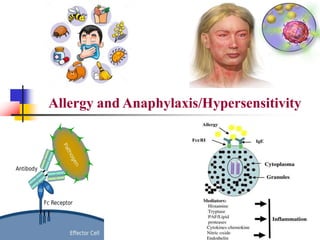
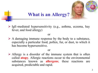
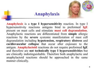
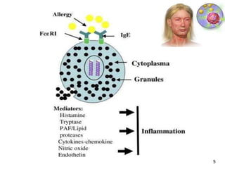
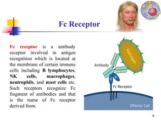
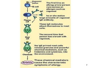
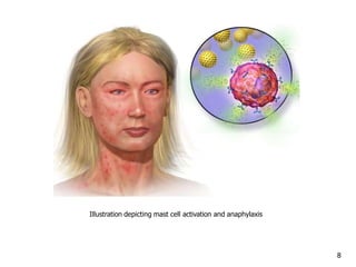
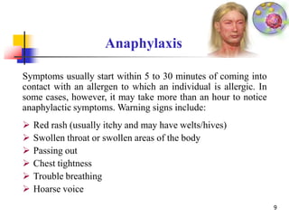
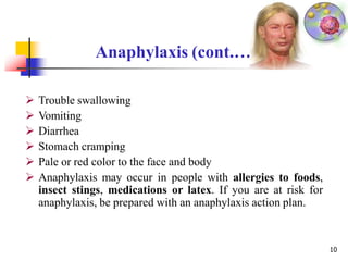
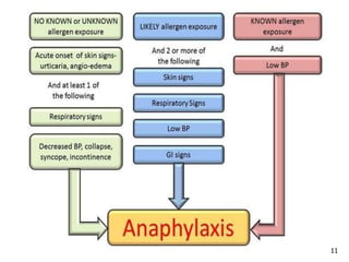
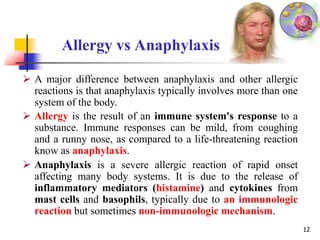
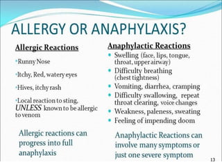
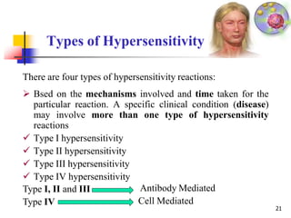
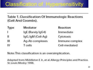
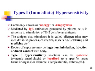
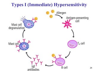
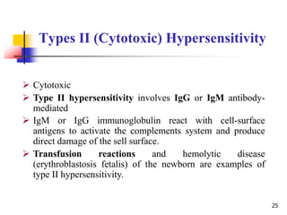
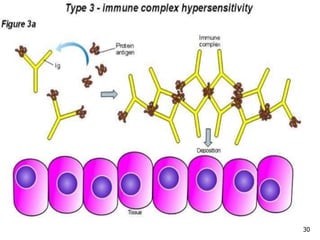
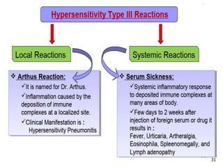
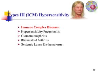
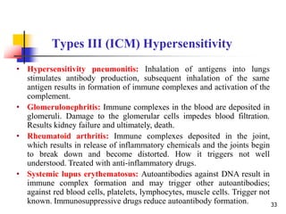
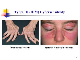
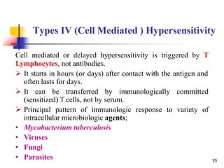
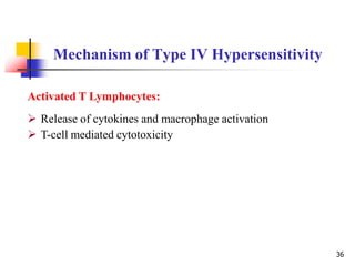
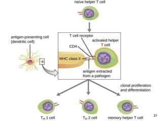
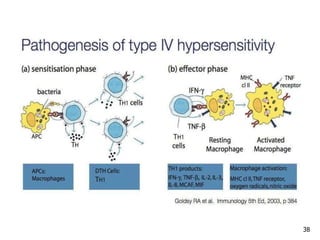
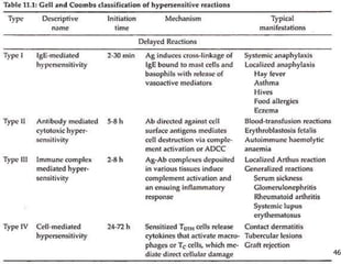
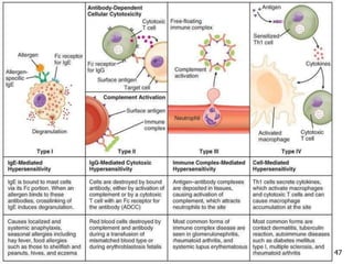
Allergy and Anaphylaxis/Hypersensitivity document discusses:
1. Allergies are immune system disorders caused by hypersensitivity to allergens like foods, pollens, or dusts. Anaphylaxis is a severe, potentially life-threatening allergic reaction affecting multiple body systems.
2. The four types of hypersensitivity reactions are: Type I involves IgE antibodies and causes immediate allergic reactions. Type II involves IgG/IgM antibodies attacking cell surfaces. Type III involves immune complex formation and complement activation causing tissue damage. Type IV is cell-mediated and involves T lymphocytes responding to antigens days later.
3. Anaphylaxis differs from other allergic reactions in affecting multiple
Normally the immune system plays an important role in protecting the body from microorganisms and other foreign substances. If the activity of the immune system is excessive or overreactive, a hypersensitivity reaction develops. The consequences of a hypersensitivity reaction may be injury to the body or death.
Immediate or Type I hypersensitivity is a rapid immunological reaction occurring in a previously sensitized individual that is triggered by the binding of an antigen to IgE antibody on the surface of mast cells.
Type II Hypersensitivity-Antibody mediated cytotoxic HypersensitivityAnup Bajracharya
Type II Hypersensitivity is antibody-mediated immune reaction in which antibodies (IgG or IgM) are directed against cellular or extracellular matrix antigens with the resultant cellular destruction, functional loss, or damage to tissues.
This presentation explores types of immune response. However under certain circumstances they can have deleterious effects on the body termed as hypersensitivity or allergy.
1. Type I Hypersensitivity:
Type I hypersensitive reactions are the commonest type among all types which is mainly induced by certain type of antigens i.e. allergens. Actually anaphylaxis means “opposite of protection” and is mediated by IgE antibodies through interaction with an allergen
Hypersensitivity can be defined as a state of altered immune response against an antigen characterized by hyper reactivity leading to immunopathology
Hypersensitivity reactions require a pre-sensitized (immune) state of the host.
NVBDCP.pptx Nation vector borne disease control programSapna Thakur
NVBDCP was launched in 2003-2004 . Vector-Borne Disease: Disease that results from an infection transmitted to humans and other animals by blood-feeding arthropods, such as mosquitoes, ticks, and fleas. Examples of vector-borne diseases include Dengue fever, West Nile Virus, Lyme disease, and malaria.
Pulmonary Thromboembolism - etilogy, types, medical- Surgical and nursing man...VarunMahajani
Disruption of blood supply to lung alveoli due to blockage of one or more pulmonary blood vessels is called as Pulmonary thromboembolism. In this presentation we will discuss its causes, types and its management in depth.
These simplified slides by Dr. Sidra Arshad present an overview of the non-respiratory functions of the respiratory tract.
Learning objectives:
1. Enlist the non-respiratory functions of the respiratory tract
2. Briefly explain how these functions are carried out
3. Discuss the significance of dead space
4. Differentiate between minute ventilation and alveolar ventilation
5. Describe the cough and sneeze reflexes
Study Resources:
1. Chapter 39, Guyton and Hall Textbook of Medical Physiology, 14th edition
2. Chapter 34, Ganong’s Review of Medical Physiology, 26th edition
3. Chapter 17, Human Physiology by Lauralee Sherwood, 9th edition
4. Non-respiratory functions of the lungs https://academic.oup.com/bjaed/article/13/3/98/278874
Acute scrotum is a general term referring to an emergency condition affecting the contents or the wall of the scrotum.
There are a number of conditions that present acutely, predominantly with pain and/or swelling
A careful and detailed history and examination, and in some cases, investigations allow differentiation between these diagnoses. A prompt diagnosis is essential as the patient may require urgent surgical intervention
Testicular torsion refers to twisting of the spermatic cord, causing ischaemia of the testicle.
Testicular torsion results from inadequate fixation of the testis to the tunica vaginalis producing ischemia from reduced arterial inflow and venous outflow obstruction.
The prevalence of testicular torsion in adult patients hospitalized with acute scrotal pain is approximately 25 to 50 percent
Lung Cancer: Artificial Intelligence, Synergetics, Complex System Analysis, S...Oleg Kshivets
RESULTS: Overall life span (LS) was 2252.1±1742.5 days and cumulative 5-year survival (5YS) reached 73.2%, 10 years – 64.8%, 20 years – 42.5%. 513 LCP lived more than 5 years (LS=3124.6±1525.6 days), 148 LCP – more than 10 years (LS=5054.4±1504.1 days).199 LCP died because of LC (LS=562.7±374.5 days). 5YS of LCP after bi/lobectomies was significantly superior in comparison with LCP after pneumonectomies (78.1% vs.63.7%, P=0.00001 by log-rank test). AT significantly improved 5YS (66.3% vs. 34.8%) (P=0.00000 by log-rank test) only for LCP with N1-2. Cox modeling displayed that 5YS of LCP significantly depended on: phase transition (PT) early-invasive LC in terms of synergetics, PT N0—N12, cell ratio factors (ratio between cancer cells- CC and blood cells subpopulations), G1-3, histology, glucose, AT, blood cell circuit, prothrombin index, heparin tolerance, recalcification time (P=0.000-0.038). Neural networks, genetic algorithm selection and bootstrap simulation revealed relationships between 5YS and PT early-invasive LC (rank=1), PT N0—N12 (rank=2), thrombocytes/CC (3), erythrocytes/CC (4), eosinophils/CC (5), healthy cells/CC (6), lymphocytes/CC (7), segmented neutrophils/CC (8), stick neutrophils/CC (9), monocytes/CC (10); leucocytes/CC (11). Correct prediction of 5YS was 100% by neural networks computing (area under ROC curve=1.0; error=0.0).
CONCLUSIONS: 5YS of LCP after radical procedures significantly depended on: 1) PT early-invasive cancer; 2) PT N0--N12; 3) cell ratio factors; 4) blood cell circuit; 5) biochemical factors; 6) hemostasis system; 7) AT; 8) LC characteristics; 9) LC cell dynamics; 10) surgery type: lobectomy/pneumonectomy; 11) anthropometric data. Optimal diagnosis and treatment strategies for LC are: 1) screening and early detection of LC; 2) availability of experienced thoracic surgeons because of complexity of radical procedures; 3) aggressive en block surgery and adequate lymph node dissection for completeness; 4) precise prediction; 5) adjuvant chemoimmunoradiotherapy for LCP with unfavorable prognosis.
Recomendações da OMS sobre cuidados maternos e neonatais para uma experiência pós-natal positiva.
Em consonância com os ODS – Objetivos do Desenvolvimento Sustentável e a Estratégia Global para a Saúde das Mulheres, Crianças e Adolescentes, e aplicando uma abordagem baseada nos direitos humanos, os esforços de cuidados pós-natais devem expandir-se para além da cobertura e da simples sobrevivência, de modo a incluir cuidados de qualidade.
Estas diretrizes visam melhorar a qualidade dos cuidados pós-natais essenciais e de rotina prestados às mulheres e aos recém-nascidos, com o objetivo final de melhorar a saúde e o bem-estar materno e neonatal.
Uma “experiência pós-natal positiva” é um resultado importante para todas as mulheres que dão à luz e para os seus recém-nascidos, estabelecendo as bases para a melhoria da saúde e do bem-estar a curto e longo prazo. Uma experiência pós-natal positiva é definida como aquela em que as mulheres, pessoas que gestam, os recém-nascidos, os casais, os pais, os cuidadores e as famílias recebem informação consistente, garantia e apoio de profissionais de saúde motivados; e onde um sistema de saúde flexível e com recursos reconheça as necessidades das mulheres e dos bebês e respeite o seu contexto cultural.
Estas diretrizes consolidadas apresentam algumas recomendações novas e já bem fundamentadas sobre cuidados pós-natais de rotina para mulheres e neonatos que recebem cuidados no pós-parto em unidades de saúde ou na comunidade, independentemente dos recursos disponíveis.
É fornecido um conjunto abrangente de recomendações para cuidados durante o período puerperal, com ênfase nos cuidados essenciais que todas as mulheres e recém-nascidos devem receber, e com a devida atenção à qualidade dos cuidados; isto é, a entrega e a experiência do cuidado recebido. Estas diretrizes atualizam e ampliam as recomendações da OMS de 2014 sobre cuidados pós-natais da mãe e do recém-nascido e complementam as atuais diretrizes da OMS sobre a gestão de complicações pós-natais.
O estabelecimento da amamentação e o manejo das principais intercorrências é contemplada.
Recomendamos muito.
Vamos discutir essas recomendações no nosso curso de pós-graduação em Aleitamento no Instituto Ciclos.
Esta publicação só está disponível em inglês até o momento.
Prof. Marcus Renato de Carvalho
www.agostodourado.com
ARTIFICIAL INTELLIGENCE IN HEALTHCARE.pdfAnujkumaranit
Artificial intelligence (AI) refers to the simulation of human intelligence processes by machines, especially computer systems. It encompasses tasks such as learning, reasoning, problem-solving, perception, and language understanding. AI technologies are revolutionizing various fields, from healthcare to finance, by enabling machines to perform tasks that typically require human intelligence.
Title: Sense of Smell
Presenter: Dr. Faiza, Assistant Professor of Physiology
Qualifications:
MBBS (Best Graduate, AIMC Lahore)
FCPS Physiology
ICMT, CHPE, DHPE (STMU)
MPH (GC University, Faisalabad)
MBA (Virtual University of Pakistan)
Learning Objectives:
Describe the primary categories of smells and the concept of odor blindness.
Explain the structure and location of the olfactory membrane and mucosa, including the types and roles of cells involved in olfaction.
Describe the pathway and mechanisms of olfactory signal transmission from the olfactory receptors to the brain.
Illustrate the biochemical cascade triggered by odorant binding to olfactory receptors, including the role of G-proteins and second messengers in generating an action potential.
Identify different types of olfactory disorders such as anosmia, hyposmia, hyperosmia, and dysosmia, including their potential causes.
Key Topics:
Olfactory Genes:
3% of the human genome accounts for olfactory genes.
400 genes for odorant receptors.
Olfactory Membrane:
Located in the superior part of the nasal cavity.
Medially: Folds downward along the superior septum.
Laterally: Folds over the superior turbinate and upper surface of the middle turbinate.
Total surface area: 5-10 square centimeters.
Olfactory Mucosa:
Olfactory Cells: Bipolar nerve cells derived from the CNS (100 million), with 4-25 olfactory cilia per cell.
Sustentacular Cells: Produce mucus and maintain ionic and molecular environment.
Basal Cells: Replace worn-out olfactory cells with an average lifespan of 1-2 months.
Bowman’s Gland: Secretes mucus.
Stimulation of Olfactory Cells:
Odorant dissolves in mucus and attaches to receptors on olfactory cilia.
Involves a cascade effect through G-proteins and second messengers, leading to depolarization and action potential generation in the olfactory nerve.
Quality of a Good Odorant:
Small (3-20 Carbon atoms), volatile, water-soluble, and lipid-soluble.
Facilitated by odorant-binding proteins in mucus.
Membrane Potential and Action Potential:
Resting membrane potential: -55mV.
Action potential frequency in the olfactory nerve increases with odorant strength.
Adaptation Towards the Sense of Smell:
Rapid adaptation within the first second, with further slow adaptation.
Psychological adaptation greater than receptor adaptation, involving feedback inhibition from the central nervous system.
Primary Sensations of Smell:
Camphoraceous, Musky, Floral, Pepperminty, Ethereal, Pungent, Putrid.
Odor Detection Threshold:
Examples: Hydrogen sulfide (0.0005 ppm), Methyl-mercaptan (0.002 ppm).
Some toxic substances are odorless at lethal concentrations.
Characteristics of Smell:
Odor blindness for single substances due to lack of appropriate receptor protein.
Behavioral and emotional influences of smell.
Transmission of Olfactory Signals:
From olfactory cells to glomeruli in the olfactory bulb, involving lateral inhibition.
Primitive, less old, and new olfactory systems with different path
- Video recording of this lecture in English language: https://youtu.be/lK81BzxMqdo
- Video recording of this lecture in Arabic language: https://youtu.be/Ve4P0COk9OI
- Link to download the book free: https://nephrotube.blogspot.com/p/nephrotube-nephrology-books.html
- Link to NephroTube website: www.NephroTube.com
- Link to NephroTube social media accounts: https://nephrotube.blogspot.com/p/join-nephrotube-on-social-media.html
These lecture slides, by Dr Sidra Arshad, offer a quick overview of physiological basis of a normal electrocardiogram.
Learning objectives:
1. Define an electrocardiogram (ECG) and electrocardiography
2. Describe how dipoles generated by the heart produce the waveforms of the ECG
3. Describe the components of a normal electrocardiogram of a typical bipolar leads (limb II)
4. Differentiate between intervals and segments
5. Enlist some common indications for obtaining an ECG
Study Resources:
1. Chapter 11, Guyton and Hall Textbook of Medical Physiology, 14th edition
2. Chapter 9, Human Physiology - From Cells to Systems, Lauralee Sherwood, 9th edition
3. Chapter 29, Ganong’s Review of Medical Physiology, 26th edition
4. Electrocardiogram, StatPearls - https://www.ncbi.nlm.nih.gov/books/NBK549803/
5. ECG in Medical Practice by ABM Abdullah, 4th edition
6. ECG Basics, http://www.nataliescasebook.com/tag/e-c-g-basics
Knee anatomy and clinical tests 2024.pdfvimalpl1234
This includes all relevant anatomy and clinical tests compiled from standard textbooks, Campbell,netter etc..It is comprehensive and best suited for orthopaedicians and orthopaedic residents.
Flu Vaccine Alert in Bangalore Karnatakaaddon Scans
As flu season approaches, health officials in Bangalore, Karnataka, are urging residents to get their flu vaccinations. The seasonal flu, while common, can lead to severe health complications, particularly for vulnerable populations such as young children, the elderly, and those with underlying health conditions.
Dr. Vidisha Kumari, a leading epidemiologist in Bangalore, emphasizes the importance of getting vaccinated. "The flu vaccine is our best defense against the influenza virus. It not only protects individuals but also helps prevent the spread of the virus in our communities," he says.
This year, the flu season is expected to coincide with a potential increase in other respiratory illnesses. The Karnataka Health Department has launched an awareness campaign highlighting the significance of flu vaccinations. They have set up multiple vaccination centers across Bangalore, making it convenient for residents to receive their shots.
To encourage widespread vaccination, the government is also collaborating with local schools, workplaces, and community centers to facilitate vaccination drives. Special attention is being given to ensuring that the vaccine is accessible to all, including marginalized communities who may have limited access to healthcare.
Residents are reminded that the flu vaccine is safe and effective. Common side effects are mild and may include soreness at the injection site, mild fever, or muscle aches. These side effects are generally short-lived and far less severe than the flu itself.
Healthcare providers are also stressing the importance of continuing COVID-19 precautions. Wearing masks, practicing good hand hygiene, and maintaining social distancing are still crucial, especially in crowded places.
Protect yourself and your loved ones by getting vaccinated. Together, we can help keep Bangalore healthy and safe this flu season. For more information on vaccination centers and schedules, residents can visit the Karnataka Health Department’s official website or follow their social media pages.
Stay informed, stay safe, and get your flu shot today!
2. What is an Allergy?
2
IgE‐mediated hypersensitivity (e.g., asthma, eczema, hay
fever, and food allergy)
or
A damaging immune response by the body to a substance,
especially a particular food, pollen, fur, or dust, to which it
has become hypersensitive.
or
Allergy is a disorder of the immune system that is often
called atopy. Allergic reactions occur to the environmental
substances known as allergens; these reactions are
acquired, predictable and rapid.
3. What is Anaphylaxis?
An often fatal hypersensitivity reaction, triggered by IgE or
anaphylatoxin‐mediated mast cell degranulation, leading to
anaphylactic shock due to vasodilatation and smooth muscle
contraction.
An acute allergic reaction to an antigen (e.g. a bee sting) to which the
body has become hypersensitive.
Anaphylaxis is an acute systemic (multi-system) and severe Type 1
Hypersensitivity allergic reactions in humans and the other mammals.
The term comes from Greek words “ava” ana (against) and phylaxis
(protection).
Atopy (Greek Atottia-placelessness) or atopic syndrome is an allergic
hypersensitivity affecting parts of the body not in direct contact with
allergen. It may involve Eczema (atopic dermatitis), Allergic
conjunctivitis, allergic rhinitis and asthma. There appears to be a strong
hereditary component. 3
4. Anaphylaxis
4
Anaphylaxis is a type I hypersensitivity reaction. In type I
hypersensitivity reactions antigens bind to preformed IgE
present on mast cells and stimulate mast cell degranulation.
Anaphylactic reactions are differentiated from simple allergic
reactions by the severe systemic manifestation of mast cell
degranulation including hypotension, respiratory distress and
cardiovascular collapse that occur after exposure to the
antigen. Anaphylactoid reactions do not require preformed IgE
and therefore are not technically type I hypersensitivities but
are clinically indistinguishable. Therefore both anaphylactic and
anaphylactoid reactions should be approached in the same
manner clinically.
6. Fc receptor
6
is a antibody
receptor involved in antigen
recognition which is located at
the membrane of certain immune
cells including B lymphocytes,
NK cells, macrophages,
neutrophils, and mast cells etc.
Such receptors recognize Fc
fragment of antibodies and that
is the name of Fc receptor
derived from.
Fc Receptor
9. Anaphylaxis
9
Symptoms usually start within 5 to 30 minutes of coming into
contact with an allergen to which an individual is allergic. In
some cases, however, it may take more than an hour to notice
anaphylactic symptoms. Warning signs include:
Red rash (usually itchy and may have welts/hives)
Swollen throat or swollen areas of the body
Passing out
Chest tightness
Trouble breathing
Hoarse voice
10. Anaphylaxis (cont.…
10
Trouble swallowing
Vomiting
Diarrhea
Stomach cramping
Pale or red color to the face and body
Anaphylaxis may occur in people with allergies to foods,
insect stings, medications or latex. If you are at risk for
anaphylaxis, be prepared with an anaphylaxis action plan.
12. Allergy vs Anaphylaxis
12
A major difference between anaphylaxis and other allergic
reactions is that anaphylaxis typically involves more than one
system of the body.
Allergy is the result of an immune system's response to a
substance. Immune responses can be mild, from coughing
and a runny nose, as compared to a life-threatening reaction
know as anaphylaxis.
Anaphylaxis is a severe allergic reaction of rapid onset
affecting many body systems. It is due to the release of
inflammatory mediators (histamine) and cytokines from
mast cells and basophils, typically due to an immunologic
reaction but sometimes non-immunologic mechanism.
14. Allergy/Sensitivity/Intolerance
14
The primary difference between an allergy, a sensitivity, and
an intolerance is that an allergy is characterized by an
immune system reaction to a substance, a sensitivity involves
non immune response and an intolerance is characterized by
the body lacking an chemical or enzyme needed to digest
certain food.
16. The Complement System
16
• The complement system is a part of the immune system that
enhances (complements) the ability of antibodies and
phagocytic cells to clear microbes and damaged cells from
an organism, promote inflammation, and attack the
pathogen's cell membrane.
• It is part of the innate immune system which is not acquired
and does not change during an individual's lifetime.
• The complement system brought into action by the
antibodies produced by the adaptive immune system.
• The complement system consists of a number of small
proteins that are synthesized by the liver, and circulate in the
blood as inactive precursors.
17. The Complement System
• When it triggers, proteases in the system cleave specific
proteins to release cytokines and initiate an amplifying
cascade of further cleavages.
• The end result of this complement activation or
complement fixation cascade is stimulation of phagocytes
to clear foreign and damaged material, inflammation to
attract additional phagocytes, and activation of the cell-
killing membrane attack complex (MAC).
• Over 30 proteins or protein fragments make up the
complement system.
• Three biochemical pathways activate the complement
system: the classical complement pathway, the
alternative complement pathway, and the lectin pathway 17
18. The Complement System
The classical complement pathway requires antigen-
antibody complexes for activation (specific immune
response), whereas the alternative pathway can be activated
by spontaneous complement component 3 (C3) hydrolysis.
The mannose-binding lectin pathway can be activated by
C3 hydrolysis or antigens without the presence of antibodies
(non-specific immune response). In all three pathways, C3-
convertase cleaves and activates component C3, creating
C3a and C3b, and causes a cascade of further cleavage and
activation events. C3b binds to the surface of pathogens,
leading to greater internalization by phagocytic cells by
opsonization (identify the invaders).
18
19. The Complement System
In the alternative pathway, C3b binds to different factors and
cleaves C5 (complement component 5 is a protein in humans
which is encoded by the C5 gene) into C5b and C5a. C5a is
an important chemotactic protein, helping recruit
inflammatory cells. C3a is the precursor of an important
cytokine. Both C3a and C5a have anaphylatoxin activity,
directly triggering degranulation of mast cells as well as
increasing vascular permeability and smooth muscle
contraction. C5b initiates the membrane attack pathway,
which results in the membrane attack complex (MAC),
consisting of C5b, C6, C7, C8, and polymeric C9. MAC is
the cytolytic end product of the complement cascade; it
forms a transmembrane channel, which causes osmotic
lysis of the target cell. 19
21. Types of Hypersensitivity
There are four types of hypersensitivity reactions:
Bsed on the mechanisms involved and time taken for the
particular reaction. A specific clinical condition (disease)
may involve more than one type of hypersensitivity
reactions
Type I hypersensitivity
Type II hypersensitivity
Type III hypersensitivity
Type IV hypersensitivity
Type I, II and III
Type IV
Antibody Mediated
Cell Mediated
21
23. Types I (Immediate) Hypersensitivity
23
Commonly known as “allergy” or Anaphylaxis
Mediated by IgE antibodies generated by plasma cells in
response to stimulation of TH2 cells by an antigens.
The antigen that stimulates it is called allergen (that may
include: dust, pollens, cosmetics, insects bite, clothing and
medicine etc.)
Routes of exposure may be ingestion, inhalation, injection
or direct contact with body.
Type I hypersensitivity reactions can be systemic
(systemic anaphylaxis) or localized to a specific target
tissue or organ (for example, allergic rhinitis, asthma etc.).
25. Types II (Cytotoxic) Hypersensitivity
25
Cytotoxic
Type II hypersensitivity involves IgG or IgM antibody-
mediated
IgM or IgG immunoglobulin react with cell-surface
antigens to activate the complements system and produce
direct damage of the sell surface.
Transfusion reactions and hemolytic disease
(erythroblastosis fetalis) of the newborn are examples of
type II hypersensitivity.
26. Types II Hypersensitivity Mechanism
In type II hypersensitivity reactions, the antibodies
produced by the immune response bind to antigens on the
patient’s own cell surfaces. The antigens recognized in this
way may either be intrinsic (“self” antigen, innately part of
the patient’s cells) or extrinsic (absorbed onto the cells
during exposure to some foreign antigen, possibly as part of
infection with a pathogen).
IgG and IgM antibodies bind to these antigens to form
complexes that activate the classical pathway of
complement activation. That is, mediators of acute
inflammation are generated at the site and membrane
attack complexes cause cell lysis and death. The reaction
takes hours to a day. 26
28. Types III (ICM) Hypersensitivity
28
Type III hypersensitivity is also called as Immune
Complex Mimics (ICM).
The reaction may take 3 - 10 hours after the exposure to the
antigen.
The reaction may be generalized (for example: serum) or
may involve individual organs including or other organs.
Antigens causing immune complex mediated injury are:
• Exogenous
• Endogenous.
29. Types III Hypersensitivity Mechanism
29
Antigens combines with antibody within circulation and
form immune complex
Wherever in the body they deposited
They activate compliment system
Polymorphonuclear cells are attracted to the site
Result in inflammation and tissue injury
33. Types III (ICM) Hypersensitivity
• Hypersensitivity pneumonitis: Inhalation of antigens into lungs
stimulates antibody production, subsequent inhalation of the same
antigen results in formation of immune complexes and activation of the
complement.
• Glomerulonephritis: Immune complexes in the blood are deposited in
glomeruli. Damage to the glomerular cells impedes blood filtration.
Results kidney failure and ultimately, death.
• Rheumatoid arthritis: Immune complexes deposited in the joint,
which results in release of inflammatory chemicals and the joints begin
to break down and become distorted. How it triggers not well
understood. Treated with anti-inflammatory drugs.
• Systemic lupus erythematosus: Autoantibodies against DNA result in
immune complex formation and may trigger other autoantibodies;
against red blood cells, platelets, lymphocytes, muscle cells. Trigger not
known. Immunosuppressive drugs reduce autoantibody formation. 33
35. Types IV (Cell Mediated ) Hypersensitivity
Cell mediated or delayed hypersensitivity is triggered by T
Lymphocytes, not antibodies.
It starts in hours (or days) after contact with the antigen and
often lasts for days.
It can be transferred by immunologically committed
(sensitized) T cells, not by serum.
Principal pattern of immunologic response to variety of
intracellular microbiologic agents;
• Mycobacterium tuberculosis
• Viruses
• Fungi
• Parasites 35
36. Mechanism of Type IV Hypersensitivity
36
Activated T Lymphocytes:
Release of cytokines and macrophage activation
T-cell mediated cytotoxicity
39. The tuberculin response:
An injection of tuberculin beneath the skin causes reaction in
individual exposed to tuberculosis or tuberculosis vaccine
Used to diagnose contact with antigens of Mycobacterium
tuberculosis
No response when individual not infected or vaccinated
Red, hard swelling develops in individuals previously
infected or immunized
39
Clinically Important Delayed
Hypersensitivity Reaction
41. Type IV (Cell Mediated) Hypersensitivity
41
Graft rejection:
Rejection of tissues or organs that have been transplanted
Grafts perceived as foreign by a recipient undergo rejection
Immune response against foreign MHC on graft cells
Rejection depends on degree to which the graft is foreign to
the recipient
• Based on the type of graft
42. Type of Grafts
42
Autograft–tissue grafted back on to the original donor.
Isograft–graft between syngeneic individuals (identical
genetic constitution) such as identical twins or mice of the
same pure inbred strain.
Allograft–graft between allogeneic individuals (members
of the same species but different genetic constitution), for
example, human to human and one mouse strain to another.
Xenograft–graft between xenogeneic individuals (different
species), for example, pig to human