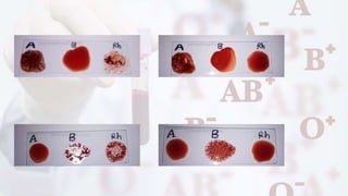
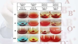
BLOOD GROUPING
&
CROSS MATCHING
Dr Sandeep singh
Dept of Pathology.
Gmc ,Knw.
Objectives:
1. Introduction.
2. Blood group system.
3. ABO system.
4. Genetics of ABO system.
Introduction
• The blood group you belong to depends on
what you have inherited from your parents.
• There are more than 20 genetically
determined blood group systems known
today
• The AB0 and Rhesus (Rh) systems are the
most important ones used for blood
transfusions.
Blood grouping system
● Major blood group system - based on
Agglutinogens on cell membrane present widely
and causes severe transfusion reaction
1. ABO
2. Rh system
● Minor blood group system – based on
Agglutinogens but present in few populations &
causes mild transfusion reaction.
1. MNS
2. P
● Familial blood group system – found in few
families
1. Kell
2. Duffy
3. Lutheran
4. Lewis
5. Diego &
6. Kidd
ABO Blood group system
The four groups are determined by the presence
or absence of blood group antigens on the RBCs
and accordingly an individual’s group is
A,B,AB or O
Karl Landsteiner's law.
1. Individuals who lack the antigens of ABO system,
have the corresponding Antibody in their serum (Table
1.0).
2. Ab’s of ABO system are usually “naturally occurring “
IgM type.
3. These can cause intravascular hemolysis in patients,
if transfused with incompatible blood. Thus , an error
in ABO grouping in donor or recipient can be fatal.
Inheritance of ABO blood groups
• The ABO system follows the mendelian laws.
• He demonstrated that everyone inherits one
ABO gene from each parent and presence of
these two gene determines the type of
antigen present on the surface of red cells.
• The genotype and phenotype of ABO group is
as follows
ABO Antigens :-
Formation of H antigen : -
• The H antigen is the foundation upon which A
and B antigens are built
• A and B genes code for enzymes that add an
immunodominant sugar to the H antigen
• Immunodominant sugars are present at the
terminal ends of the chains and confer the ABO
antigen specificity.
• The basic precursor substance is converted to H
substance by enzyme .
Formation of Precursor substance
Formation of H antigen
Formation of A and B Antigen : -
• A gene codes for an enzyme (transferase)
that adds N-acetyl galactosamine to the
terminal sugar of the H antigen – N-
acetylglucosaminyltransferase
• B gene cods for an enzyme that adds D
galactose to the terminal sugar of the H
antigen – D-galactosyltransferase
Formation of A antigen
Formation of B antigen
ABO Antibodies :-
• ABO antibodies are generally IgM in nature.
• They are naturally occurring complete and
cold reacting antibodies, which do not cross
placental barrier and
• They are capable of binding complement.
• The anti – AB of O blood group carries a
higher titer than anti – A or anti – B.
• The IgM anti – A and anti –B are not produced
up to the age of 3 to 6months.
ABO subgroups:-
The subgroups of A and AB are of clinical significance :
1. Subgroups of A :
• The group A has subclassified as A1 and A2
a. A1 : - the A red cells which react with both anti – A
and anti – A1 are designated as A1 subgroup. A1 has
more antigenic sites foe A antigen and less for H.
b. A2 :- the A red cells which react with only anti – A and
not with anti – A1 are called A2. this is weak A
subgroup and carries more H substance.
Bombay blood group : -
• Bhende YM, et al in year 1952, first discovered th
is blood group in the city of Bombay, India, from
where it got its name.
• O blood group individuals do not carry either A or
B antigen but have maximum amount of H
antigen on their red cells. Some individuals lack
even H antigen along with A and B. these
individuals are called Oh phenotype.
• Since there is no H antigen on the surface of red cells of
Oh, the anti-H antibody develops in their serum, along
with all the other antibodies found in any O blood group.
anti-H present in Oh is clinically significant, warm
antibody reactive at 37°C.
• Bombay blood group is not compatible with any ABO blo
od group and the choice of blood for these individuals
remains only Bombay itself.
ABO Grouping : -
The ABO grouping can be performed by the
following methods
1. Slide Method
2. Tube Method or Technique
3. Micro typing System (Gel System)
4. Microplate Technique
5. Glass Microbeads Method
1. Slide method
It is a simple technique, which should be employed
in cases of emergency or outdoor camps. The
technique is less sensitive and not capable of
detecting weak antigens.
REAGENTS REQUIRED : -
• Monoclonal anti-A , anti-B sera.
•Monoclonal
•anti-A ,
• anti-B sera.
Procedure : -
1. the test can be performed either on glass slides or
on ceramic tiles.
2. Place one drop of Anti – A , Anti – B and Anti – D each
on slide in 3 labelled circles.
3. Add one drop of blood (preferably 20% red
cell suspension) on each circle of the slide.
4. Mix properly by a clean glass stick or the corner
of another slide.
5. Rock the slides in order to mix the cells and sera
and leave at room temperature for 2 minutes.
6. Record the results.
Interpretation of results : -
• The agglutination appears like granular
precipitate resembling yogurt.
• Agglutination (+) in one or both antisera is interpr
eted as follows
2. Tube method : -
This is a more sensitive technique which can detect weak antigens
and antibodies. the cell grouping and serum groupings are
performed separately and are complimentary to each other.
Washing of red cells :-
1. Before going for any procedure in the blood bank, the red
cells must be washed properly.
2. 0.5 ml of red cells are mixed with normal saline filling 2/3rd of
test tube.
3. The mixture is centrifuged at 3000 rpm for 1 minute. the
supernatant is discarded.
4. Refill the tube with same amount of normal saline and centrifuge
again.
5. Repeat the procedure three times and discard the supernatant
every time. the remaining cells are washed cells.
Preparation of 5% red cell suspension : -
1. Mix the washed red cells and the normal
saline in one of the following ratios as per
requirement:
• 0.1 ml of cells + 1.9 ml of normal saline, 0.2
ml of cells + 3.8 ml of normal saline, 0.5 ml
of cells + 9.5 ml of normal saline .
2. Centrifuge the mixture at 1000 rpm for 1
minute.
A. Forward grouping
Cell grouping (forward grouping)
1. Prepare 5% red cell suspension in normal saline.
2. Add 1 drop of anti-A in the tube labelled A, anti-B in
the tube labelled B and anti-AB in the tube labelled
AB.
3. Add 1 drop of the cell suspension in each tube.
4. Mix properly, incubate the mixture at
room temperature (RT) for 5-10minutes and
then centrifuge at 1000 rpm for 1 minute.
5. If no hemolysis is observed in the supernatant,
disperse the cell button.
6. Check for agglutination. If no clump is seen by
naked eyes, examine under microscope for
weak agglutination.
7. Record the results.
B. Reverse grouping
Serum grouping (reverse grouping)
1. The serum of the donor/patient is tested
against known cells of group A, B and O. these cells
are either prepared in the lab by pooling or can be
acquired from manufacturers.
2. Arrange three test tubes and label them A, B and
O.
3. Place 2 drops of the serum to be tested in each
tube.
4. Add 1 drop of A group cells to the tube A, B group cells
to tube B and O group cells to the tube labelled O.
5. Shake the contents gently. Incubate at RT for 5-10
minutes and centrifuge at 1000 rpm for 1 minute.
6. If the supernatant shows no signs of hemolysis, disperse
the cell button and observe for agglutination.
7. If no agglutination is observed through naked eyes use
microscope to examine.
8. Record the results
Interpretation
3. Micro typing System (Gel System)
• The gel technique by its ID card
system has made the blood grouping
a very convenient procedure.
• Forward and reverse ABO grouping a
nd Rh grouping are done with
a single card in a very short time.
Principle :-
• The basic principle is that instead of test tube,
the serum and cell reaction takes place in a
microtube consisting of a reaction chamber that
narrows to become a column Red cells or
mixture of cells & serum (as appropriate) are
added into the gel The cells are always added
prior to the serum so that the serum does not
come into contact with the gel
Reagents
The “ID-card ABO/D + reverse grouping for patients”.
the microtubes contain the following reagent.
1. Microtube 1 - anti-A
2. Microtube 2 - anti-B
3. Microtube 3 - anti-D
4. Microtube 4 - negative Rh control
5. Microtube 5 - A1 cells
6. Microtube 6 - B cells.
Sample required
• Freshly drawn blood samples in EDTA or citrate. For
reverse grouping, either plasma or serum can be used.
Preparation of blood samples
• Prepare 5% cell suspension.
• Bring the ID-diluent 2 at room temperature
• Dispense 0.5 ml of ID-diluent 2 into a clean tube
• Add 50 µL of whole blood or 25 µL of packed
cells, mix gently.
Procedure : -
1. Write the patient or donor’s name and
registration number on the ID card.
2. Remove the aluminum foil from the top of
the microtubes by holding the card in the upright
position.
3. Pipette 10 µL of patient’s 5% red cell suspension to
the microtubes 1-4 (A, B, D and control (ctl)).
4. Place 50 µL of ID-cell A1 in microtube 5 (A1).
5. Place 50 µL ID-cell B in microtube 6 (B).
6. Centrifuge the ID card for 10 minutes in the ID-
centrifuge.
7. Read and record the results.
Result: -
• Positive – Agglutinated cells forming a red line
on the surface of the gel.
• Negative – Compact button of cells on the
bottom of the microtube.
Interpretation of results:-
• The result of the ID card is also interpreted as the
results of the test tube method, considering the
microtubes 1 and 2 as forward grouping, 3 as Rh,4
as negative Rh control, 5 and 6 as reverse grouping.
• The negative control must not show any agglutinatio
n. If it shows agglutination, your procedure is invalid.
• Repeat the whole procedure.
Transfusion matching
2 Methods for cross matching
Major Cross match
Minor Cross Match
Other methods crossmatch
THANK YOU!!

ABO blood.pptx

  • 1.
    BLOOD GROUPING & CROSS MATCHING DrSandeep singh Dept of Pathology. Gmc ,Knw.
  • 2.
    Objectives: 1. Introduction. 2. Bloodgroup system. 3. ABO system. 4. Genetics of ABO system.
  • 3.
    Introduction • The bloodgroup you belong to depends on what you have inherited from your parents. • There are more than 20 genetically determined blood group systems known today • The AB0 and Rhesus (Rh) systems are the most important ones used for blood transfusions.
  • 4.
    Blood grouping system ●Major blood group system - based on Agglutinogens on cell membrane present widely and causes severe transfusion reaction 1. ABO 2. Rh system ● Minor blood group system – based on Agglutinogens but present in few populations & causes mild transfusion reaction. 1. MNS 2. P
  • 5.
    ● Familial bloodgroup system – found in few families 1. Kell 2. Duffy 3. Lutheran 4. Lewis 5. Diego & 6. Kidd
  • 6.
    ABO Blood groupsystem The four groups are determined by the presence or absence of blood group antigens on the RBCs and accordingly an individual’s group is A,B,AB or O
  • 7.
    Karl Landsteiner's law. 1.Individuals who lack the antigens of ABO system, have the corresponding Antibody in their serum (Table 1.0). 2. Ab’s of ABO system are usually “naturally occurring “ IgM type. 3. These can cause intravascular hemolysis in patients, if transfused with incompatible blood. Thus , an error in ABO grouping in donor or recipient can be fatal.
  • 9.
    Inheritance of ABOblood groups • The ABO system follows the mendelian laws. • He demonstrated that everyone inherits one ABO gene from each parent and presence of these two gene determines the type of antigen present on the surface of red cells. • The genotype and phenotype of ABO group is as follows
  • 11.
    ABO Antigens :- Formationof H antigen : - • The H antigen is the foundation upon which A and B antigens are built • A and B genes code for enzymes that add an immunodominant sugar to the H antigen • Immunodominant sugars are present at the terminal ends of the chains and confer the ABO antigen specificity. • The basic precursor substance is converted to H substance by enzyme .
  • 12.
  • 13.
  • 14.
    Formation of Aand B Antigen : - • A gene codes for an enzyme (transferase) that adds N-acetyl galactosamine to the terminal sugar of the H antigen – N- acetylglucosaminyltransferase • B gene cods for an enzyme that adds D galactose to the terminal sugar of the H antigen – D-galactosyltransferase
  • 15.
  • 16.
  • 17.
    ABO Antibodies :- •ABO antibodies are generally IgM in nature. • They are naturally occurring complete and cold reacting antibodies, which do not cross placental barrier and • They are capable of binding complement. • The anti – AB of O blood group carries a higher titer than anti – A or anti – B. • The IgM anti – A and anti –B are not produced up to the age of 3 to 6months.
  • 18.
    ABO subgroups:- The subgroupsof A and AB are of clinical significance : 1. Subgroups of A : • The group A has subclassified as A1 and A2 a. A1 : - the A red cells which react with both anti – A and anti – A1 are designated as A1 subgroup. A1 has more antigenic sites foe A antigen and less for H. b. A2 :- the A red cells which react with only anti – A and not with anti – A1 are called A2. this is weak A subgroup and carries more H substance.
  • 19.
    Bombay blood group: - • Bhende YM, et al in year 1952, first discovered th is blood group in the city of Bombay, India, from where it got its name. • O blood group individuals do not carry either A or B antigen but have maximum amount of H antigen on their red cells. Some individuals lack even H antigen along with A and B. these individuals are called Oh phenotype.
  • 20.
    • Since thereis no H antigen on the surface of red cells of Oh, the anti-H antibody develops in their serum, along with all the other antibodies found in any O blood group. anti-H present in Oh is clinically significant, warm antibody reactive at 37°C. • Bombay blood group is not compatible with any ABO blo od group and the choice of blood for these individuals remains only Bombay itself.
  • 21.
    ABO Grouping :- The ABO grouping can be performed by the following methods 1. Slide Method 2. Tube Method or Technique 3. Micro typing System (Gel System) 4. Microplate Technique 5. Glass Microbeads Method
  • 22.
    1. Slide method Itis a simple technique, which should be employed in cases of emergency or outdoor camps. The technique is less sensitive and not capable of detecting weak antigens. REAGENTS REQUIRED : - • Monoclonal anti-A , anti-B sera.
  • 23.
  • 24.
    Procedure : - 1.the test can be performed either on glass slides or on ceramic tiles. 2. Place one drop of Anti – A , Anti – B and Anti – D each on slide in 3 labelled circles. 3. Add one drop of blood (preferably 20% red cell suspension) on each circle of the slide. 4. Mix properly by a clean glass stick or the corner of another slide. 5. Rock the slides in order to mix the cells and sera and leave at room temperature for 2 minutes. 6. Record the results.
  • 25.
    Interpretation of results: - • The agglutination appears like granular precipitate resembling yogurt. • Agglutination (+) in one or both antisera is interpr eted as follows
  • 28.
    2. Tube method: - This is a more sensitive technique which can detect weak antigens and antibodies. the cell grouping and serum groupings are performed separately and are complimentary to each other. Washing of red cells :- 1. Before going for any procedure in the blood bank, the red cells must be washed properly. 2. 0.5 ml of red cells are mixed with normal saline filling 2/3rd of test tube. 3. The mixture is centrifuged at 3000 rpm for 1 minute. the supernatant is discarded. 4. Refill the tube with same amount of normal saline and centrifuge again. 5. Repeat the procedure three times and discard the supernatant every time. the remaining cells are washed cells.
  • 29.
    Preparation of 5%red cell suspension : - 1. Mix the washed red cells and the normal saline in one of the following ratios as per requirement: • 0.1 ml of cells + 1.9 ml of normal saline, 0.2 ml of cells + 3.8 ml of normal saline, 0.5 ml of cells + 9.5 ml of normal saline . 2. Centrifuge the mixture at 1000 rpm for 1 minute.
  • 30.
    A. Forward grouping Cellgrouping (forward grouping) 1. Prepare 5% red cell suspension in normal saline. 2. Add 1 drop of anti-A in the tube labelled A, anti-B in the tube labelled B and anti-AB in the tube labelled AB. 3. Add 1 drop of the cell suspension in each tube.
  • 31.
    4. Mix properly,incubate the mixture at room temperature (RT) for 5-10minutes and then centrifuge at 1000 rpm for 1 minute. 5. If no hemolysis is observed in the supernatant, disperse the cell button. 6. Check for agglutination. If no clump is seen by naked eyes, examine under microscope for weak agglutination. 7. Record the results.
  • 32.
    B. Reverse grouping Serumgrouping (reverse grouping) 1. The serum of the donor/patient is tested against known cells of group A, B and O. these cells are either prepared in the lab by pooling or can be acquired from manufacturers. 2. Arrange three test tubes and label them A, B and O. 3. Place 2 drops of the serum to be tested in each tube.
  • 33.
    4. Add 1drop of A group cells to the tube A, B group cells to tube B and O group cells to the tube labelled O. 5. Shake the contents gently. Incubate at RT for 5-10 minutes and centrifuge at 1000 rpm for 1 minute. 6. If the supernatant shows no signs of hemolysis, disperse the cell button and observe for agglutination. 7. If no agglutination is observed through naked eyes use microscope to examine. 8. Record the results
  • 34.
  • 36.
    3. Micro typingSystem (Gel System) • The gel technique by its ID card system has made the blood grouping a very convenient procedure. • Forward and reverse ABO grouping a nd Rh grouping are done with a single card in a very short time.
  • 37.
    Principle :- • Thebasic principle is that instead of test tube, the serum and cell reaction takes place in a microtube consisting of a reaction chamber that narrows to become a column Red cells or mixture of cells & serum (as appropriate) are added into the gel The cells are always added prior to the serum so that the serum does not come into contact with the gel
  • 39.
    Reagents The “ID-card ABO/D+ reverse grouping for patients”. the microtubes contain the following reagent. 1. Microtube 1 - anti-A 2. Microtube 2 - anti-B 3. Microtube 3 - anti-D 4. Microtube 4 - negative Rh control 5. Microtube 5 - A1 cells 6. Microtube 6 - B cells.
  • 40.
    Sample required • Freshlydrawn blood samples in EDTA or citrate. For reverse grouping, either plasma or serum can be used. Preparation of blood samples • Prepare 5% cell suspension. • Bring the ID-diluent 2 at room temperature • Dispense 0.5 ml of ID-diluent 2 into a clean tube • Add 50 µL of whole blood or 25 µL of packed cells, mix gently.
  • 41.
    Procedure : - 1.Write the patient or donor’s name and registration number on the ID card. 2. Remove the aluminum foil from the top of the microtubes by holding the card in the upright position. 3. Pipette 10 µL of patient’s 5% red cell suspension to the microtubes 1-4 (A, B, D and control (ctl)). 4. Place 50 µL of ID-cell A1 in microtube 5 (A1). 5. Place 50 µL ID-cell B in microtube 6 (B). 6. Centrifuge the ID card for 10 minutes in the ID- centrifuge. 7. Read and record the results.
  • 42.
    Result: - • Positive– Agglutinated cells forming a red line on the surface of the gel. • Negative – Compact button of cells on the bottom of the microtube.
  • 43.
    Interpretation of results:- •The result of the ID card is also interpreted as the results of the test tube method, considering the microtubes 1 and 2 as forward grouping, 3 as Rh,4 as negative Rh control, 5 and 6 as reverse grouping. • The negative control must not show any agglutinatio n. If it shows agglutination, your procedure is invalid. • Repeat the whole procedure.
  • 46.
  • 48.
    2 Methods forcross matching
  • 49.
  • 50.
  • 51.
  • 56.