Downloaded 128 times

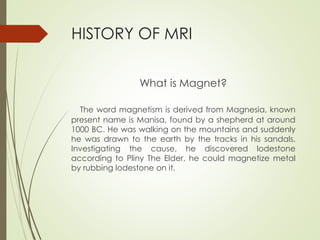


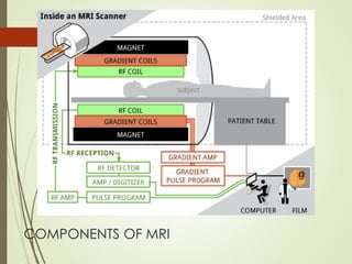

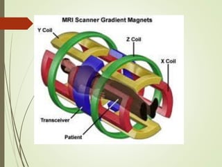
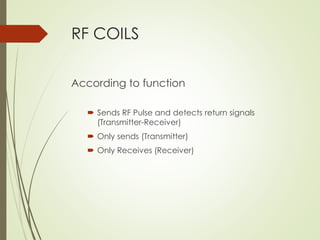
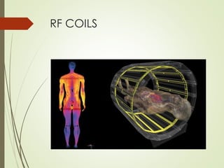
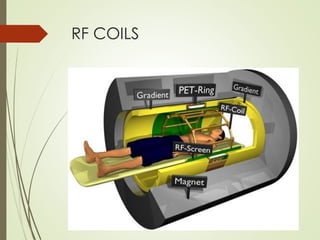
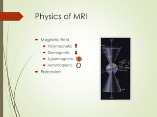
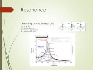
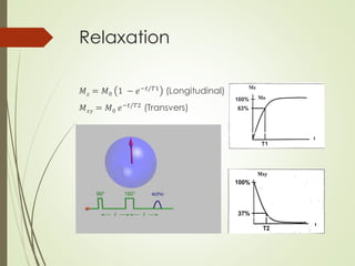


This document provides an overview of magnetic resonance imaging (MRI). It discusses the history of MRI, including key contributors like Felix Bloch and Edward Purcell. It describes the main components of an MRI machine, including the main magnet, gradient coils, RF coils, shielding, and computers. It explains the physics principles behind MRI such as magnetic fields, precession, relaxation, and gradients. It also covers MRI signal types, sequencing, tissue contrast, image quality, artifacts, and safety considerations.


![MAGNETIC_RESONANCE.._IMAGING[MRI][1].pptx](https://cdn.slidesharecdn.com/ss_thumbnails/magneticresonanceimagingmri1-240903182728-4f857936-thumbnail.jpg?width=640&height=640&fit=bounds)


