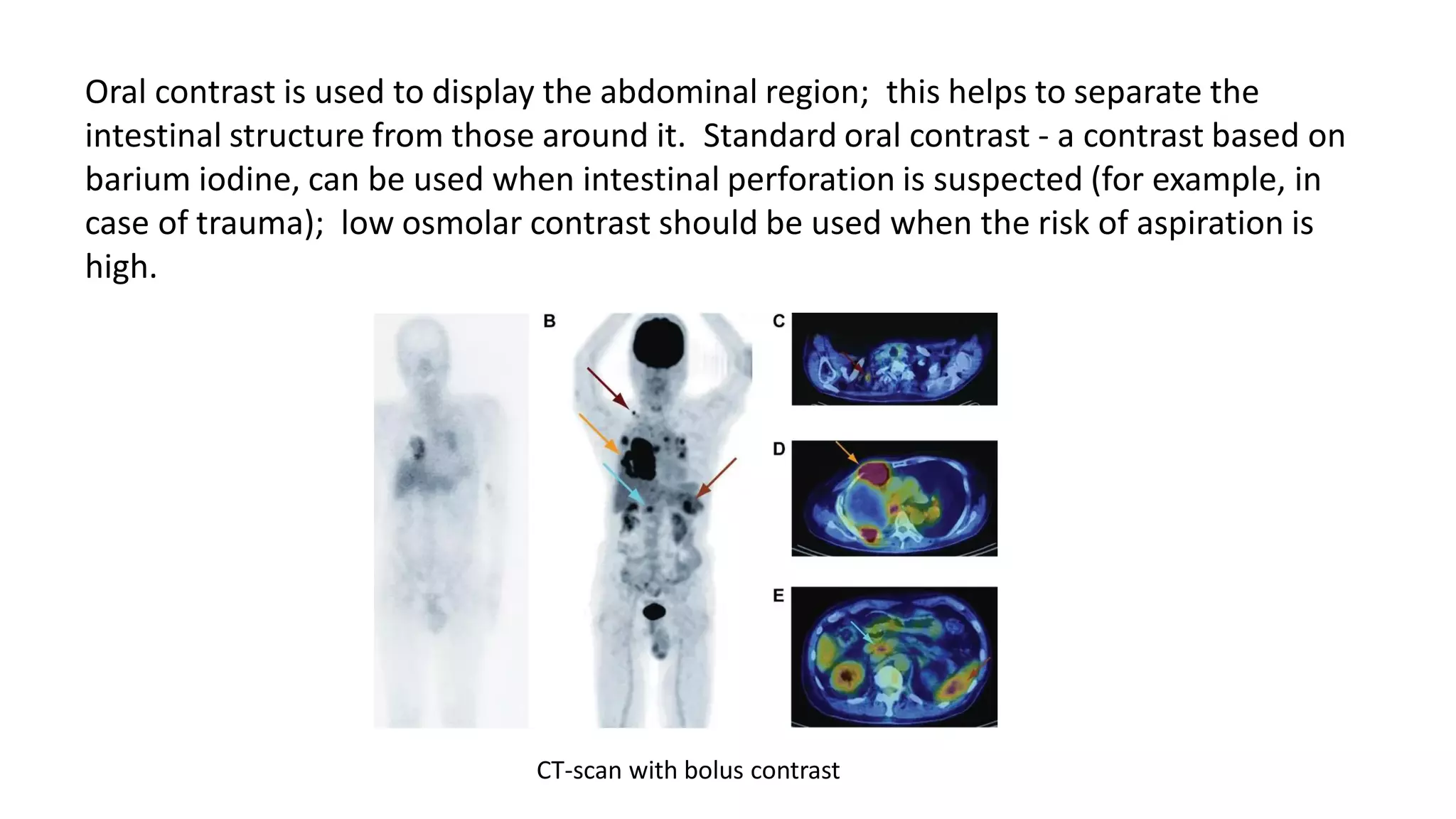
COMPUTED
TOMOGRAPHY
Assistant Turatbekkyzy Ch.
The emergence of computed tomography, as a method of
scanning the human body, became possible only thanks to
the discovery by Wilhelm Roentgen, a German physicist, of
X-rays with a unique ability to penetrate solid objects.
Some time after this discovery, the rays were called X-rays,
and the scientific and medical world has found an
unprecedented way to explore the internal state of the human
body without open surgical interventions - scanning with X-rays.
Radiography, as a method of obtaining images of body parts in one plane, in fact, became
the first step towards the emergence of computed tomography - already at the beginning
of the 20th century, radiography began to be used in medical institutions. And thanks to
the achievements of scientific and technological progress in the 20th century, the results
of which were the first computers (electronic computers), computed tomography was first
introduced to the medical community around the world in the 70s.
Godfrey Hounsfield
A start was made. In 1895, the discovery of penetrating X-rays occurs. At the beginning of the 20th
century, Johann Radon, an Austrian mathematician, deduced a law justifying the ability of X-rays to be
absorbed in different ways by media of different densities. It is this property of X-ray radiation that
underlies the entire method of computed tomography (CT). American and Austrian physicists Cormack
and Hounsfield, based on the Radon theory, independently continue to work in this direction, and at the
end of the 60s present the first prototypes of computed tomographs to the world. Since 1972, these
devices have been used to diagnose patients around the world.
Allan Cormack Godfrey HounsfieldJohann Radon
Computed tomography is a
special type of X-ray
examination that is
performed by indirectly
measuring the attenuation
or attenuation of X-rays
from various positions
defined around the patient
being examined.
TYPES OF COMPUTER TOMOGRAPHS
The process of development of computer tomographs
has 5 stages, respectively, during this time 5 types of
tomographs have been developed.
The first generation tomographs or "EMI-scanner"
were designed in the likeness of the Hounsfield
apparatus. The scientist used a crystal detector with a
photomultiplier tube in his device. A tube connected
to a detector was used as a radiation source. The tube
alternately made translational and rotational
movements with constantly broadcast X-ray
radiation. Such devices were used only for examining
the brain, since the diameter of the translucent zone
did not exceed 24-25 centimeters, in addition, the
scanning lasted for a long time, and it was problematic
to ensure complete immobility of the patient for the
entire duration of the scan.
The second generation of computed tomographs appeared in 1974, when devices with multiple
detectors were first introduced to the world. The difference from the devices of the previous type was
that the translational movements of the tube were made faster, and after this movement, the tube
made a turn of 3-10 degrees. Due to this, the images obtained were clearer, and the radiation load on
the body decreased. However, the duration of the tomography using such a device was still long - up to
60 minutes.
TYPES OF COMPUTER TOMOGRAPHS ​
The third stage in the development of tomographic devices for the first time excluded the translational
movement of the tube. The diameter of the investigated area increased to 40-50 centimeters, in
addition, the computer equipment used became much more powerful: more modern primary matrices
began to be used in it.
The fourth generation of tomographs appeared at the turn of the seventies and eighties. They provided
for the presence of 1100-1200 fixed detectors located in a ring. Only the X-ray tube was set in motion,
due to which the image acquisition time was significantly reduced.
ESSENCE OF THE METHOD OF COMPUTER
TOMOGRAPHY
Diagnostics by means of CT is the process of
obtaining an image of a layer of tissue of small
thickness by processing the data obtained from
X-ray detectors, by means of transmission of
this layer in different projections. During
scanning, the tube rotates around the
object. Differences in the density of different
parts of the research object, which radiation
meets on its way, cause changes in its intensity,
which are recorded by the detector. The
received signal is processed by a computer
program that constructs a layer-by-layer image
on its basis.
CLASSIFICATIONS OF COMPUTER TOMOGRAPHY FOR DIFFERENT SIGNS
One of the reasons for dividing the procedure into types is the amount of image that it allows to obtain in one
rotation of the tube:
• a single-slice CT scan gives one image in one projection in one rotation;
• multi-slice CT scans can scan from 2 to 640 slices in one tube rotation cycle.
Depending on the use of a contrast agent in the process, there are:
• CT without contrast;
• CT scan with contrast - when a coloring agent is injected intravenously or orally to the patient during the
procedure.
The use of computed tomography with contrast is due to the need:
• increasing the information content of the obtained images:
• enhancing the differentiation of closely located organs in the image;
• separation of pathological and normal structures in the images;
• clarification of the nature of the detected pathological changes.
By the number of detectors and tube revolutions per unit time, the following types of computed tomography are
distinguished:
• sequential CT;
• spiral tomography;
• multilayer multislice computed tomography.
Spiral step
The spiral pitch characterizes the degree of movement of the table in mm per rotation and the thickness
of the cut. Slow table advance forms a compressed spiral. Accelerating the movement of the table
without changing the slice thickness or rotation speed creates space between slices on the resulting
spiral.
Most often, the pitch of the spiral is understood as the ratio of the movement (feed) of the table during
the rotation of the gantry, expressed in mm, to the collimation, also expressed in mm.
Since the dimensions (mm) in the numerator and denominator are balanced, the pitch of the spiral is a
dimensionless quantity. For MSCT for the so-called. spiral volumetric pitch is usually taken as the ratio
of the table feed to a single cut rather than the total set of cuts along the Z axis. For the example used
above, the helix volumetric pitch is 16 (24 mm / 1.5 mm). However, there is a tendency to return to the
first definition of the pitch of the spiral.
New scanners enable the choice of craniocaudal (Z-axis) expansion of the study area by topogram. Also,
as needed, the tube turnover time, slice collimation (thin or thick slice) and examination time (breath
holding interval) are adjusted. Software such as SureView calculates the appropriate spiral pitch,
typically setting a value between 0.5 and 2.0.
Axial projection
Coronary projection​
Sagittal projection​​
CT projection
ESSENCE OF THE
METHOD OF
COMPUTER TO
MOGRAPH
• Most CT scans are oriented vertically to the body
axis. These are commonly referred to as axial or cross-
sections. For each slice, the X-ray tube is rotated around the
patient, the slice thickness is pre-selected. Most CT scanners
work on the principle of constant rotation with a fan-shaped
beam spread. In this case, the X-ray tube and the detector
are rigidly paired, and their rotational movements around
the scanned area occur simultaneously with the emission
and capture of X-rays. Thus, X-rays, passing through the
patient, reach the detectors located on the opposite
side. The fan-shaped divergence occurs in the range from 40
° to 60 °, depending on the device device, and is determined
by the angle starting from the focal spot of the X-ray tube
and expanding in the form of a sector to the outer
boundaries of the row of detectors. Usually, the image is
formed with every 360 ° rotation, the data obtained is
sufficient for this. During the scanning process, the
attenuation coefficients are measured at many points,
forming an attenuation profile. In fact, the attenuation
profiles are nothing more than a set of received signals from
all detector channels from a given angle of the tube-detector
system.
ESSENCE OF THE METHOD
OF COMPUTER TOMOGRAPH​
• Modern CT scanners are capable of emitting and collecting data from approximately
1400 positions of the detector-tube system in a circle of 360 °, or about 4 positions
per degree. Each attenuation profile includes measurements from 1500 detector
channels, i.e. approximately 30 channels per degree, assuming a 50 ° beam spread. At
the beginning of the study, while advancing the patient's table with a constant speed
inside the gantry, a digital radiograph ("scan" or "topogram") is obtained, on which
the required sections can be planned later. In a CT scan of the spine or head, the
gantry is rotated at the desired angle, thereby achieving the optimal orientation of the
sections).
Computed tomography uses the complex readings of an X-ray sensor that rotates
around the patient in order to obtain a large number of different images of a certain
depth (tomogram), which are digitized and converted into cross images. CT provides
2- and 3-dimensional information that cannot be obtained with a simple X-ray and
with much higher contrast resolution. As a result, CT has become the new standard
for displaying most of the intracranial, head and cervical, intrathoracic and intra-
abdominal structures.
ESSENCE OF THE METHOD OF
COMPUTER TOMOGRAPHY
Radiation exposure is an important issue when using
CT. The radiation dose from a conventional abdominal
CT scan is 200 to 300 times higher than the radiation
dose received from a typical chest x-ray. CT today is
the most common source of artificial radiation for the
majority of the population and accounts for more
than 2/3 of the total medical exposure. This degree of
human exposure to radiation is not trivial; the lifetime
risk of children exposed to CT radiation today is
estimated to be much higher than that of
adults. Therefore, the need for CT examination must
be carefully weighed against the possible risk for each
individual patient.
SEQUENTIAL COMPUTER TOMOGRAPHY
This type of CT implies that after each revolution, the X-ray
tube stops to return to its original position before starting
the next cycle. While the tube is stationary, the tomograph
table with the patient moves forward a certain distance (the
so-called “table step”) in order to take a picture of the next
slice. The slice thickness, and, accordingly, the step, is
selected depending on the objectives of the survey.
When examining the chest and abdominal cavity, the patient
uses the time the tube is still in order to exhale or inhale, and
hold the breath for the next X-ray. This scanning process is
fragmented, discrete. It is divided into cycles equal to one
revolution of the tube around the scanned object.
Sequential CT is practically not used today. It was used to examine various organs and parts of the body,
but it has a number of drawbacks (significant duration, shift and inconsistency of tomographic sections
as a result of patient movements), due to which it was replaced by other types of computed tomography
- spiral and multilayer multispiral.
SPIRAL TOMOGRAPHY
This type of CT was first proposed in medical
practice in 1988.
Its essence lies in the continuity of two actions:
rotation of the X-ray tube around the object of study,
and continuous translational motion of the table with
the patient along the longitudinal scanning axis
through the gantry aperture.
Gantry includes a radiation source, signal detectors,
and a system that ensures their continuous movement.
The diameter of the gantry aperture is the depth of the
area of ​​the object to which the scanning capabilities extend.
During this type of tomography, the movement of the X-ray
tube has a spiral trajectory. In this case, the speed of movement
of the table with the patient can take arbitrary values ​​necessary
to achieve the objectives of the study. This technology made it
possible to reduce the duration of the procedure, and consequently,
the radiation exposure to the subject.
SPIRAL TOMOGRAPHY
MULTI-SPIRAL MULTILAYER COMPUTER TOMOGRAPHY
The fundamental difference between this type of computed
tomography lies in the number of detectors - at least 2 rows
of them can be located around the circumference of the
gantry, up to 1100-1200 pieces in total.
For the first time, multislice or multislice scanning technology
was proposed in 1992. Initially, it meant the production of
two slices during one cycle of rotation of the X-ray tube,
which significantly increased the productivity of the
tomograph. Today, devices allow you to obtain up to 640
slices of an object in one rotation, resulting in not only a high-
precision and high-quality picture in the images, but also the
ability to monitor the state of organs in real time. The time of
the procedure has also been significantly reduced -
multispiral computed tomography, or MSCT, lasts only 5-7
minutes. This type of tomography is preferable for examining
bone tissue.
MULTI-SPIRAL MULTILAYER COMPUTER TOMOGRAPHY​
Computed tomography with two radiation sources. DSCT - Dual Source Computed
Tomography.
In 2005 Siemens Medical Solutions introduced the first device with two X-ray sources. The theoretical
prerequisites for its creation were back in 1979, but technically its implementation at that time was
impossible. In fact, it is one of the logical continuation of the MSCT technology. The fact is that when
examining the heart (CT coronary angiography), it is necessary to obtain images of objects in constant and
rapid movement, which requires a very short scanning period. In MSCT, this was achieved by
synchronizing the ECG and conventional examination with rapid tube rotation. But the minimum time
interval required to register a relatively stationary cut for MSCT at a tube revolution time of 0.33 s (≈3
revolutions per second) is 173 ms, that is, the tube half-revolution time. This temporal resolution is quite
sufficient for a normal heart rate (studies have shown efficiency at frequencies of less than 65 beats per
minute and about 80, with a gap of low efficiency between these indicators and at high values). For some
time they tried to increase the speed of rotation of the tube in the gantry of the tomograph.
At present, the limit of technical possibilities for its increase has been reached, since with a tube rotation
of 0.33 s, its weight increases by 28 times (overload 28 g). To obtain a temporal resolution of less than
100 ms, overloads of more than 75 g are required. The use of two X-ray tubes, located at an angle of 90 °,
gives a temporal resolution equal to a quarter of the tube revolution period (83 ms at a revolution in 0.33
s). This made it possible to obtain images of the heart regardless of the frequency of contractions. Also,
such a device has another significant advantage: each tube can operate in its own mode (at different
values ​​of voltage and current, kV and mA, respectively). This allows for better differentiation of closely
spaced objects of different densities in the image. This is especially important when contrasting vessels
and formations located close to bones or metal structures. This effect is based on different absorption of
radiation when its parameters change in a mixture of blood + iodine-containing contrast agent, while this
parameter remains unchanged in hydroxyapatite (bone base) or metals. The rest of the devices are
conventional MSCT devices and have all their advantages.​
Computed tomography with two radiation sources. DSCT - Dual Source Computed
Tomography.
Computed tomography with two radiation sources. DSCT - Dual
Source Computed Tomography.​
single photon emission CT SEQUENTIAL COMPUTER TOMOGRAPHY
MULTI-SPIRAL MULTILAYER
COMPUTER TOMOGRAPHY​
Computedtomography with two radiation
sources. DSCT - Dual Source
ComputedTomography.​​
CT scans can be obtained with / or without contrast. Low-contrast CT scans can show acute hemorrhage
(which appears bright white) and characterize bone fractures. Contrast CT uses intravenous or oral
contrast, or both. Intravenous contrast, similar to that used in plain X-ray, is used to display tumors,
infection, inflammation, and trauma in soft tissues and to assess vascular health, as in cases of suspected
pulmonary embolism, aortic aneurysm, or aortic dissection. Excretion of contrast through the kidneys
allows assessment of the genitourinary system. For information on contrast reactions.
Intravenous contrast
Oral contrast is used to display the abdominal region; this helps to separate the
intestinal structure from those around it. Standard oral contrast - a contrast based on
barium iodine, can be used when intestinal perforation is suspected (for example, in
case of trauma); low osmolar contrast should be used when the risk of aspiration is
high.
CT-scan with bolus contrast
Training:
Special preparation of the patient for CT scan of the head, neck, chest cavity and
extremities is not required. When examining the aorta, inferior vena cava, liver,
spleen, kidneys, the patient is advised to limit himself to a light breakfast. The
patient must appear on an empty stomach for examination of the
gallbladder. Before a CT scan of the pancreas and liver, measures should be taken
to reduce flatulence. For a clearer differentiation of the stomach and intestines
during CT of the abdominal cavity, they are contrasted by fractional ingestion by
the patient before examination of about 500 ml of a 2.5% solution of a water-
soluble iodine contrast agent. It should also be noted that if the patient
underwent an X-ray examination of the stomach or intestines on the eve of the CT
scan, the barium accumulated in them will create artifacts in the image. In this
regard, CT scan should not be prescribed until the digestive canal is completely
emptied of this contrast agent.
An additional CT technique has been developed - enhanced CT. It consists in conducting tomography
after intravenous administration of a water-soluble contrast agent to the patient (perfusion). This
technique increases the absorption of X-ray radiation due to the appearance of a contrast solution in
the vascular system and organ parenchyma. At the same time, on the one hand, the contrast of the
image increases, and on the other hand, strongly vascularized formations, for example, vascular
tumors, metastases of some tumors, stand out. Naturally, against the background of an enhanced
shadow image of the organ parenchyma, low-vascular or completely non-vascular zones (cysts,
tumors) are better detected in it.
Training:
Indications:
-Headache
-Head trauma without loss of consciousness
-Fainting
-Exclusion of lung cancer. In the case of using computed tomography for screening, the study
is done routinely.
-Severe injuries
-Suspected cerebral hemorrhage
-Suspected vascular injury (eg, dissecting aortic aneurysm)
-Suspicion of some other acute damage to hollow and parenchymal organs (complications of
both the underlying disease and as a result of the treatment)
-Most CT scans are done routinely, as directed by a doctor, to confirm the diagnosis. As a
rule, before the computed tomography, simpler studies are done - X-rays, ultrasound,
analyzes, etc.
-To monitor the results of treatment.
-For medical and diagnostic procedures, for example, puncture under the control of
computed tomography, etc.
Disadvantages:
-The relative disadvantage of CT is the high cost of the study compared to
conventional X-ray methods. This limits the widespread use of CT to strict
indications.
-The presence of ionizing radiation and the use of X-ray contrast media
-Some absolute and relative contraindications:
-No contrast
-Pregnancy
-Body weight more than maximum for the device
-With contrast
-Allergy to contrast agent
-Renal failure
-Severe diabetes mellitus
-Pregnancy (teratogenic effects of X-rays)
-Severe general condition of the patient
-Body weight more than maximum for the device
-Diseases of the thyroid gland
-Multiple myeloma
The use of contrast agents
The use of contrast agents inside
With computed tomography of the abdominal cavity and pelvic organs, it is very important to
clearly differentiate the intestinal loops from adjacent muscles and other organs. Contrasting
of the intestinal lumen after oral administration of contrast medium will help to solve this
problem. For example, without a contrast agent, it is difficult to distinguish the duodenum
from the head of the pancreas.
The rest of the gastrointestinal tract is also very similar to nearby structures. After taking oral
contrast media, the duodenum and pancreas are clearly visible. To obtain the optimal image
quality, the contrast agent is taken orally on an empty stomach.
Choosing the right contrast agent
Better mucosal envelopment is achieved with barium sulfate, but it is insoluble in water. Therefore,
this contrast agent for oral administration should not be used if surgery is planned to open the
intestinal lumen, for example, partial resection with anastomosis, or if there is a risk of damage to the
intestine. Also, barium suspension cannot be used if a fistula or perforation of intestinal loops is
suspected. In these situations, it is necessary to use a water-soluble contrast agent such as
gastrografin, since it is easily absorbed when it enters the abdominal cavity.
For a better assessment of the walls of the stomach, ordinary water is often used as a hypodense
contrast agent, while buscopan is administered intravenously in order to relax the smooth muscles. If
the bladder is removed and a reservoir is created from the ileum, the abdomen is first examined with
intravenous contrast medium. which is excreted in the urine into the reservoir and does not enter
other parts of the intestine. If it is necessary to study other parts of the gastrointestinal tract, an
additional scan is performed after taking a contrast agent inside.
Time factor
It takes 20-30 minutes to fill the proximal gastrointestinal tract. The patient drinks the contrast agent
on an empty stomach in small portions in several doses. If it is necessary to fill the colon and
especially the rectum with barium sulfate, it may take at least 45-60 minutes. A water-soluble
contrast agent (such as gastrografin) moves slightly faster through the intestines. When examining
the pelvic organs (bladder, cervix, ovaries), rectal injection of 100-200 ml of contrast agent
guarantees their clear delimitation from the rectum.
Dosage
To contrast the entire gastrointestinal tract, it is necessary to thoroughly mix 250 - 300 ml of a
suspension of barium sulfate with water, bringing the volume to 1000 ml. If it is necessary to use a
water-soluble drug, 10 - 20 ml of gastrografin (in 1000 ml of water) is sufficient for a full study of the
gastrointestinal tract. If contrasting of only the upper GI tract is required, 500 ml of any oral contrast
agent will suffice
Intravenous use of contrast agents
Increasing the density of blood vessels allows not only to better differentiate them from the
surrounding structures, but also helps to assess the perfusion (accumulation of contrast agent) of
pathologically altered tissues. This is important in violation of the blood-brain barrier, assessment of
the boundaries of an abscess or inhomogeneous accumulation of contrast agent in tumor-like
formations. This phenomenon is called contrast enhancement. In this case, the amplification of the
signal occurs due to the accumulation of the contrast agent in the tissues and the associated increase
in their density.
Depending on the clinical task, a scan of the region of interest without contrast enhancement is
usually performed before IV administration of a contrast agent - a native scan. Comparison of normal
and enhanced images simplifies the assessment of vascular grafts, inflammatory changes in bones and
abscess capsule. The same technique is used in the traditional CT examination of focal lesions in the
liver. If spiral CT of the liver is used, the venous phase of the contrast agent perfusion can be used as
an imaging analogue without amplification for comparison with the early arterial phase. This makes it
possible to identify even small focal formations.
Intravenous contrast agent
Contrast preparations are injected intravenously in such a way that the bolus (high concentration)
in the vessels remains as long as possible before it is diluted in the pulmonary
circulation. Therefore, in order to achieve a sufficient degree of vascular enhancement, the
administration of contrast agents should be carried out quickly (2 - 6 ml / s). Intravenous cannulas
with an outer diameter of at least 1.0 mm (20G), but better - 1.2 - 1.4 mm (18G, 17G) are used. In
this case, it is very important to make sure that the cannula is correctly placed in the vessel
lumen. Before the administration of the contrast agent, an intravenous test injection of sterile
saline is carried out at the same rate. The absence of subcutaneous swelling at the puncture site
confirms the correct positioning of the cannula. This also confirms the possibility of passing the
required amount of contrast agent through the punctured vein.
Dosage
The dose of the contrast agent is calculated based on the patient's body weight and the assigned
diagnostic task. For example, the concentration of contrast agent in the study of the neck or aortic
aneurysm (to exclude its dissection) should be higher than in the CT scan of the head. In most
cases, a good contrast quality is obtained with the introduction of 1.2 ml of the drug per 1 kg of the
patient's body weight with a concentration of 0.623 g / ml iopromide. In this case, it is possible to
achieve a combination of optimal vascular contrast and good contrast medium tolerance.
Adverse reactions to the administration of contrast agents
Adverse reactions to the administration of contrast agents are rare. Most of them appear
within 30 minutes after injection, and in 70% of cases - in the first 5 minutes. The need to
observe a patient for more than 30 minutes arises only if he has risk factors. Usually,
information about the possible occurrence of adverse reactions is available in patients in the
medical history, and before the study they receive appropriate premedication.
If, despite all the precautions, after IV administration of a contrast agent, the patient develops
erythema, urticaria, itching, nausea, vomiting or, in severe cases, a drop in blood pressure,
shock, loss of consciousness, then treatment measures should be started immediately according
to below the tables below. It should be remembered that the effect of antihistamines after
intravenous administration does not occur immediately, but after a certain latency
period. Severe reactions (pulmonary edema, convulsions, anaphylactic shock) when using
modern X-ray contrast agents are very rare and, if they occur, require urgent intensive care.
Any reactions to contrast agents observed in the patient should be recorded in his medical
history. Thus, the radiologist, planning future studies, will be warned in advance about the
patient's increased sensitivity to contrast agents.
computed tomography

computed tomography

  • 1.
  • 2.
    The emergence ofcomputed tomography, as a method of scanning the human body, became possible only thanks to the discovery by Wilhelm Roentgen, a German physicist, of X-rays with a unique ability to penetrate solid objects. Some time after this discovery, the rays were called X-rays, and the scientific and medical world has found an unprecedented way to explore the internal state of the human body without open surgical interventions - scanning with X-rays. Radiography, as a method of obtaining images of body parts in one plane, in fact, became the first step towards the emergence of computed tomography - already at the beginning of the 20th century, radiography began to be used in medical institutions. And thanks to the achievements of scientific and technological progress in the 20th century, the results of which were the first computers (electronic computers), computed tomography was first introduced to the medical community around the world in the 70s. Godfrey Hounsfield
  • 3.
    A start wasmade. In 1895, the discovery of penetrating X-rays occurs. At the beginning of the 20th century, Johann Radon, an Austrian mathematician, deduced a law justifying the ability of X-rays to be absorbed in different ways by media of different densities. It is this property of X-ray radiation that underlies the entire method of computed tomography (CT). American and Austrian physicists Cormack and Hounsfield, based on the Radon theory, independently continue to work in this direction, and at the end of the 60s present the first prototypes of computed tomographs to the world. Since 1972, these devices have been used to diagnose patients around the world. Allan Cormack Godfrey HounsfieldJohann Radon
  • 4.
    Computed tomography isa special type of X-ray examination that is performed by indirectly measuring the attenuation or attenuation of X-rays from various positions defined around the patient being examined.
  • 5.
    TYPES OF COMPUTERTOMOGRAPHS The process of development of computer tomographs has 5 stages, respectively, during this time 5 types of tomographs have been developed. The first generation tomographs or "EMI-scanner" were designed in the likeness of the Hounsfield apparatus. The scientist used a crystal detector with a photomultiplier tube in his device. A tube connected to a detector was used as a radiation source. The tube alternately made translational and rotational movements with constantly broadcast X-ray radiation. Such devices were used only for examining the brain, since the diameter of the translucent zone did not exceed 24-25 centimeters, in addition, the scanning lasted for a long time, and it was problematic to ensure complete immobility of the patient for the entire duration of the scan.
  • 6.
    The second generationof computed tomographs appeared in 1974, when devices with multiple detectors were first introduced to the world. The difference from the devices of the previous type was that the translational movements of the tube were made faster, and after this movement, the tube made a turn of 3-10 degrees. Due to this, the images obtained were clearer, and the radiation load on the body decreased. However, the duration of the tomography using such a device was still long - up to 60 minutes. TYPES OF COMPUTER TOMOGRAPHS ​ The third stage in the development of tomographic devices for the first time excluded the translational movement of the tube. The diameter of the investigated area increased to 40-50 centimeters, in addition, the computer equipment used became much more powerful: more modern primary matrices began to be used in it. The fourth generation of tomographs appeared at the turn of the seventies and eighties. They provided for the presence of 1100-1200 fixed detectors located in a ring. Only the X-ray tube was set in motion, due to which the image acquisition time was significantly reduced.
  • 7.
    ESSENCE OF THEMETHOD OF COMPUTER TOMOGRAPHY Diagnostics by means of CT is the process of obtaining an image of a layer of tissue of small thickness by processing the data obtained from X-ray detectors, by means of transmission of this layer in different projections. During scanning, the tube rotates around the object. Differences in the density of different parts of the research object, which radiation meets on its way, cause changes in its intensity, which are recorded by the detector. The received signal is processed by a computer program that constructs a layer-by-layer image on its basis.
  • 8.
    CLASSIFICATIONS OF COMPUTERTOMOGRAPHY FOR DIFFERENT SIGNS One of the reasons for dividing the procedure into types is the amount of image that it allows to obtain in one rotation of the tube: • a single-slice CT scan gives one image in one projection in one rotation; • multi-slice CT scans can scan from 2 to 640 slices in one tube rotation cycle. Depending on the use of a contrast agent in the process, there are: • CT without contrast; • CT scan with contrast - when a coloring agent is injected intravenously or orally to the patient during the procedure. The use of computed tomography with contrast is due to the need: • increasing the information content of the obtained images: • enhancing the differentiation of closely located organs in the image; • separation of pathological and normal structures in the images; • clarification of the nature of the detected pathological changes. By the number of detectors and tube revolutions per unit time, the following types of computed tomography are distinguished: • sequential CT; • spiral tomography; • multilayer multislice computed tomography.
  • 9.
    Spiral step The spiralpitch characterizes the degree of movement of the table in mm per rotation and the thickness of the cut. Slow table advance forms a compressed spiral. Accelerating the movement of the table without changing the slice thickness or rotation speed creates space between slices on the resulting spiral. Most often, the pitch of the spiral is understood as the ratio of the movement (feed) of the table during the rotation of the gantry, expressed in mm, to the collimation, also expressed in mm. Since the dimensions (mm) in the numerator and denominator are balanced, the pitch of the spiral is a dimensionless quantity. For MSCT for the so-called. spiral volumetric pitch is usually taken as the ratio of the table feed to a single cut rather than the total set of cuts along the Z axis. For the example used above, the helix volumetric pitch is 16 (24 mm / 1.5 mm). However, there is a tendency to return to the first definition of the pitch of the spiral. New scanners enable the choice of craniocaudal (Z-axis) expansion of the study area by topogram. Also, as needed, the tube turnover time, slice collimation (thin or thick slice) and examination time (breath holding interval) are adjusted. Software such as SureView calculates the appropriate spiral pitch, typically setting a value between 0.5 and 2.0.
  • 10.
  • 11.
    ESSENCE OF THE METHODOF COMPUTER TO MOGRAPH • Most CT scans are oriented vertically to the body axis. These are commonly referred to as axial or cross- sections. For each slice, the X-ray tube is rotated around the patient, the slice thickness is pre-selected. Most CT scanners work on the principle of constant rotation with a fan-shaped beam spread. In this case, the X-ray tube and the detector are rigidly paired, and their rotational movements around the scanned area occur simultaneously with the emission and capture of X-rays. Thus, X-rays, passing through the patient, reach the detectors located on the opposite side. The fan-shaped divergence occurs in the range from 40 ° to 60 °, depending on the device device, and is determined by the angle starting from the focal spot of the X-ray tube and expanding in the form of a sector to the outer boundaries of the row of detectors. Usually, the image is formed with every 360 ° rotation, the data obtained is sufficient for this. During the scanning process, the attenuation coefficients are measured at many points, forming an attenuation profile. In fact, the attenuation profiles are nothing more than a set of received signals from all detector channels from a given angle of the tube-detector system.
  • 12.
    ESSENCE OF THEMETHOD OF COMPUTER TOMOGRAPH​ • Modern CT scanners are capable of emitting and collecting data from approximately 1400 positions of the detector-tube system in a circle of 360 °, or about 4 positions per degree. Each attenuation profile includes measurements from 1500 detector channels, i.e. approximately 30 channels per degree, assuming a 50 ° beam spread. At the beginning of the study, while advancing the patient's table with a constant speed inside the gantry, a digital radiograph ("scan" or "topogram") is obtained, on which the required sections can be planned later. In a CT scan of the spine or head, the gantry is rotated at the desired angle, thereby achieving the optimal orientation of the sections).
  • 13.
    Computed tomography usesthe complex readings of an X-ray sensor that rotates around the patient in order to obtain a large number of different images of a certain depth (tomogram), which are digitized and converted into cross images. CT provides 2- and 3-dimensional information that cannot be obtained with a simple X-ray and with much higher contrast resolution. As a result, CT has become the new standard for displaying most of the intracranial, head and cervical, intrathoracic and intra- abdominal structures. ESSENCE OF THE METHOD OF COMPUTER TOMOGRAPHY
  • 14.
    Radiation exposure isan important issue when using CT. The radiation dose from a conventional abdominal CT scan is 200 to 300 times higher than the radiation dose received from a typical chest x-ray. CT today is the most common source of artificial radiation for the majority of the population and accounts for more than 2/3 of the total medical exposure. This degree of human exposure to radiation is not trivial; the lifetime risk of children exposed to CT radiation today is estimated to be much higher than that of adults. Therefore, the need for CT examination must be carefully weighed against the possible risk for each individual patient.
  • 16.
    SEQUENTIAL COMPUTER TOMOGRAPHY Thistype of CT implies that after each revolution, the X-ray tube stops to return to its original position before starting the next cycle. While the tube is stationary, the tomograph table with the patient moves forward a certain distance (the so-called “table step”) in order to take a picture of the next slice. The slice thickness, and, accordingly, the step, is selected depending on the objectives of the survey. When examining the chest and abdominal cavity, the patient uses the time the tube is still in order to exhale or inhale, and hold the breath for the next X-ray. This scanning process is fragmented, discrete. It is divided into cycles equal to one revolution of the tube around the scanned object. Sequential CT is practically not used today. It was used to examine various organs and parts of the body, but it has a number of drawbacks (significant duration, shift and inconsistency of tomographic sections as a result of patient movements), due to which it was replaced by other types of computed tomography - spiral and multilayer multispiral.
  • 17.
    SPIRAL TOMOGRAPHY This typeof CT was first proposed in medical practice in 1988. Its essence lies in the continuity of two actions: rotation of the X-ray tube around the object of study, and continuous translational motion of the table with the patient along the longitudinal scanning axis through the gantry aperture. Gantry includes a radiation source, signal detectors, and a system that ensures their continuous movement. The diameter of the gantry aperture is the depth of the area of ​​the object to which the scanning capabilities extend. During this type of tomography, the movement of the X-ray tube has a spiral trajectory. In this case, the speed of movement of the table with the patient can take arbitrary values ​​necessary to achieve the objectives of the study. This technology made it possible to reduce the duration of the procedure, and consequently, the radiation exposure to the subject.
  • 18.
  • 19.
    MULTI-SPIRAL MULTILAYER COMPUTERTOMOGRAPHY The fundamental difference between this type of computed tomography lies in the number of detectors - at least 2 rows of them can be located around the circumference of the gantry, up to 1100-1200 pieces in total. For the first time, multislice or multislice scanning technology was proposed in 1992. Initially, it meant the production of two slices during one cycle of rotation of the X-ray tube, which significantly increased the productivity of the tomograph. Today, devices allow you to obtain up to 640 slices of an object in one rotation, resulting in not only a high- precision and high-quality picture in the images, but also the ability to monitor the state of organs in real time. The time of the procedure has also been significantly reduced - multispiral computed tomography, or MSCT, lasts only 5-7 minutes. This type of tomography is preferable for examining bone tissue.
  • 20.
  • 21.
    Computed tomography withtwo radiation sources. DSCT - Dual Source Computed Tomography. In 2005 Siemens Medical Solutions introduced the first device with two X-ray sources. The theoretical prerequisites for its creation were back in 1979, but technically its implementation at that time was impossible. In fact, it is one of the logical continuation of the MSCT technology. The fact is that when examining the heart (CT coronary angiography), it is necessary to obtain images of objects in constant and rapid movement, which requires a very short scanning period. In MSCT, this was achieved by synchronizing the ECG and conventional examination with rapid tube rotation. But the minimum time interval required to register a relatively stationary cut for MSCT at a tube revolution time of 0.33 s (≈3 revolutions per second) is 173 ms, that is, the tube half-revolution time. This temporal resolution is quite sufficient for a normal heart rate (studies have shown efficiency at frequencies of less than 65 beats per minute and about 80, with a gap of low efficiency between these indicators and at high values). For some time they tried to increase the speed of rotation of the tube in the gantry of the tomograph.
  • 22.
    At present, thelimit of technical possibilities for its increase has been reached, since with a tube rotation of 0.33 s, its weight increases by 28 times (overload 28 g). To obtain a temporal resolution of less than 100 ms, overloads of more than 75 g are required. The use of two X-ray tubes, located at an angle of 90 °, gives a temporal resolution equal to a quarter of the tube revolution period (83 ms at a revolution in 0.33 s). This made it possible to obtain images of the heart regardless of the frequency of contractions. Also, such a device has another significant advantage: each tube can operate in its own mode (at different values ​​of voltage and current, kV and mA, respectively). This allows for better differentiation of closely spaced objects of different densities in the image. This is especially important when contrasting vessels and formations located close to bones or metal structures. This effect is based on different absorption of radiation when its parameters change in a mixture of blood + iodine-containing contrast agent, while this parameter remains unchanged in hydroxyapatite (bone base) or metals. The rest of the devices are conventional MSCT devices and have all their advantages.​ Computed tomography with two radiation sources. DSCT - Dual Source Computed Tomography.
  • 23.
    Computed tomography withtwo radiation sources. DSCT - Dual Source Computed Tomography.​
  • 24.
    single photon emissionCT SEQUENTIAL COMPUTER TOMOGRAPHY MULTI-SPIRAL MULTILAYER COMPUTER TOMOGRAPHY​ Computedtomography with two radiation sources. DSCT - Dual Source ComputedTomography.​​
  • 25.
    CT scans canbe obtained with / or without contrast. Low-contrast CT scans can show acute hemorrhage (which appears bright white) and characterize bone fractures. Contrast CT uses intravenous or oral contrast, or both. Intravenous contrast, similar to that used in plain X-ray, is used to display tumors, infection, inflammation, and trauma in soft tissues and to assess vascular health, as in cases of suspected pulmonary embolism, aortic aneurysm, or aortic dissection. Excretion of contrast through the kidneys allows assessment of the genitourinary system. For information on contrast reactions. Intravenous contrast
  • 26.
    Oral contrast isused to display the abdominal region; this helps to separate the intestinal structure from those around it. Standard oral contrast - a contrast based on barium iodine, can be used when intestinal perforation is suspected (for example, in case of trauma); low osmolar contrast should be used when the risk of aspiration is high. CT-scan with bolus contrast
  • 27.
    Training: Special preparation ofthe patient for CT scan of the head, neck, chest cavity and extremities is not required. When examining the aorta, inferior vena cava, liver, spleen, kidneys, the patient is advised to limit himself to a light breakfast. The patient must appear on an empty stomach for examination of the gallbladder. Before a CT scan of the pancreas and liver, measures should be taken to reduce flatulence. For a clearer differentiation of the stomach and intestines during CT of the abdominal cavity, they are contrasted by fractional ingestion by the patient before examination of about 500 ml of a 2.5% solution of a water- soluble iodine contrast agent. It should also be noted that if the patient underwent an X-ray examination of the stomach or intestines on the eve of the CT scan, the barium accumulated in them will create artifacts in the image. In this regard, CT scan should not be prescribed until the digestive canal is completely emptied of this contrast agent.
  • 28.
    An additional CTtechnique has been developed - enhanced CT. It consists in conducting tomography after intravenous administration of a water-soluble contrast agent to the patient (perfusion). This technique increases the absorption of X-ray radiation due to the appearance of a contrast solution in the vascular system and organ parenchyma. At the same time, on the one hand, the contrast of the image increases, and on the other hand, strongly vascularized formations, for example, vascular tumors, metastases of some tumors, stand out. Naturally, against the background of an enhanced shadow image of the organ parenchyma, low-vascular or completely non-vascular zones (cysts, tumors) are better detected in it. Training:
  • 29.
    Indications: -Headache -Head trauma withoutloss of consciousness -Fainting -Exclusion of lung cancer. In the case of using computed tomography for screening, the study is done routinely. -Severe injuries -Suspected cerebral hemorrhage -Suspected vascular injury (eg, dissecting aortic aneurysm) -Suspicion of some other acute damage to hollow and parenchymal organs (complications of both the underlying disease and as a result of the treatment) -Most CT scans are done routinely, as directed by a doctor, to confirm the diagnosis. As a rule, before the computed tomography, simpler studies are done - X-rays, ultrasound, analyzes, etc. -To monitor the results of treatment. -For medical and diagnostic procedures, for example, puncture under the control of computed tomography, etc.
  • 30.
    Disadvantages: -The relative disadvantageof CT is the high cost of the study compared to conventional X-ray methods. This limits the widespread use of CT to strict indications. -The presence of ionizing radiation and the use of X-ray contrast media -Some absolute and relative contraindications: -No contrast -Pregnancy -Body weight more than maximum for the device -With contrast -Allergy to contrast agent -Renal failure -Severe diabetes mellitus -Pregnancy (teratogenic effects of X-rays) -Severe general condition of the patient -Body weight more than maximum for the device -Diseases of the thyroid gland -Multiple myeloma
  • 31.
    The use ofcontrast agents The use of contrast agents inside With computed tomography of the abdominal cavity and pelvic organs, it is very important to clearly differentiate the intestinal loops from adjacent muscles and other organs. Contrasting of the intestinal lumen after oral administration of contrast medium will help to solve this problem. For example, without a contrast agent, it is difficult to distinguish the duodenum from the head of the pancreas. The rest of the gastrointestinal tract is also very similar to nearby structures. After taking oral contrast media, the duodenum and pancreas are clearly visible. To obtain the optimal image quality, the contrast agent is taken orally on an empty stomach.
  • 32.
    Choosing the rightcontrast agent Better mucosal envelopment is achieved with barium sulfate, but it is insoluble in water. Therefore, this contrast agent for oral administration should not be used if surgery is planned to open the intestinal lumen, for example, partial resection with anastomosis, or if there is a risk of damage to the intestine. Also, barium suspension cannot be used if a fistula or perforation of intestinal loops is suspected. In these situations, it is necessary to use a water-soluble contrast agent such as gastrografin, since it is easily absorbed when it enters the abdominal cavity. For a better assessment of the walls of the stomach, ordinary water is often used as a hypodense contrast agent, while buscopan is administered intravenously in order to relax the smooth muscles. If the bladder is removed and a reservoir is created from the ileum, the abdomen is first examined with intravenous contrast medium. which is excreted in the urine into the reservoir and does not enter other parts of the intestine. If it is necessary to study other parts of the gastrointestinal tract, an additional scan is performed after taking a contrast agent inside.
  • 33.
    Time factor It takes20-30 minutes to fill the proximal gastrointestinal tract. The patient drinks the contrast agent on an empty stomach in small portions in several doses. If it is necessary to fill the colon and especially the rectum with barium sulfate, it may take at least 45-60 minutes. A water-soluble contrast agent (such as gastrografin) moves slightly faster through the intestines. When examining the pelvic organs (bladder, cervix, ovaries), rectal injection of 100-200 ml of contrast agent guarantees their clear delimitation from the rectum. Dosage To contrast the entire gastrointestinal tract, it is necessary to thoroughly mix 250 - 300 ml of a suspension of barium sulfate with water, bringing the volume to 1000 ml. If it is necessary to use a water-soluble drug, 10 - 20 ml of gastrografin (in 1000 ml of water) is sufficient for a full study of the gastrointestinal tract. If contrasting of only the upper GI tract is required, 500 ml of any oral contrast agent will suffice
  • 34.
    Intravenous use ofcontrast agents Increasing the density of blood vessels allows not only to better differentiate them from the surrounding structures, but also helps to assess the perfusion (accumulation of contrast agent) of pathologically altered tissues. This is important in violation of the blood-brain barrier, assessment of the boundaries of an abscess or inhomogeneous accumulation of contrast agent in tumor-like formations. This phenomenon is called contrast enhancement. In this case, the amplification of the signal occurs due to the accumulation of the contrast agent in the tissues and the associated increase in their density. Depending on the clinical task, a scan of the region of interest without contrast enhancement is usually performed before IV administration of a contrast agent - a native scan. Comparison of normal and enhanced images simplifies the assessment of vascular grafts, inflammatory changes in bones and abscess capsule. The same technique is used in the traditional CT examination of focal lesions in the liver. If spiral CT of the liver is used, the venous phase of the contrast agent perfusion can be used as an imaging analogue without amplification for comparison with the early arterial phase. This makes it possible to identify even small focal formations.
  • 35.
    Intravenous contrast agent Contrastpreparations are injected intravenously in such a way that the bolus (high concentration) in the vessels remains as long as possible before it is diluted in the pulmonary circulation. Therefore, in order to achieve a sufficient degree of vascular enhancement, the administration of contrast agents should be carried out quickly (2 - 6 ml / s). Intravenous cannulas with an outer diameter of at least 1.0 mm (20G), but better - 1.2 - 1.4 mm (18G, 17G) are used. In this case, it is very important to make sure that the cannula is correctly placed in the vessel lumen. Before the administration of the contrast agent, an intravenous test injection of sterile saline is carried out at the same rate. The absence of subcutaneous swelling at the puncture site confirms the correct positioning of the cannula. This also confirms the possibility of passing the required amount of contrast agent through the punctured vein.
  • 36.
    Dosage The dose ofthe contrast agent is calculated based on the patient's body weight and the assigned diagnostic task. For example, the concentration of contrast agent in the study of the neck or aortic aneurysm (to exclude its dissection) should be higher than in the CT scan of the head. In most cases, a good contrast quality is obtained with the introduction of 1.2 ml of the drug per 1 kg of the patient's body weight with a concentration of 0.623 g / ml iopromide. In this case, it is possible to achieve a combination of optimal vascular contrast and good contrast medium tolerance.
  • 37.
    Adverse reactions tothe administration of contrast agents Adverse reactions to the administration of contrast agents are rare. Most of them appear within 30 minutes after injection, and in 70% of cases - in the first 5 minutes. The need to observe a patient for more than 30 minutes arises only if he has risk factors. Usually, information about the possible occurrence of adverse reactions is available in patients in the medical history, and before the study they receive appropriate premedication. If, despite all the precautions, after IV administration of a contrast agent, the patient develops erythema, urticaria, itching, nausea, vomiting or, in severe cases, a drop in blood pressure, shock, loss of consciousness, then treatment measures should be started immediately according to below the tables below. It should be remembered that the effect of antihistamines after intravenous administration does not occur immediately, but after a certain latency period. Severe reactions (pulmonary edema, convulsions, anaphylactic shock) when using modern X-ray contrast agents are very rare and, if they occur, require urgent intensive care. Any reactions to contrast agents observed in the patient should be recorded in his medical history. Thus, the radiologist, planning future studies, will be warned in advance about the patient's increased sensitivity to contrast agents.