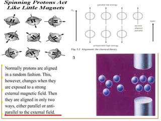
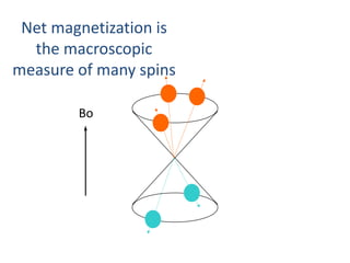
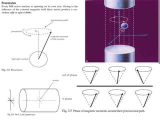
1. MRI physics involves the behavior of spins under magnetic fields and the use of radiofrequency pulses to manipulate spin alignment and generate signals.
2. Different pulse sequences such as spin echo, gradient echo, and inversion recovery use varying combinations of excitation pulses, rephasing gradients, and timing delays to produce images weighted towards T1, T2, proton density or other tissue contrasts.
3. Sequence parameters like repetition time (TR), echo time (TE), and inversion time (TI) determine the relative contributions of T1, T2 and other factors to image contrast.
Everything regarding the physics of MRA is given along with flow charts and images. Also have covered new advances and refrences taken from MR made easy and some articles related to MRI
Mri spin echo pulse sequences its variations andYashawant Yadav
MRI spin echo pulse sequences its variation and applications , in this slide collection principle of spine echo pulse sequences is described with physic behind it ,, this slide also coves the inverse recovery pulse sequences and types ,,,, image weighting and parameters are explained .. hope it may be help ful.
A pulse sequence is a sequence of events, which are
needed to acquire MRI images. These events are: RF
pulses, gradient switches and signal collecting.
A pulse sequence is a sequence of events, which we need to acquire MRI images.
Basic pulse sequences
1. Spin echo sequence(SE)
2. Gradient echo sequence(GRE)
3. Inversion Recovery Sequence(IR)
4. Echo Planar Imaging(EPI)
The gradient echo pulse sequence is the simplest type of MRI sequence.
The major purposes behind the gradient technique is a significant reduction in scan time. Small variable flip angle are employed , usually less than 90 degrees. which in turn allow very short repetition time thus decreasing the scan time.
Gradient echo pulse sequence differ from spin echo pulse sequence . There is no 180 degree pulse in GRE. T2 relaxation in GRE is called as T2* relaxation. Gradient can be used to either dephase or rephase the magnetic moments of nuclei.
MRI spin echo sequences are a fundamental imaging technique in magnetic resonance imaging (MRI). They work by manipulating the spin of hydrogen nuclei in the body's tissues to create detailed images. In a spin echo sequence: Spin echo sequences are versatile and used in various MRI applications, including anatomical imaging, quantitative measurements, and lesion characterization. They can produce T1-weighted and T2-weighted images, depending on the specific sequence parameters chosen, offering valuable diagnostic information in medical imaging.
MRI PHYSICS PART 3 Susceptibility-weighted images BY GKM .pptxGulshan Verma
Susceptibility-weighted imaging (SWI) is based on a fully flow compensated, high-resolution, 3D gradient echo method by integrating both magnitude and phase information.
It was previously referred to as high resolution blood oxygen level– dependent (BOLD) venography (HRBV), but because of its broader application than evaluating venous structures, it is now referred to as SWI.
Susceptibility-weighted imaging (SWI) allows detection and characterization of tissue components based on differences in their susceptibilities.
SWI sequences are typically acquired in 3D (rather than 2D) mode,
Allowing thinner slices, and Use Smaller voxel sizes,
Flow compensation in all three directions is used to reduce artifacts,
Parallel imaging is employed to reduce imaging time.
Either single or multiple echoes may be acquired in a given TR interval.
A key feature of SWI is that magnitude and phase information are independently processed/displayed as well as combined for diagnostic purposes
Here I discussed about the concept,types, types of images obtained, the advantages and disadvantages of MRI shortly...anyone who wants to know about MRI just go through it. I just prepared it in very simple language for the convenience of the readers all over the world. Thank you.
Introduction to AI for Nonprofits with Tapp NetworkTechSoup
Dive into the world of AI! Experts Jon Hill and Tareq Monaur will guide you through AI's role in enhancing nonprofit websites and basic marketing strategies, making it easy to understand and apply.
Read| The latest issue of The Challenger is here! We are thrilled to announce that our school paper has qualified for the NATIONAL SCHOOLS PRESS CONFERENCE (NSPC) 2024. Thank you for your unwavering support and trust. Dive into the stories that made us stand out!
Executive Directors Chat Leveraging AI for Diversity, Equity, and InclusionTechSoup
Let’s explore the intersection of technology and equity in the final session of our DEI series. Discover how AI tools, like ChatGPT, can be used to support and enhance your nonprofit's DEI initiatives. Participants will gain insights into practical AI applications and get tips for leveraging technology to advance their DEI goals.
How to Add Chatter in the odoo 17 ERP ModuleCeline George
In Odoo, the chatter is like a chat tool that helps you work together on records. You can leave notes and track things, making it easier to talk with your team and partners. Inside chatter, all communication history, activity, and changes will be displayed.
Thinking of getting a dog? Be aware that breeds like Pit Bulls, Rottweilers, and German Shepherds can be loyal and dangerous. Proper training and socialization are crucial to preventing aggressive behaviors. Ensure safety by understanding their needs and always supervising interactions. Stay safe, and enjoy your furry friends!
How to Build a Module in Odoo 17 Using the Scaffold MethodCeline George
Odoo provides an option for creating a module by using a single line command. By using this command the user can make a whole structure of a module. It is very easy for a beginner to make a module. There is no need to make each file manually. This slide will show how to create a module using the scaffold method.
Acetabularia Information For Class 9 .docxvaibhavrinwa19
Acetabularia acetabulum is a single-celled green alga that in its vegetative state is morphologically differentiated into a basal rhizoid and an axially elongated stalk, which bears whorls of branching hairs. The single diploid nucleus resides in the rhizoid.
Exploiting Artificial Intelligence for Empowering Researchers and Faculty, In...Dr. Vinod Kumar Kanvaria
Exploiting Artificial Intelligence for Empowering Researchers and Faculty,
International FDP on Fundamentals of Research in Social Sciences
at Integral University, Lucknow, 06.06.2024
By Dr. Vinod Kumar Kanvaria
A Strategic Approach: GenAI in EducationPeter Windle
Artificial Intelligence (AI) technologies such as Generative AI, Image Generators and Large Language Models have had a dramatic impact on teaching, learning and assessment over the past 18 months. The most immediate threat AI posed was to Academic Integrity with Higher Education Institutes (HEIs) focusing their efforts on combating the use of GenAI in assessment. Guidelines were developed for staff and students, policies put in place too. Innovative educators have forged paths in the use of Generative AI for teaching, learning and assessments leading to pockets of transformation springing up across HEIs, often with little or no top-down guidance, support or direction.
This Gasta posits a strategic approach to integrating AI into HEIs to prepare staff, students and the curriculum for an evolving world and workplace. We will highlight the advantages of working with these technologies beyond the realm of teaching, learning and assessment by considering prompt engineering skills, industry impact, curriculum changes, and the need for staff upskilling. In contrast, not engaging strategically with Generative AI poses risks, including falling behind peers, missed opportunities and failing to ensure our graduates remain employable. The rapid evolution of AI technologies necessitates a proactive and strategic approach if we are to remain relevant.
5. Resonance
Resonance is an energy transition that occurs when an object is subjected to a frequency the
same as its own. In MR, resonance is induced by applying a radiofrequency (RF) pulse:
• at the same frequency as the precessing hydrogen nuclei;
• at 90° to B0.
6.
7.
8.
9.
10. T1 recovery
The T1 time is defined as the time it takes for 63% of the longitudinal
magnetization to recover. The period of time during which this occurs is the
time between one excitation pulse and the next or the TR.
11. T1 recovery in fat
• T1 relaxation occurs as a result of nuclei exchanging the energy given to them by the RF pulse
to their surrounding environment. The efficiencyof this process determines the T1 time of the
tissue in which they are situated.
• Owing to the fact that fat is able to absorb energy quickly the T1 time of fat is very short, i.e.
nuclei dispose of their energy to the surrounding fat tissue and return to B0 in a short time.
T1 recovery in water
• Water is very inefficient at receiving energy from nuclei. The T1 time of water is therefore
quite long, i.e. nuclei take a lot longer to dispose of their energy to the surrounding water tissue
andreturn to B0.
12. • The TR controls how much of the NMV in fat or water has recovered before the application of
the next RF pulse.
13. T2 decay
T2 decay is caused by the exchange of energy from one nucleus to another. It is called spin–spin
energy transfer. It occurs as a result of the intrinsic magnetic fields of the nuclei interacting with
each other. This energy exchange produces a loss of phase coherence or dephasing, and results
in decay of the NMV in the transverse plane. T2 decay time is the time it takes for
63% of the transverse magnetization to be lost due to dephasing, i.e. 37% remains. The period
of time over which this occurs is the time between the excitation pulse and the MR signal or the
TE. The TE therefore determines how much T2 decay occurs in a particular tissue.
14. Short TEs do not permit full dephasing in either fat or water so their transverse components are
similar. There is little contrast difference between fat and water due to differences in T2 decay
using short TEs.
Long TEs allow dephasing of the transverse components in fat and water. There is a contrast
difference between fat and water due to differences in T2 decay times when using long TEs.
15. T1 weighting
A TR is selected that is short enough to ensure that the NMV in neither
fat nor water has had time to relax back to B0 before the application of
the next excitation pulse. If the TR is long, the NMV in both fat and
water recovers and their respective T1 relaxation times can no longer be
distinguished. A T1 weighted image is an image whose contrast is
predominantly due to the differences in T1 recovery times of tissues.
In T1 weighted images, tissues with short T1 relaxation times such as fat, are bright
(high signal), because they recover most of their longitudinal magnetization during the
TR and therefore more magnetization is available to be flipped into the transverse
plane by the next RF pulse.
Tissues with long T1 relaxation times such as water, are
dark (low signal) because they do not recover much of
their longitudinal magnetization during the TR and
therefore less magnetization is available to be flipped
into the transverse plane by the next RF pulse.
16. T2 weighting
A TE is selected that is long enough to ensure that the NMV in both fat and water have
time to decay. If the TE is too short, the NMV in neither fat nor water has had time to
decay and their respective T2 times cannot be distinguished. A T2 weighted image is
an image whose contrast is predominantly due to the differences in T2 recovery times
of tissues.
For T2 weighting the
differences between
the T2 times of tissues
is exaggerated,
therefore the TE must
be long. T1 effects are
diminished by selecting
a long TR.
• Tissues with a short T2 decay time such as fat are dark (low signal) because they
lose most of their coherent transverse magnetization during the TE period.
• Tissues with a long T2 decay time such as water are bright (high signal), because
they retain most of their transverse coherence during the TE period.
• T2 weighted images best demonstrate pathology as most pathology has an increased
water content and is therefore bright on T2 weighted images.
17.
18. Proton density weighting
In a PD weighted image differences in the proton densities (number of hydrogen
protons in the tissue) must be demonstrated. To achieve this both T1 and T2 effects
are diminished. T1 effects are reduced by selecting a long TR and T2 effects are
diminished by selecting a short TE.
• Tissues with a low proton density are dark (low signal) because the low number of protons
result in a small component of transverse magnetization.
• Tissues with a high proton density are bright (high signal) because the high number of protons
result in a large component of transverse magnetization.
19. Conventional spin echo pulse
sequence (SE)
Conventional spin echo pulse sequences are used to produce T1, T2 or proton density weighted
images and are one of the most basic pulse sequences used in MRI. In a spin echo pulse
sequence there is a 90° excitation pulse followed by a 180° rephasing pulse followed by an
Echo.
22. Fast or turbo spin echo (FSE/ TSE)
• FSE employs a train of 180° rephasing pulses, each one producing a spin echo. This train of
spin echoes is called an echo train. The number of 180° RF pulses and resultant echoes is called
the echo train length (ETL) or turbo factor. The spacing between each echo is called the
echo spacing.
• After each rephasing, a phase encoding step is performed and data from the resultant echo
are stored in K space . Therefore several lines of K space are filled with every TR instead of one
line as in conventional spin echo. As K space is filled more rapidly, the scan time decreases.
Uses
FSE produces T1, T2 or proton density scans
in a fraction of the time of Conventional
spin echo. Because the scan times are reduced,
matrix size can be increased to improve spatial
resolution. FSE is usually used for brains, spines,
joints, extremities and the pelvis
23.
24. Inversion recovery (IR)
• Inversion recovery is a spin echo sequence that begins with a 180° inverting pulse. This inverts
the NMV through 180°.
• When the pulse is removed the NMV begins to relax back to B0. A 90° pulse is then applied at
time interval TI (time from inversion) after the 180° inverting pulse.
• A further 180° RF pulse is applied which rephases spins in the transverse plane and produces
an echo at time TE after the excitation pulse.
25.
26.
27. STIR (short TI inversion recovery)
It uses short TIs such as 100– 180 ms, depending on field strength . TIs of this magnitude
place the 90° excitation pulse at the time that NMV of fat is passing exactly through the
transverse plane. At this point (called the null point) there is no longitudinal component in fat.
Therefore the 90° excitation pulse produces no transverse component in fat and therefore no
signal. In this way a fat suppressed image results .
FLAIR (fluid attenuated inversion recovery)
It uses long TIs such as 1700–2200 ms, to null the signal from CSF in exactly the same way as the
STIR sequence . Because CSF has a long T1 recovery time, the TI must be longer to correspond
with its null point.
28.
29. Gradient echo pulse sequences ( FFE / GRE /GE)
Gradient echo pulse sequences are sequences that use a gradient to reduce magnetic
homogeneity effects, as opposed to a 180° RF pulse used in spin echo sequences.
The gradient echo sequence differs from the spin echo sequence in regard to:
• the flip angle usually below 90°
• the absence of a 180° RF rephasing pulse
A flip angle lower than 90° (partial flip angle) decreases the amount of magnetization tipped
into the transverse plane. The consequence of a low-flip angle excitation is a faster recovery of
longitudinal magnetization that allows shorter TR/TE and decreases scan time.
30. • After the RF pulse is withdrawn, the FID is immediately produced as a result of
inhomogeneities in the magnetic field. T2* dephasing therefore occurs before T1 and T2
processes have had time to develop.
• The magnetic moments within the
transverse component of magnetization
dephase and are then rephased by a
gradient.
• A gradient causes a change in the
magnetic field strength that changes the
precessional frequency and phase of spins.
This effect rephases the magnetic moments
so that a signal is received by the coil that
contains T1 and T2 information. This signal
is called a gradient echo.
31. In a gradient echo pulse sequence ,rephasing is performed by the frequency encoding
gradient .
• The nuclei are dephased with a negative gradient pulse. The negative gradient slows
down the slow nuclei even further and speeds up the fast ones. This accelerates the
dephasing process.
• The gradient polarity is then reversed to positive. The positive gradient speeds up the
slow nuclei and slows down the fast ones. The nuclei rephase and produce a gradient echo.
32. T1 weighting (GRE/FFE/GE)
• The TR must not permit nuclei in the majority of tissues to recover to the longitudinal axis
prior to the repetition of the next RF excitation pulse. Therefore the TR must be short.
• The flip angle must shift the majority of the NMV towards the transverse plane
to produce saturation. The flip angle must be large.
• Use a TE to produce minimum T2* effects. The TE should limit the amount of dephasing that
occurs before the echo is regenerated. Therefore the TE must be short.
T2* weighting (GRE/FFE/GE)
• The TE should permit maximum dephasing to occur before the signal is generated to produce
maximum T2* effects. The TE must be long.
• The flip angle must shift only a minimum of the NMV towards the transverse plane. Small flip
angles ensure that the majority of the net magnetization components remain in the longitudinal
axis to prevent saturation. The flip angle must be small.
• The TR must be long enough to prevent saturation but can be reduced without producing
significant saturation because of the small flip angle.
Proton density weighting(GRE/FFE/GE)
Select TR and flip angle to produce minimum T1 effects and a TE to
produce minimum T2*/T2 effects. As a result proton density predominates.
• The flip angle must be small.
• The TR must be long to minimize saturation and T1 effects as well.
• The TE must be short to minimize the T2*/T2 effects.
33. Advantages of Gradient echo (GRE / FFE)
sequences
1. To detect haemorrhage, calcification
2. Abdominal breath hold studies
3. 3 D imaging
4. Time of flight (TOF) or inflow
angiography