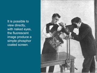
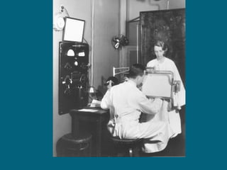
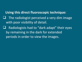
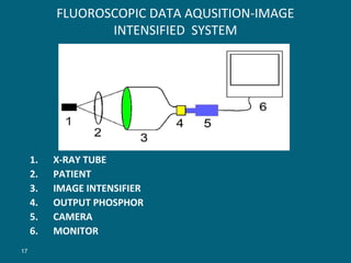
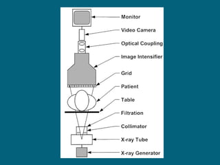
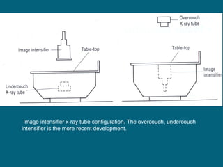
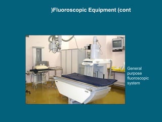
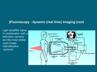
This document discusses fluoroscopy and image intensifiers. It begins with a brief history of fluoroscopy, describing early techniques where radiologists viewed dim, fluorescent images directly. It then explains how modern image intensifiers work, increasing image brightness by converting x-ray photons to electrons that excite a phosphor, producing a brighter light image. The key components of an image intensifier - including input phosphor, photocathode, electron acceleration, and output phosphor - are identified. Factors affecting image quality such as unsharpness, noise, resolution and distortion are also outlined.