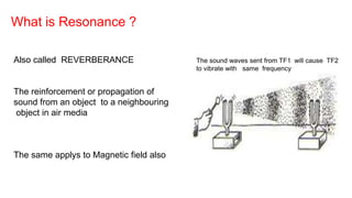
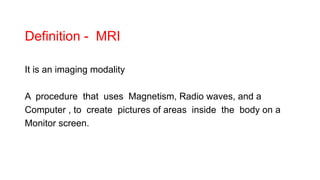
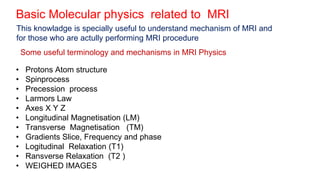
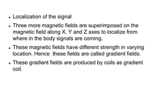
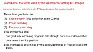
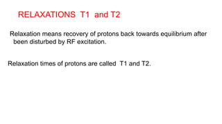
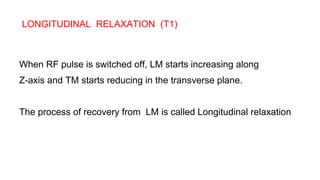
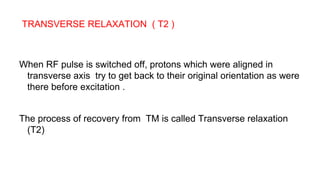
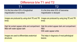
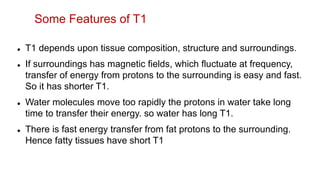
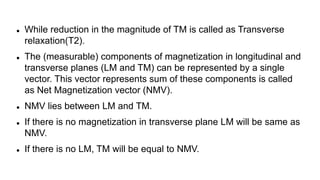
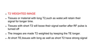
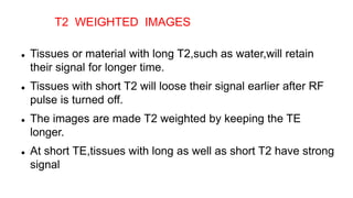
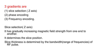
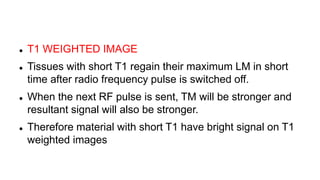
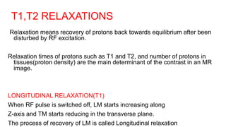
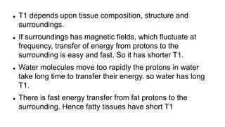
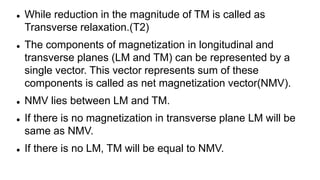
MRI in Obstetrics PART I BASIC PHYSICS
( ALSO PL SEE PART 2 FOR COMPLETION )
Dr Shivamurthy H M Prof
Dr Ayesha Sultana Rajgoli, Resident
Dept of OBG
SNMC , Bagalkot
Karnataka , India .
Viewers please note this topic of MRI in OBSTETRICS is uploaded
in two parts as the whole topic could not uploaded in single ppt
sesssion becaus ethe size of the file is big
This is part 1 presentation which contins only BASIC PHYSICS
Next part 2 presentation which contains only OBSTETRIC
aspects of MRI is uploaded in a separately .
The main idea of this ppt presentation is only to share knowladge
and give basic idea of MRI to students of both OBG and
Radiology
What is Resonance ?
Also called REVERBERANCE
The reinforcement or propagation of
sound from an object to a neighbouring
object in air media
The same applys to Magnetic field also
The sound waves sent from TF1 will cause TF2
to vibrate with same frequency
Definition - MRI
It is an imaging modality
A procedure that uses Magnetism, Radio waves, and a
Computer , to create pictures of areas inside the body on a
Monitor screen.
 1977- Raymond Damadian is credited as the first person to use MRI
for diagnosis
 1980- MRI machines became commercially available
 1983- In obstetrics MRI was first described by smith FW, et al.
 2003- For invention of MRI nobel prize in physiology was given to Paul
Lauterbur and Peter Mansfield
Historical
Dr. Raymond Damadian with his MRI system
Principles in M R I
Four basic steps are involved
1) Placing the patient in the magnetic field.
2) Transmitting Radio frequency pulse by coil on to the subject.
3) Receiving signals from the patient by coil.
4) Transformation of signals into image by complex processing in the
computers
(1) Placing the patient in the magnetic core
(2) Propagation MRI Pulse (produced by the external
magnetic Source) which delivered to pt body
• These Pulses from external magnetic coil are also called Resonance
Frequency (R F) Pulses
• The Protons in pts body will pick up energy in stepwise manner from
RF source and precess and they upgrade in their own energy also.
• The movement of protons in the patients body are under the
influence of external magnetic field .
• Under this external magnetic influence , all randomly moving
protons in patients body will align and precess along the external
magnetic field.(z axis)
• The magnetic field in the patient body will process and
generates current .
• This current is received as signal by Radio frequency-coil.
(3) MR Signals are received
(4) Formationof image from the signlas
MR signals which are received by the coil which is below the
patient , are transformed into image by computers by complex
processing.
Basic Molecular physics related to MRI
• Protons Atom structure
• Spinprocess
• Precession process
• Larmors Law
• Axes X Y Z
• Longitudinal Magnetisation (LM)
• Transverse Magnetisation (TM)
• Gradients Slice, Frequency and phase
• Logitudinal Relaxation (T1)
• Ransverse Relaxation (T2 )
• WEIGHED IMAGES
This knowladge is specially useful to understand mechanism of MRI and
for those who are actully performing MRI procedure
Some useful terminology and mechanisms in MRI Physics
Atom is made of a nucleus (which contains protons and neutrons) and
outer electrons which are revolving around the nucleus
Structure of an ‘atom’
Movement of protons under the effect of magnetic field is a
very important process, in MRI.
Two phenomena used in MRI procedure, Spin and Precession
• Spinning -rotation of something (in this case proton) around it’s
own axis.
• Precession -is the rotation of axis along with spin effect.
When a proton aligns along EMF, they spin around
their axis but the axis also rotates forming a cone
which is called Precession which has a frequency
designated as WO
Phenmenon of Spin
Larmors equation
WO = Ɣ B O
 Where wo = precession frequency in HZ.
 BO = Strength of external magnetic field in tesla.
 Ɣ = Gyromagnetic ratio, which is specific to particular
nuclues.
• In other words the movement of Protons is directly proportional to the
strength of External Magnetic Field(EMF)
 External magnetic field is directed along the z-axis of the patient.
 Z-axis is the long axis of the patient as well as bore of the
magnet.
 Always more protons spinning on the positive side or parallel to
Z-axis than negative side.
 Forces of these protons add up together to form a magnetic
vector along the Z axis called as LM.
 Thus formed along the external magnetic field cant measured
directly it has to be transverse
Longitudinal Magnetization
• The first step is to send Radio frequency pulses.
• The precessing protons pick up some energy from the Radio
frequency pulse.
• Some of these protons go to higher energy level and start
precessing anti-parallel.
• The imbalance results in tilting of the magnetization into the
transverse plane.
Transverse Magnetization
 Forces of these protons add up together to form a magnetic vector along the Z axis called as
LM.
 Thus formed along the external magnetic field cant measured directly it has to be transverse
RF pulse not only causes higher energy, but also makes protons to
precess in phase manner and in synchrony.
 External magnetic field by convention is directed along the positive, z-axis
 Z-axis is the long axis of the patient as well as bore of the magnet.
 Always more protons will spin on the positive side ( along or parallel to Z-axis).
 These protons - magnetic forces add up together along the Longitudinal
axis called as Longitudinal Magnetism (Z axis) (also called L vector).
 Some of them will go for Transverse magnetism(TM) (Y-axis also called T
vector.)
 Measurement of Net Magnetism( NMV) is the difference between these 2
vectors LMV and TLV. ie TLV=LMV-TLV
Longitudinal / Transverse Magnetization AXES
 Localization of the signal
 Three more magnetic fields are superimposed on the
magnetic field along X, Y and Z axes to localize from
where in the body signals are coming.
 These magnetic fields have different strength in varying
location. Hence these fields are called gradient fields.
 These gradient fields are produced by coils as gradient
coil.
These three gradients are
(1) Slice selection (also called her again Z axis)
(2) Phase encoding
(3) Frequency encoding.
Slice selection( Z axis)
It has gradually increasing magnetic field strength from one end to another.
It determines the slice position.
Slice thickness is determined by the bandwidth(range of frequencies) of RF
pulse.
3 gradients, the terms used by the Operator for getting MR Images
( remember these have nothing to do with Z YX axes of magnetic fiels explained already )
RELAXATIONS T1 and T2
Relaxation means recovery of protons back towards equilibrium after
been disturbed by RF excitation.
Relaxation times of protons are called T1 and T2.
LONGITUDINAL RELAXATION (T1)
When RF pulse is switched off, LM starts increasing along
Z-axis and TM starts reducing in the transverse plane.
The process of recovery from LM is called Longitudinal relaxation
TRANSVERSE RELAXATION ( T2 )
When RF pulse is switched off, protons which were aligned in
transverse axis try to get back to their original orientation as were
there before excitation .
The process of recovery from TM is called Transverse relaxation
(T2)
Difference b/w T1 and T2
T1 T2
It is the time when 63% of longitudinal
magnetization is recovered
It is the time when 63% of transverse
magnetization is decayed
Images are produced by using short TE and TR
times
Images are produced by using long TE and TR
times
High fat content appear white and compartment
filled with water appear dark
High fat content appear dark and compartment
filled
With water appear white
Images are used to differentiate anatomical
structures
This helps in diagnosis of most pathological
lesions
The number of protons in tissues(proton density) are the
main determinant of the contrast in an MR image.
 T1 depends upon tissue composition, structure and surroundings.
 If surroundings has magnetic fields, which fluctuate at frequency,
transfer of energy from protons to the surrounding is easy and fast.
So it has shorter T1.
 Water molecules move too rapidly the protons in water take long
time to transfer their energy. so water has long T1.
 There is fast energy transfer from fat protons to the surrounding.
Hence fatty tissues have short T1
Some Features of T1
 While reduction in the magnitude of TM is called as Transverse
relaxation(T2).
 The (measurable) components of magnetization in longitudinal and
transverse planes (LM and TM) can be represented by a single
vector. This vector represents sum of these components is called
as Net Magnetization vector (NMV).
 NMV lies between LM and TM.
 If there is no magnetization in transverse plane LM will be same as
NMV.
 If there is no LM, TM will be equal to NMV.
 T2 is the time taken by TM to disappear
 T2 depends on in homogenity of local magnetic fields within the
tissue
 As water molecules move very fast,their magnetic fields fluctuate
fast
 If liquid is impure or the tissue has larger molecules, molecules
move at slower rate
 Impure liquids or tissues with larger molecules have short T2.Fat
has shorter T2
Features of T2
TYPES OF IMAGES WEIGHTED IMAGES
 T2 WEIGHTED IMAGE
 Tissues or material with long T2,such as water,will retain their
signal for longer time.
 Tissues with short T2 will loose their signal earlier after RF pulse is
turned off
 The images are made T2 weighted by keeping the TE longer.
 At short TE,tissues with long as well as short T2 have strong signal
 Tissues or material with long T2,such as water,will retain
their signal for longer time.
 Tissues with short T2 will loose their signal earlier after RF
pulse is turned off.
 The images are made T2 weighted by keeping the TE
longer.
 At short TE,tissues with long as well as short T2 have strong
signal
T2 WEIGHTED IMAGES
3 gradients are
(1) slice selection ( Z axis)
(2) phase encoding
(3) Frequency encoding.
Slice selection( Z axis)
It has gradually increasing magnetic field strength from one end to
another.
It determines the slice position.
Slice thickness is determined by the bandwidth(range of frequencies) of
RF pulse.
IMAGE AFTER SETTING SLICE SELECTION FREQUENCY AND PHASE CODING
FREQUENCY
GRADIENT
PHASE GRADIENT
SLICE SELECTION GRADIENT ( Z axis)
MRI TERMS
(1) Signal intensity
 Low, intermediate, and high signal intensity are commonly used
terms in MRI.
 When tissue is imaged as white, it is described as high signal
intensity, while tissue is imaged with gray tone is called as
intermediate signal intensity and if it is imaged as dark gray or
black, it is low signal intensity.
( 2) Sequence
 Here, a particular setting of radio frequency pulses and gradients
is used, which results in a particular image.
 Each tissue returns to its equilibrium state after excitation by
independent relaxation process of T1 and T2
 The most common MRI sequences are T1 weighted and T2
weighted scans
 TIME TO REPEAT(TR)
 Time interval between start of one RF pulse and start of
the next RF pulse.
 For a spin-echo sequence time interval between
beginnings of 90degree pulses is the TR.
 TIME TO ECHO(TE)
 Time interval between start of RF pulse and reception of
the signal(echo)
 T1 WEIGHTED IMAGE
 Tissues with short T1 regain their maximum LM in short
time after radio frequency pulse is switched off.
 When the next RF pulse is sent, TM will be stronger and
resultant signal will also be stronger.
 Therefore material with short T1 have bright signal on T1
weighted images
• Gadolinium is classified as category C
• It crosses the placenta and is excreted by the fetal kidneys.
• It should be used in pregnancy, if judged absolutely essential.
• After I.V administration of contrast to the postnatal patient very
minute(only 0.1%) dose is excreted in breast milk.
• Only 1% of the amount excreted in breast milk is subsequently
absorbed by the baby.
Use of contrast agents in obstetrical mri
T1,T2 RELAXATIONS
Relaxation means recovery of protons back towards equilibrium after been
disturbed by RF excitation.
Relaxation times of protons such as T1 and T2, and number of protons in
tissues(proton density) are the main determinant of the contrast in an MR
image.
LONGITUDINAL RELAXATION(T1)
When RF pulse is switched off, LM starts increasing along
Z-axis and TM starts reducing in the transverse plane.
The process of recovery of LM is called Longitudinal relaxation
 T1 depends upon tissue composition, structure and
surroundings.
 If surroundings has magnetic fields, which fluctuate at
frequency, transfer of energy from protons to the
surrounding is easy and fast. So it has shorter T1.
 Water molecules move too rapidly the protons in water
take long time to transfer their energy. so water has long
T1.
 There is fast energy transfer from fat protons to the
surrounding. Hence fatty tissues have short T1
 While reduction in the magnitude of TM is called as
Transverse relaxation.(T2)
 The components of magnetization in longitudinal and
transverse planes (LM and TM) can be represented by a
single vector. This vector represents sum of these
components is called as net magnetization vector(NMV).
 NMV lies between LM and TM.
 If there is no magnetization in transverse plane LM will be
same as NMV.
 If there is no LM, TM will be equal to NMV.
 Localization of the signal
 Three more magnetic fields are superimposed on the
magnetic field along X, Y and Z axes to localize from
where in the body signals are coming.
 These magnetic fields have different strength in varying
location. Hence these fields are called gradient fields.
 These gradient fields are produced by coils as gradient
coil.
 T2 is the time taken by TM to disappear
 T2 depends on in homogenity of local magnetic fields
within the tissue
 As water molecules move very fast,their magnetic fields
fluctuate fast
 If liquid is impure or the tissue has larger molecules,
molecules move at slower rate
 Impure liquids or tissues with larger molecules have short
T2.Fat has shorter T2
 Short TR and short TE gives T1 weighted images.
 Long TR and short TE gives T2 weighted images.
THE END of part 1
GO TO PART 2

MRI obstetric practice part 1 Basic Physics

  • 1.
    MRI in ObstetricsPART I BASIC PHYSICS ( ALSO PL SEE PART 2 FOR COMPLETION ) Dr Shivamurthy H M Prof Dr Ayesha Sultana Rajgoli, Resident Dept of OBG SNMC , Bagalkot Karnataka , India .
  • 2.
    Viewers please notethis topic of MRI in OBSTETRICS is uploaded in two parts as the whole topic could not uploaded in single ppt sesssion becaus ethe size of the file is big This is part 1 presentation which contins only BASIC PHYSICS Next part 2 presentation which contains only OBSTETRIC aspects of MRI is uploaded in a separately . The main idea of this ppt presentation is only to share knowladge and give basic idea of MRI to students of both OBG and Radiology
  • 3.
    What is Resonance? Also called REVERBERANCE The reinforcement or propagation of sound from an object to a neighbouring object in air media The same applys to Magnetic field also The sound waves sent from TF1 will cause TF2 to vibrate with same frequency
  • 4.
    Definition - MRI Itis an imaging modality A procedure that uses Magnetism, Radio waves, and a Computer , to create pictures of areas inside the body on a Monitor screen.
  • 5.
     1977- RaymondDamadian is credited as the first person to use MRI for diagnosis  1980- MRI machines became commercially available  1983- In obstetrics MRI was first described by smith FW, et al.  2003- For invention of MRI nobel prize in physiology was given to Paul Lauterbur and Peter Mansfield Historical
  • 6.
    Dr. Raymond Damadianwith his MRI system
  • 7.
    Principles in MR I Four basic steps are involved 1) Placing the patient in the magnetic field. 2) Transmitting Radio frequency pulse by coil on to the subject. 3) Receiving signals from the patient by coil. 4) Transformation of signals into image by complex processing in the computers
  • 8.
    (1) Placing thepatient in the magnetic core
  • 9.
    (2) Propagation MRIPulse (produced by the external magnetic Source) which delivered to pt body • These Pulses from external magnetic coil are also called Resonance Frequency (R F) Pulses • The Protons in pts body will pick up energy in stepwise manner from RF source and precess and they upgrade in their own energy also.
  • 10.
    • The movementof protons in the patients body are under the influence of external magnetic field . • Under this external magnetic influence , all randomly moving protons in patients body will align and precess along the external magnetic field.(z axis)
  • 11.
    • The magneticfield in the patient body will process and generates current . • This current is received as signal by Radio frequency-coil. (3) MR Signals are received
  • 12.
    (4) Formationof imagefrom the signlas MR signals which are received by the coil which is below the patient , are transformed into image by computers by complex processing.
  • 13.
    Basic Molecular physicsrelated to MRI • Protons Atom structure • Spinprocess • Precession process • Larmors Law • Axes X Y Z • Longitudinal Magnetisation (LM) • Transverse Magnetisation (TM) • Gradients Slice, Frequency and phase • Logitudinal Relaxation (T1) • Ransverse Relaxation (T2 ) • WEIGHED IMAGES This knowladge is specially useful to understand mechanism of MRI and for those who are actully performing MRI procedure Some useful terminology and mechanisms in MRI Physics
  • 14.
    Atom is madeof a nucleus (which contains protons and neutrons) and outer electrons which are revolving around the nucleus Structure of an ‘atom’
  • 15.
    Movement of protonsunder the effect of magnetic field is a very important process, in MRI. Two phenomena used in MRI procedure, Spin and Precession • Spinning -rotation of something (in this case proton) around it’s own axis. • Precession -is the rotation of axis along with spin effect.
  • 16.
    When a protonaligns along EMF, they spin around their axis but the axis also rotates forming a cone which is called Precession which has a frequency designated as WO Phenmenon of Spin
  • 17.
    Larmors equation WO =Ɣ B O  Where wo = precession frequency in HZ.  BO = Strength of external magnetic field in tesla.  Ɣ = Gyromagnetic ratio, which is specific to particular nuclues. • In other words the movement of Protons is directly proportional to the strength of External Magnetic Field(EMF)
  • 18.
     External magneticfield is directed along the z-axis of the patient.  Z-axis is the long axis of the patient as well as bore of the magnet.  Always more protons spinning on the positive side or parallel to Z-axis than negative side.  Forces of these protons add up together to form a magnetic vector along the Z axis called as LM.  Thus formed along the external magnetic field cant measured directly it has to be transverse Longitudinal Magnetization
  • 19.
    • The firststep is to send Radio frequency pulses. • The precessing protons pick up some energy from the Radio frequency pulse. • Some of these protons go to higher energy level and start precessing anti-parallel. • The imbalance results in tilting of the magnetization into the transverse plane. Transverse Magnetization
  • 20.
     Forces ofthese protons add up together to form a magnetic vector along the Z axis called as LM.  Thus formed along the external magnetic field cant measured directly it has to be transverse
  • 21.
    RF pulse notonly causes higher energy, but also makes protons to precess in phase manner and in synchrony.
  • 22.
     External magneticfield by convention is directed along the positive, z-axis  Z-axis is the long axis of the patient as well as bore of the magnet.  Always more protons will spin on the positive side ( along or parallel to Z-axis).  These protons - magnetic forces add up together along the Longitudinal axis called as Longitudinal Magnetism (Z axis) (also called L vector).  Some of them will go for Transverse magnetism(TM) (Y-axis also called T vector.)  Measurement of Net Magnetism( NMV) is the difference between these 2 vectors LMV and TLV. ie TLV=LMV-TLV Longitudinal / Transverse Magnetization AXES
  • 23.
     Localization ofthe signal  Three more magnetic fields are superimposed on the magnetic field along X, Y and Z axes to localize from where in the body signals are coming.  These magnetic fields have different strength in varying location. Hence these fields are called gradient fields.  These gradient fields are produced by coils as gradient coil.
  • 24.
    These three gradientsare (1) Slice selection (also called her again Z axis) (2) Phase encoding (3) Frequency encoding. Slice selection( Z axis) It has gradually increasing magnetic field strength from one end to another. It determines the slice position. Slice thickness is determined by the bandwidth(range of frequencies) of RF pulse. 3 gradients, the terms used by the Operator for getting MR Images ( remember these have nothing to do with Z YX axes of magnetic fiels explained already )
  • 25.
    RELAXATIONS T1 andT2 Relaxation means recovery of protons back towards equilibrium after been disturbed by RF excitation. Relaxation times of protons are called T1 and T2.
  • 26.
    LONGITUDINAL RELAXATION (T1) WhenRF pulse is switched off, LM starts increasing along Z-axis and TM starts reducing in the transverse plane. The process of recovery from LM is called Longitudinal relaxation
  • 27.
    TRANSVERSE RELAXATION (T2 ) When RF pulse is switched off, protons which were aligned in transverse axis try to get back to their original orientation as were there before excitation . The process of recovery from TM is called Transverse relaxation (T2)
  • 28.
    Difference b/w T1and T2 T1 T2 It is the time when 63% of longitudinal magnetization is recovered It is the time when 63% of transverse magnetization is decayed Images are produced by using short TE and TR times Images are produced by using long TE and TR times High fat content appear white and compartment filled with water appear dark High fat content appear dark and compartment filled With water appear white Images are used to differentiate anatomical structures This helps in diagnosis of most pathological lesions
  • 29.
    The number ofprotons in tissues(proton density) are the main determinant of the contrast in an MR image.
  • 30.
     T1 dependsupon tissue composition, structure and surroundings.  If surroundings has magnetic fields, which fluctuate at frequency, transfer of energy from protons to the surrounding is easy and fast. So it has shorter T1.  Water molecules move too rapidly the protons in water take long time to transfer their energy. so water has long T1.  There is fast energy transfer from fat protons to the surrounding. Hence fatty tissues have short T1 Some Features of T1
  • 31.
     While reductionin the magnitude of TM is called as Transverse relaxation(T2).  The (measurable) components of magnetization in longitudinal and transverse planes (LM and TM) can be represented by a single vector. This vector represents sum of these components is called as Net Magnetization vector (NMV).  NMV lies between LM and TM.  If there is no magnetization in transverse plane LM will be same as NMV.  If there is no LM, TM will be equal to NMV.
  • 32.
     T2 isthe time taken by TM to disappear  T2 depends on in homogenity of local magnetic fields within the tissue  As water molecules move very fast,their magnetic fields fluctuate fast  If liquid is impure or the tissue has larger molecules, molecules move at slower rate  Impure liquids or tissues with larger molecules have short T2.Fat has shorter T2 Features of T2
  • 33.
    TYPES OF IMAGESWEIGHTED IMAGES
  • 34.
     T2 WEIGHTEDIMAGE  Tissues or material with long T2,such as water,will retain their signal for longer time.  Tissues with short T2 will loose their signal earlier after RF pulse is turned off  The images are made T2 weighted by keeping the TE longer.  At short TE,tissues with long as well as short T2 have strong signal
  • 35.
     Tissues ormaterial with long T2,such as water,will retain their signal for longer time.  Tissues with short T2 will loose their signal earlier after RF pulse is turned off.  The images are made T2 weighted by keeping the TE longer.  At short TE,tissues with long as well as short T2 have strong signal T2 WEIGHTED IMAGES
  • 36.
    3 gradients are (1)slice selection ( Z axis) (2) phase encoding (3) Frequency encoding. Slice selection( Z axis) It has gradually increasing magnetic field strength from one end to another. It determines the slice position. Slice thickness is determined by the bandwidth(range of frequencies) of RF pulse.
  • 37.
    IMAGE AFTER SETTINGSLICE SELECTION FREQUENCY AND PHASE CODING FREQUENCY GRADIENT PHASE GRADIENT SLICE SELECTION GRADIENT ( Z axis)
  • 38.
    MRI TERMS (1) Signalintensity  Low, intermediate, and high signal intensity are commonly used terms in MRI.  When tissue is imaged as white, it is described as high signal intensity, while tissue is imaged with gray tone is called as intermediate signal intensity and if it is imaged as dark gray or black, it is low signal intensity. ( 2) Sequence  Here, a particular setting of radio frequency pulses and gradients is used, which results in a particular image.
  • 39.
     Each tissuereturns to its equilibrium state after excitation by independent relaxation process of T1 and T2  The most common MRI sequences are T1 weighted and T2 weighted scans
  • 40.
     TIME TOREPEAT(TR)  Time interval between start of one RF pulse and start of the next RF pulse.  For a spin-echo sequence time interval between beginnings of 90degree pulses is the TR.  TIME TO ECHO(TE)  Time interval between start of RF pulse and reception of the signal(echo)
  • 41.
     T1 WEIGHTEDIMAGE  Tissues with short T1 regain their maximum LM in short time after radio frequency pulse is switched off.  When the next RF pulse is sent, TM will be stronger and resultant signal will also be stronger.  Therefore material with short T1 have bright signal on T1 weighted images
  • 42.
    • Gadolinium isclassified as category C • It crosses the placenta and is excreted by the fetal kidneys. • It should be used in pregnancy, if judged absolutely essential. • After I.V administration of contrast to the postnatal patient very minute(only 0.1%) dose is excreted in breast milk. • Only 1% of the amount excreted in breast milk is subsequently absorbed by the baby. Use of contrast agents in obstetrical mri
  • 43.
    T1,T2 RELAXATIONS Relaxation meansrecovery of protons back towards equilibrium after been disturbed by RF excitation. Relaxation times of protons such as T1 and T2, and number of protons in tissues(proton density) are the main determinant of the contrast in an MR image. LONGITUDINAL RELAXATION(T1) When RF pulse is switched off, LM starts increasing along Z-axis and TM starts reducing in the transverse plane. The process of recovery of LM is called Longitudinal relaxation
  • 44.
     T1 dependsupon tissue composition, structure and surroundings.  If surroundings has magnetic fields, which fluctuate at frequency, transfer of energy from protons to the surrounding is easy and fast. So it has shorter T1.  Water molecules move too rapidly the protons in water take long time to transfer their energy. so water has long T1.  There is fast energy transfer from fat protons to the surrounding. Hence fatty tissues have short T1
  • 45.
     While reductionin the magnitude of TM is called as Transverse relaxation.(T2)  The components of magnetization in longitudinal and transverse planes (LM and TM) can be represented by a single vector. This vector represents sum of these components is called as net magnetization vector(NMV).  NMV lies between LM and TM.  If there is no magnetization in transverse plane LM will be same as NMV.  If there is no LM, TM will be equal to NMV.
  • 46.
     Localization ofthe signal  Three more magnetic fields are superimposed on the magnetic field along X, Y and Z axes to localize from where in the body signals are coming.  These magnetic fields have different strength in varying location. Hence these fields are called gradient fields.  These gradient fields are produced by coils as gradient coil.
  • 47.
     T2 isthe time taken by TM to disappear  T2 depends on in homogenity of local magnetic fields within the tissue  As water molecules move very fast,their magnetic fields fluctuate fast  If liquid is impure or the tissue has larger molecules, molecules move at slower rate  Impure liquids or tissues with larger molecules have short T2.Fat has shorter T2
  • 48.
     Short TRand short TE gives T1 weighted images.  Long TR and short TE gives T2 weighted images.
  • 49.
    THE END ofpart 1 GO TO PART 2