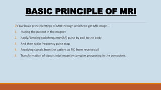
Magnetic resonance imaging (MRI) uses strong magnetic fields and radio waves to generate images of organs and tissues in the body. It does not use ionizing radiation like CT scans. MRI works by aligning hydrogen protons in the body under a magnetic field, then using radio waves to induce precession and generate signals that are processed into images. Key components of an MRI machine include large superconducting magnets, gradient coils to spatially encode signals, and RF coils for transmitting and receiving radio signals. MRI provides excellent soft tissue contrast and is used to diagnose many medical conditions.
Quality Assurance Programme in Computed TomographyRamzee Small
Introduction to Computed Tomography
Basic description of the components of a CT System
Introduction to Quality Assurance
Quality Assurance and Quality Control Tests in Computed Tomography base on frequency
Objective of QA/QC Test
Quality Assurance Programme in Computed TomographyRamzee Small
Introduction to Computed Tomography
Basic description of the components of a CT System
Introduction to Quality Assurance
Quality Assurance and Quality Control Tests in Computed Tomography base on frequency
Objective of QA/QC Test
this slide sharer contents are basic principle of CT fluoroscopy , software and hardware parts of equipment and image aqua cation and radiation dose comparison and videos related to equipment .
Single photon emission computed tomography (spect)Syed Hammad .
brief but informative knowledge about what basically SPECT is and what is the phenomenon behind this machine ... easy to understand as well as presenting during lectures and in classes . share it
MAGNETIC RESONANCE IMAGING QUESTION AND ANSWER 3 MARKS.pdfGanesan Yogananthem
Dear B.Sc MIT Students,
Attached is an essential document featuring comprehensive Questions & Answers for MAGNETIC RESONANCE IMAGING. We encourage you to utilize this resource to deepen your understanding and excel in your studies. Wishing you all the success in your academic endeavors and future careers.
Best regards,
MRI Scanner, Instrumentation. MDIRT ST. Louis Bamenda, Nchanji Nkeh KenethNchanji Nkeh Keneth
The MRI Scanner; historical facts, Understanding Nuclear Magnetic Resonance, The Scanner componenets, Radiofrequency coils, the casing, types of MRI magnets, understanding the principle of superconductivity. MRI applications
Magnetic resonance imaging (MRI) is an imaging technique used primarily in medical settings to produce high quality images of the soft tissues of the human body.
Magnetic Resonance Imaging ( MRI ) PresentationReduan Ahmad
MRI :
Big magnet and radio waves take picture inside our body , no radiation involved.
Super clear body pictures, helps doctors see problems with out hunting us .
Secret sauce of MRI , makes detailed pictures possible .
TEST BANK for Operations Management, 14th Edition by William J. Stevenson, Ve...kevinkariuki227
TEST BANK for Operations Management, 14th Edition by William J. Stevenson, Verified Chapters 1 - 19, Complete Newest Version.pdf
Recomendações da OMS sobre cuidados maternos e neonatais para uma experiência pós-natal positiva.
Em consonância com os ODS – Objetivos do Desenvolvimento Sustentável e a Estratégia Global para a Saúde das Mulheres, Crianças e Adolescentes, e aplicando uma abordagem baseada nos direitos humanos, os esforços de cuidados pós-natais devem expandir-se para além da cobertura e da simples sobrevivência, de modo a incluir cuidados de qualidade.
Estas diretrizes visam melhorar a qualidade dos cuidados pós-natais essenciais e de rotina prestados às mulheres e aos recém-nascidos, com o objetivo final de melhorar a saúde e o bem-estar materno e neonatal.
Uma “experiência pós-natal positiva” é um resultado importante para todas as mulheres que dão à luz e para os seus recém-nascidos, estabelecendo as bases para a melhoria da saúde e do bem-estar a curto e longo prazo. Uma experiência pós-natal positiva é definida como aquela em que as mulheres, pessoas que gestam, os recém-nascidos, os casais, os pais, os cuidadores e as famílias recebem informação consistente, garantia e apoio de profissionais de saúde motivados; e onde um sistema de saúde flexível e com recursos reconheça as necessidades das mulheres e dos bebês e respeite o seu contexto cultural.
Estas diretrizes consolidadas apresentam algumas recomendações novas e já bem fundamentadas sobre cuidados pós-natais de rotina para mulheres e neonatos que recebem cuidados no pós-parto em unidades de saúde ou na comunidade, independentemente dos recursos disponíveis.
É fornecido um conjunto abrangente de recomendações para cuidados durante o período puerperal, com ênfase nos cuidados essenciais que todas as mulheres e recém-nascidos devem receber, e com a devida atenção à qualidade dos cuidados; isto é, a entrega e a experiência do cuidado recebido. Estas diretrizes atualizam e ampliam as recomendações da OMS de 2014 sobre cuidados pós-natais da mãe e do recém-nascido e complementam as atuais diretrizes da OMS sobre a gestão de complicações pós-natais.
O estabelecimento da amamentação e o manejo das principais intercorrências é contemplada.
Recomendamos muito.
Vamos discutir essas recomendações no nosso curso de pós-graduação em Aleitamento no Instituto Ciclos.
Esta publicação só está disponível em inglês até o momento.
Prof. Marcus Renato de Carvalho
www.agostodourado.com
Acute scrotum is a general term referring to an emergency condition affecting the contents or the wall of the scrotum.
There are a number of conditions that present acutely, predominantly with pain and/or swelling
A careful and detailed history and examination, and in some cases, investigations allow differentiation between these diagnoses. A prompt diagnosis is essential as the patient may require urgent surgical intervention
Testicular torsion refers to twisting of the spermatic cord, causing ischaemia of the testicle.
Testicular torsion results from inadequate fixation of the testis to the tunica vaginalis producing ischemia from reduced arterial inflow and venous outflow obstruction.
The prevalence of testicular torsion in adult patients hospitalized with acute scrotal pain is approximately 25 to 50 percent
Tom Selleck Health: A Comprehensive Look at the Iconic Actor’s Wellness Journeygreendigital
Tom Selleck, an enduring figure in Hollywood. has captivated audiences for decades with his rugged charm, iconic moustache. and memorable roles in television and film. From his breakout role as Thomas Magnum in Magnum P.I. to his current portrayal of Frank Reagan in Blue Bloods. Selleck's career has spanned over 50 years. But beyond his professional achievements. fans have often been curious about Tom Selleck Health. especially as he has aged in the public eye.
Follow us on: Pinterest
Introduction
Many have been interested in Tom Selleck health. not only because of his enduring presence on screen but also because of the challenges. and lifestyle choices he has faced and made over the years. This article delves into the various aspects of Tom Selleck health. exploring his fitness regimen, diet, mental health. and the challenges he has encountered as he ages. We'll look at how he maintains his well-being. the health issues he has faced, and his approach to ageing .
Early Life and Career
Childhood and Athletic Beginnings
Tom Selleck was born on January 29, 1945, in Detroit, Michigan, and grew up in Sherman Oaks, California. From an early age, he was involved in sports, particularly basketball. which played a significant role in his physical development. His athletic pursuits continued into college. where he attended the University of Southern California (USC) on a basketball scholarship. This early involvement in sports laid a strong foundation for his physical health and disciplined lifestyle.
Transition to Acting
Selleck's transition from an athlete to an actor came with its physical demands. His first significant role in "Magnum P.I." required him to perform various stunts and maintain a fit appearance. This role, which he played from 1980 to 1988. necessitated a rigorous fitness routine to meet the show's demands. setting the stage for his long-term commitment to health and wellness.
Fitness Regimen
Workout Routine
Tom Selleck health and fitness regimen has evolved. adapting to his changing roles and age. During his "Magnum, P.I." days. Selleck's workouts were intense and focused on building and maintaining muscle mass. His routine included weightlifting, cardiovascular exercises. and specific training for the stunts he performed on the show.
Selleck adjusted his fitness routine as he aged to suit his body's needs. Today, his workouts focus on maintaining flexibility, strength, and cardiovascular health. He incorporates low-impact exercises such as swimming, walking, and light weightlifting. This balanced approach helps him stay fit without putting undue strain on his joints and muscles.
Importance of Flexibility and Mobility
In recent years, Selleck has emphasized the importance of flexibility and mobility in his fitness regimen. Understanding the natural decline in muscle mass and joint flexibility with age. he includes stretching and yoga in his routine. These practices help prevent injuries, improve posture, and maintain mobilit
- Video recording of this lecture in English language: https://youtu.be/lK81BzxMqdo
- Video recording of this lecture in Arabic language: https://youtu.be/Ve4P0COk9OI
- Link to download the book free: https://nephrotube.blogspot.com/p/nephrotube-nephrology-books.html
- Link to NephroTube website: www.NephroTube.com
- Link to NephroTube social media accounts: https://nephrotube.blogspot.com/p/join-nephrotube-on-social-media.html
Title: Sense of Taste
Presenter: Dr. Faiza, Assistant Professor of Physiology
Qualifications:
MBBS (Best Graduate, AIMC Lahore)
FCPS Physiology
ICMT, CHPE, DHPE (STMU)
MPH (GC University, Faisalabad)
MBA (Virtual University of Pakistan)
Learning Objectives:
Describe the structure and function of taste buds.
Describe the relationship between the taste threshold and taste index of common substances.
Explain the chemical basis and signal transduction of taste perception for each type of primary taste sensation.
Recognize different abnormalities of taste perception and their causes.
Key Topics:
Significance of Taste Sensation:
Differentiation between pleasant and harmful food
Influence on behavior
Selection of food based on metabolic needs
Receptors of Taste:
Taste buds on the tongue
Influence of sense of smell, texture of food, and pain stimulation (e.g., by pepper)
Primary and Secondary Taste Sensations:
Primary taste sensations: Sweet, Sour, Salty, Bitter, Umami
Chemical basis and signal transduction mechanisms for each taste
Taste Threshold and Index:
Taste threshold values for Sweet (sucrose), Salty (NaCl), Sour (HCl), and Bitter (Quinine)
Taste index relationship: Inversely proportional to taste threshold
Taste Blindness:
Inability to taste certain substances, particularly thiourea compounds
Example: Phenylthiocarbamide
Structure and Function of Taste Buds:
Composition: Epithelial cells, Sustentacular/Supporting cells, Taste cells, Basal cells
Features: Taste pores, Taste hairs/microvilli, and Taste nerve fibers
Location of Taste Buds:
Found in papillae of the tongue (Fungiform, Circumvallate, Foliate)
Also present on the palate, tonsillar pillars, epiglottis, and proximal esophagus
Mechanism of Taste Stimulation:
Interaction of taste substances with receptors on microvilli
Signal transduction pathways for Umami, Sweet, Bitter, Sour, and Salty tastes
Taste Sensitivity and Adaptation:
Decrease in sensitivity with age
Rapid adaptation of taste sensation
Role of Saliva in Taste:
Dissolution of tastants to reach receptors
Washing away the stimulus
Taste Preferences and Aversions:
Mechanisms behind taste preference and aversion
Influence of receptors and neural pathways
Impact of Sensory Nerve Damage:
Degeneration of taste buds if the sensory nerve fiber is cut
Abnormalities of Taste Detection:
Conditions: Ageusia, Hypogeusia, Dysgeusia (parageusia)
Causes: Nerve damage, neurological disorders, infections, poor oral hygiene, adverse drug effects, deficiencies, aging, tobacco use, altered neurotransmitter levels
Neurotransmitters and Taste Threshold:
Effects of serotonin (5-HT) and norepinephrine (NE) on taste sensitivity
Supertasters:
25% of the population with heightened sensitivity to taste, especially bitterness
Increased number of fungiform papillae
These simplified slides by Dr. Sidra Arshad present an overview of the non-respiratory functions of the respiratory tract.
Learning objectives:
1. Enlist the non-respiratory functions of the respiratory tract
2. Briefly explain how these functions are carried out
3. Discuss the significance of dead space
4. Differentiate between minute ventilation and alveolar ventilation
5. Describe the cough and sneeze reflexes
Study Resources:
1. Chapter 39, Guyton and Hall Textbook of Medical Physiology, 14th edition
2. Chapter 34, Ganong’s Review of Medical Physiology, 26th edition
3. Chapter 17, Human Physiology by Lauralee Sherwood, 9th edition
4. Non-respiratory functions of the lungs https://academic.oup.com/bjaed/article/13/3/98/278874
New Directions in Targeted Therapeutic Approaches for Older Adults With Mantl...i3 Health
i3 Health is pleased to make the speaker slides from this activity available for use as a non-accredited self-study or teaching resource.
This slide deck presented by Dr. Kami Maddocks, Professor-Clinical in the Division of Hematology and
Associate Division Director for Ambulatory Operations
The Ohio State University Comprehensive Cancer Center, will provide insight into new directions in targeted therapeutic approaches for older adults with mantle cell lymphoma.
STATEMENT OF NEED
Mantle cell lymphoma (MCL) is a rare, aggressive B-cell non-Hodgkin lymphoma (NHL) accounting for 5% to 7% of all lymphomas. Its prognosis ranges from indolent disease that does not require treatment for years to very aggressive disease, which is associated with poor survival (Silkenstedt et al, 2021). Typically, MCL is diagnosed at advanced stage and in older patients who cannot tolerate intensive therapy (NCCN, 2022). Although recent advances have slightly increased remission rates, recurrence and relapse remain very common, leading to a median overall survival between 3 and 6 years (LLS, 2021). Though there are several effective options, progress is still needed towards establishing an accepted frontline approach for MCL (Castellino et al, 2022). Treatment selection and management of MCL are complicated by the heterogeneity of prognosis, advanced age and comorbidities of patients, and lack of an established standard approach for treatment, making it vital that clinicians be familiar with the latest research and advances in this area. In this activity chaired by Michael Wang, MD, Professor in the Department of Lymphoma & Myeloma at MD Anderson Cancer Center, expert faculty will discuss prognostic factors informing treatment, the promising results of recent trials in new therapeutic approaches, and the implications of treatment resistance in therapeutic selection for MCL.
Target Audience
Hematology/oncology fellows, attending faculty, and other health care professionals involved in the treatment of patients with mantle cell lymphoma (MCL).
Learning Objectives
1.) Identify clinical and biological prognostic factors that can guide treatment decision making for older adults with MCL
2.) Evaluate emerging data on targeted therapeutic approaches for treatment-naive and relapsed/refractory MCL and their applicability to older adults
3.) Assess mechanisms of resistance to targeted therapies for MCL and their implications for treatment selection
Pulmonary Thromboembolism - etilogy, types, medical- Surgical and nursing man...VarunMahajani
Disruption of blood supply to lung alveoli due to blockage of one or more pulmonary blood vessels is called as Pulmonary thromboembolism. In this presentation we will discuss its causes, types and its management in depth.
Title: Sense of Smell
Presenter: Dr. Faiza, Assistant Professor of Physiology
Qualifications:
MBBS (Best Graduate, AIMC Lahore)
FCPS Physiology
ICMT, CHPE, DHPE (STMU)
MPH (GC University, Faisalabad)
MBA (Virtual University of Pakistan)
Learning Objectives:
Describe the primary categories of smells and the concept of odor blindness.
Explain the structure and location of the olfactory membrane and mucosa, including the types and roles of cells involved in olfaction.
Describe the pathway and mechanisms of olfactory signal transmission from the olfactory receptors to the brain.
Illustrate the biochemical cascade triggered by odorant binding to olfactory receptors, including the role of G-proteins and second messengers in generating an action potential.
Identify different types of olfactory disorders such as anosmia, hyposmia, hyperosmia, and dysosmia, including their potential causes.
Key Topics:
Olfactory Genes:
3% of the human genome accounts for olfactory genes.
400 genes for odorant receptors.
Olfactory Membrane:
Located in the superior part of the nasal cavity.
Medially: Folds downward along the superior septum.
Laterally: Folds over the superior turbinate and upper surface of the middle turbinate.
Total surface area: 5-10 square centimeters.
Olfactory Mucosa:
Olfactory Cells: Bipolar nerve cells derived from the CNS (100 million), with 4-25 olfactory cilia per cell.
Sustentacular Cells: Produce mucus and maintain ionic and molecular environment.
Basal Cells: Replace worn-out olfactory cells with an average lifespan of 1-2 months.
Bowman’s Gland: Secretes mucus.
Stimulation of Olfactory Cells:
Odorant dissolves in mucus and attaches to receptors on olfactory cilia.
Involves a cascade effect through G-proteins and second messengers, leading to depolarization and action potential generation in the olfactory nerve.
Quality of a Good Odorant:
Small (3-20 Carbon atoms), volatile, water-soluble, and lipid-soluble.
Facilitated by odorant-binding proteins in mucus.
Membrane Potential and Action Potential:
Resting membrane potential: -55mV.
Action potential frequency in the olfactory nerve increases with odorant strength.
Adaptation Towards the Sense of Smell:
Rapid adaptation within the first second, with further slow adaptation.
Psychological adaptation greater than receptor adaptation, involving feedback inhibition from the central nervous system.
Primary Sensations of Smell:
Camphoraceous, Musky, Floral, Pepperminty, Ethereal, Pungent, Putrid.
Odor Detection Threshold:
Examples: Hydrogen sulfide (0.0005 ppm), Methyl-mercaptan (0.002 ppm).
Some toxic substances are odorless at lethal concentrations.
Characteristics of Smell:
Odor blindness for single substances due to lack of appropriate receptor protein.
Behavioral and emotional influences of smell.
Transmission of Olfactory Signals:
From olfactory cells to glomeruli in the olfactory bulb, involving lateral inhibition.
Primitive, less old, and new olfactory systems with different path
NVBDCP.pptx Nation vector borne disease control programSapna Thakur
NVBDCP was launched in 2003-2004 . Vector-Borne Disease: Disease that results from an infection transmitted to humans and other animals by blood-feeding arthropods, such as mosquitoes, ticks, and fleas. Examples of vector-borne diseases include Dengue fever, West Nile Virus, Lyme disease, and malaria.
ARTIFICIAL INTELLIGENCE IN HEALTHCARE.pdfAnujkumaranit
Artificial intelligence (AI) refers to the simulation of human intelligence processes by machines, especially computer systems. It encompasses tasks such as learning, reasoning, problem-solving, perception, and language understanding. AI technologies are revolutionizing various fields, from healthcare to finance, by enabling machines to perform tasks that typically require human intelligence.
Lung Cancer: Artificial Intelligence, Synergetics, Complex System Analysis, S...Oleg Kshivets
RESULTS: Overall life span (LS) was 2252.1±1742.5 days and cumulative 5-year survival (5YS) reached 73.2%, 10 years – 64.8%, 20 years – 42.5%. 513 LCP lived more than 5 years (LS=3124.6±1525.6 days), 148 LCP – more than 10 years (LS=5054.4±1504.1 days).199 LCP died because of LC (LS=562.7±374.5 days). 5YS of LCP after bi/lobectomies was significantly superior in comparison with LCP after pneumonectomies (78.1% vs.63.7%, P=0.00001 by log-rank test). AT significantly improved 5YS (66.3% vs. 34.8%) (P=0.00000 by log-rank test) only for LCP with N1-2. Cox modeling displayed that 5YS of LCP significantly depended on: phase transition (PT) early-invasive LC in terms of synergetics, PT N0—N12, cell ratio factors (ratio between cancer cells- CC and blood cells subpopulations), G1-3, histology, glucose, AT, blood cell circuit, prothrombin index, heparin tolerance, recalcification time (P=0.000-0.038). Neural networks, genetic algorithm selection and bootstrap simulation revealed relationships between 5YS and PT early-invasive LC (rank=1), PT N0—N12 (rank=2), thrombocytes/CC (3), erythrocytes/CC (4), eosinophils/CC (5), healthy cells/CC (6), lymphocytes/CC (7), segmented neutrophils/CC (8), stick neutrophils/CC (9), monocytes/CC (10); leucocytes/CC (11). Correct prediction of 5YS was 100% by neural networks computing (area under ROC curve=1.0; error=0.0).
CONCLUSIONS: 5YS of LCP after radical procedures significantly depended on: 1) PT early-invasive cancer; 2) PT N0--N12; 3) cell ratio factors; 4) blood cell circuit; 5) biochemical factors; 6) hemostasis system; 7) AT; 8) LC characteristics; 9) LC cell dynamics; 10) surgery type: lobectomy/pneumonectomy; 11) anthropometric data. Optimal diagnosis and treatment strategies for LC are: 1) screening and early detection of LC; 2) availability of experienced thoracic surgeons because of complexity of radical procedures; 3) aggressive en block surgery and adequate lymph node dissection for completeness; 4) precise prediction; 5) adjuvant chemoimmunoradiotherapy for LCP with unfavorable prognosis.
Couples presenting to the infertility clinic- Do they really have infertility...Sujoy Dasgupta
Dr Sujoy Dasgupta presented the study on "Couples presenting to the infertility clinic- Do they really have infertility? – The unexplored stories of non-consummation" in the 13th Congress of the Asia Pacific Initiative on Reproduction (ASPIRE 2024) at Manila on 24 May, 2024.
3. INTRODUCTION
MRI stands for magnetic resonance imaging
MRI technique used in radiology to form picture of anatomy and physiological process of the
body.
MRI scanners use strong magnetic fields, magnetic field gradient and radiowaves to generate
images of the organs.
Do not involve X-ray or use of ionizing radiation --- like CT Scan and PET scans.
Uses the application NMR – (investigation of chemical and physical properties at molecular
level)
Based on the principle of NMR
4. HISTORY
In 1997, the first MRI exam was performed on
human being.
FATHER OF MRI
Magnetic resonance imaging inventor ----- RAYMOND
VAHAN DAMADIAN
{ Before talking about MRI principle first look types of
magnets }
5. Types of Magnet
[1] Permanent magnets
A permanent magnet consists of a material, which
has been magnetized such that it won‟t loose its
magnetic field The field strength is usually very low
and ranges between 0.064T ~ 0.3T, Permanent
magnets have usually an open design, which is
more comfortable for the patient.
Toshiba‟s Access 0.064Tesla system. The Access
was the worlds first open MRI scanner.
6. Resistive magnets are very large electro magnets ,The magnetic
field is generated by a current, which runs through loops of
wire. The field strength can be up to 0.3 Tesla. They produce a
lot of heat, which requires watercooling. They need a lot of
power to run, and are usually switched off when not in use to
conserve power. They usually have an open design, which
reduces claustrophobia.
[2] Resistive Magnets
Hitachi‟s Airis 0.3 Tesla system.
7. [3] Superconducting magnets
Today‟s most commonly used magnets are
superconducting magnets. The magnetic field is
generated by a current, which runs through a loop of
wire. The wire is surrounded with a coolant, such as
liquid helium, to reduce the electric resistance of the
wire. At 4 Kelvin (-269º C) electric wire looses its
resistance. Once a system is energized, it won‟t loose
its magnetic field. Superconductivity allows for
systems with very high field strengths up to 12 Tesla.
The ones that are most used in clinical environments
run at 1.5 Tesla. Most superconducting magnets are
bore type magnets
examples of bore type magnets
8. • RF Coils
RF coils are needed to transmit and receive radio-frequency waves used in MRI scanners. RF coils are one of the
most important components that affect image quality. Current MRI scanners have a range of RF coils suitable to
acquire images of all body parts. There are two types of RF coils: volume coils and surface coils
[1] Volume RF Coils OR Quadrature Coils
The design of a volume coil is usually a saddle
shape, which guarantees a uniform RF field
inside the coil. Volume coils need to have the
area of examination inside the coil. They can
be used for transmit and receive, although
sometimes they are used for receive only the
knee coil is receive only
[2] Surface coils
As the name already implies, surface coils are placed close to the
area under examination such as the temporo - mandibular joint, the
orbits or the shoulder. The coil consists of a single or double loop of
copper wire. They have a high Signal to Noise Ratio (SNR) and allow
for very high resolution imaging. The disadvantage is that they loose
signal uniformity very quickly when you move away from the coil.
9. [3] Gradient Coils
Gradient coils are a set of wires in the magnet, which enable us
to create additional magnetic fields, which are, in a way,
superimposed on the main magnetic field B0.
Sounds complicated, but it's not really.
There are 3 sets of wires. Each set can create a magnetic field
in a specific direction: Z, X or Y. When a current is fed into the Z
gradient, then a magnetic field is generated in the Z direction .
The same goes for the other gradients
Interesting detail: Everyone knows that MRI can make a lot of
noise during acquisition. The magnetic field, which is
generated, is very strong. Although the gradient coils are very
tightly fixed in a kind of resin, the forces, exhibited by the
gradient coil, are enough to make them vibrate, hence the
noise.
1. The Gz gradient selected an axial slice.
2. The Gy gradient created rows with different phases.
3. The Gx gradient created columns with different frequencies.
10. [4] Phased Array Coils
Phased array coils consist of multiple surface coils.
Surface coils have the highest SNR but have a limited
sensitive area. By combining 4 or 6 surface coils it is
possible to create a coil with a large sensitive area.
The QD Body Array coil is a volume coil, while the Spine
Array coil is a surface coil. Today most MRI systems come
with Quadrature and phased array coils.
• Other Hardware
A very important part is the Radio Frequency (RF)
chain, which produces the RF signal transmitted into
the patient, and receives the RF signal from the patient.
Actually, the receive coil is a part of the RF chain. The
frequency range used in MRI is the same as used for
radio transmissions. That‟s why MRI scanners are
placed in a Faraday cage to prevent radio waves to
enter the scanner room .
11. BASIC PRINCIPLE OF MRI
Four basic principle/steps of MRI through which we get MR image---
1. Placing the patient in the magnet
2. Apply/Sending radiofrequency(RF) pulse by coil to the body
3. And then radio frequency pulse stop
4. Receiving signals from the patient as FID from receive coil
5. Transformation of signals into image by complex processing in the computers.
12. At molecular level about Protons -
Today’s MR imaging is based on PROTON IMAGING.
PROTON -
• Positively charged particle
• Present in nucleus of atom
• Example hydrogen having one proton (H¹⁺)
Proton help in MR imaging
o Positively charged
o Having rotatory movement called spin.
o Randomly moving in human body
o As moving charge generate current and induce small M.F. around It, called Magnetic dipole moment.
In Human body, proton(H⁺) moves randomly ---
When patient is place in magnet and external magnetic field is applied, protons align themselves
and spin in the direction of Ext. M.F. 1st step
13. Some proton align parallel and some anti-parallel to Ext. M.F.
Proton align parallel to M.F. , not only rotate around itself ( called spin),
but also its axis of rotation form cone called precession (as shown in diagram )
No. of precession of proton per second called precession frequency(P.F.) ,(hertz)
Therefore, precession frequency is directly proportion to Ext. M.F.
Hence larmour equation is given as, W₀ α B₀
W₀ = γ B₀
where, W₀ - P.F.
B₀ - M.F.
γ - Gyroscopic ratio, specific to particular nuclei
14. P.F. of hydrogen at ---
LONGITUDINAL MAGNETIZATION ( L.M.)
What happen when proton align under the influence of Ext. M.F. ?
• For orientation in space consider X,Y,Z axis.
• Ext. M.F. along Z-axis and Z-axes Is toward patient as well as bore of the magnet.
• As we discuss earlier proton is parallel ( positive) and anti-parallel (negative) to Ext. M.F.
• Force of parallel (positive) and anti-parallel (negative) cancel each other,
but proton spinning to positive side are always more than negative side.
• Force at positive side add up together to form magnetic vector to Z-axis called Longitudinal
magnetization
ds
1 T
1.5 T
3 T
42 MHz
64 MHz
128 MHz
16. TRASNVERSE MAGNETIZATION ( T.M.)
• As L.M. formed along Z-axes
• Next step send RF pulse 2nd step
• Precessing proton pick up and go to higher energy level and start precessing antiparallel.
• Imbalance results in tilting of magnetization into transverse plane. Called Transverse magnetization.
In short, RF pulse cause tilting of magnetization into transverse plane.
• Therefore precessing frequency is same as that of RF pulse
• As RF pulse and proton have higher similar frequency protons can pick some energy from RF pulse . This is called
resonance R of MRI.
17. MR Signal
• Transverse Magnetization has precession frequency.
• It constantly rotates at larmour frequency in transverse plane and induce electric current.
• Receiver RF pulse receive current as MR signal. 3rd step
Strength of signal α Magnitude of T.M.
• MR signals are transformed into MRI image by computers using mathematical methods such as
Fourier Transfromation. 4th step
18. REVISION
Basic four steps of MR imaging includes—
1. Patient is placed in the magnet-
• All randomly moving protons in patients body align and precess along the external M.F.
• L.M. is formed long the Z-axes.
2. RF pulse is sent---
• Precessing proton pick up energy from RF pulse to go to higher energy level
• Results in reduction in L.M. and formation of T.M. in X-Y plane.
3. MR signal is received---
• T.M. vector precess in transverse plane and generates current.
• Current is received as signal by the RF coil.
4. Image formation ----
• MR signal received by the coil transformed into image by complex mathematical process
such as Fourier Transformation by computers.
19. Why proton only?
• Other substances can also utilize in MR imaging.
• Requirement are --- nuclei have spin and have odd number of proton.
• Example 13C , 19F, 23Na, 31P can be used for MR imaging.
• HYDROGEN atom has only on proton
• Hydrogen is present in abundance in body water and fat
• H⁺ gives best and most intense signal among all nuclei.
20. WHAT CAN BE DIAGNOSED BY AN MRI SCAN ?
Most ailments of brain, including tumor
Sport injuries
Most spinal injuries
Pelvic problems
Prostrate problems
Soft tissue and bone pathology
WHO CAN’T HAVE AN MRI SCAN
A cardiac pacemaker
Brain aneurysm clips
A cochlear implant
Eye implants
IUD
Dental fillings and bridge
Tattoos
21.
22. In medical imaging, artifacts are misrepresentations of tissue structures produced by
imaging techniques such as ultrasound, X-ray, CT scan, and magnetic resonance imaging
(MRI).
In this section I‟ll discuss the following artifacts:
• Motion related artifacts
• Para-magnetic artifacts
• Phase Wrap artifacts
• Frequency artifacts
• Susceptibility artifacts
• Clipping artifact
• Chemical Shift Artifact
• Spike artifact
• “Zebra” artifac
Image Artifacts
23. Motion artifacts are caused by phase mis-
mapping of the protons. protons may have
moved, due to respiration, pulsation or
motion, through the gradient magnetic field,
thus acquiring an additional phase
• Motion artifacts • Para-Magnetic Artifacts
Para-magnetic artifacts are caused by
metal (~ iron). Metal deflects the magnetic
field, thus changing the resonance frequency
beyond the range, which is used in MRI.
Image Artifacts
24. •Phase Wrap Artifacts
Phase Wrap Artifacts Phase wrap artifacts
are caused by mis-mapping of phase.
Phase wrap occurs when the Field Of View
is smaller than the object. The part of the
body outside the FOV will be „wrapped
around‟ into the image
•Frequency Artifacts
Frequency artifacts are caused by „dirty‟ frequencies. Faulty
electronics, external transmitters, RFcage leak, non-shielded
equipment in the scanner room, metal in the patient, or when
the door to the scanner room is left open can generate „dirty‟
frequencies. It usually requires an engineer to solve this kind of
artifact, although, the door to the scanner room can be closed
by non-qualified people as well
Image Artifacts
25. •Susceptibility Artifacts
Susceptibility is the ability of substances to be
magnetized, for example iron in blood.
Susceptibility artifacts are caused by local
magnetic field inhomogeneity. The different
bonding properties of hydrogen protons in fat and
muscle will cause local magnetic field
inhomogeneity at the tissue boundaries
•Clipping Artifact
Signal clipping or „over flow‟ occurs when
the receiver gain is set to high during the
pre-scan. The maximum received signal is
higher than the value set in the receiver gain
Image Artifacts
26. •Chemical Shift Artifact
Chemical shift artifacts are caused by different
resonance frequencies of hydrogen in lipids
and hydrogen in water. Chemical shift happens
in the frequency direction.
•Spike Artifact
A spike artifact is caused by one „bad‟ data point in
k-space. one data point in k-space, which is out of
the ordinary. The resulting image show diagonal
lines throughout the image.
Image Artifacts
27. •“Zebra” Artifact
The “Zebra” artifact
may occur when the
patient touches the coil,
or as a result of phase
wrap. To avoid this
problem you have to
make sure that the
patient is not touching
the receive coil, or use
No-Wrap option.
Image Artifacts