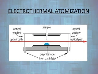
ATOMIC ABSORPTION
SPECTROSCOPY
GROUP # 6
INTRODUCTION
BY: SEENAM IFTIKHAR
SPECTROSCOPY
• Spectroscopy is the branch of science that
deals with the study of interaction of
electromagnetic radiations with matter i.e.
atoms or molecules of drugs.
• Spectroscopy is of two types, Emission
spectroscopy and absorption spectroscopy.
TYPES OF SPECTROSCOPY
EMISSION SPECTROSCOPY
Examples include:
• Fluorimetry
• Flame photometry
• Atomic emission
spectroscopy.
ABSORPTION SPECTROSCOPY
Examples include:
• UV/Visible spectroscopy
• IR spectroscopy
• Nuclear Magnetic
Resonance Spectroscopy
(NMR)
• Atomic Absorption
Spectroscopy
ATOMIC SPECTROSCOPY
• Atomic Spectroscopy is the result of
phenomenon of absorption, emission or
fluorescence by atoms or elementary ions
mostly in ultraviolet region.
• The spectra are obtained by converting the
component into gaseous atoms or elementary
ions by suitable heat treatments.
TYPES OF ATOMIC ABSORPTION
SPECTROSCOPY
AAS
Atomic
Absorption
Spectra
Atomic
Emission
Spectra
Atomic
Fluorescence
Spectra
ATOMIC LINE WIDTH AND PRINCIPLE
OF ATOMIC ABSORPTION
SPECTROSCOPY
BY: MAZNA SALEEM
ATOMIC LINE WIDTH
• The width of atomic lines are of considerable
importance in atomic absorption or atomic
emission spectroscopy.
• Narrow lines are highly desirable for both
absorption and emission spectroscopy because
they reduce the possibility of interferences due to
overlapping spectra.
PRINCIPLE OF AAS:
 The technique uses basically the principle that
free atoms (gas) generated in an atomizer can
absorb radiation at specific frequency.
 Atomic-absorption spectroscopy quantifies the
absorption of ground state atoms in the gaseous
state .
 The atoms absorb ultraviolet or visible light and
make transitions to higher electronic energy
levels.
 The analyte concentration is determined from
the amount of absorption.
SPECTROPHOTOMETER
BY: AISHA AMJAD
DEFINITION
• Spectrophotometer is an instrument that
measures the amount of Light absorbed by a
sample as function of wavelength.
PROPERTIES
• It is a techniques are used to measure the
concentration of solutes in a solution.
• It is the quantitative measurement of the reflection
or transmission properties of a material.
• It is use in various fields such as chemistry, physics,
biochemistry, and chemical engineering and clinical
applications.
CONSTRUCTION OF ATOMIC
ABSORPTION
SPECTROPHOTOMETER
HOW DOES IT WORK?
RADIATION
SOURCES
BY: HIBA MUSHTAQ
RADIATION SOURCES
• Absorption Lines are very much narrow (0.002
to 0.005)
• This limited lines creates problem in AAS.
• The problem's solution is nothing but the
Radiation Sources.
LAMP SOURCES
A separate lamp source is needed for each
element or sometimes group of element.
• Hollow Cathode lamp
• Electrodeless discharge lamp.
HOLLOW CATHODE TUBE
Apparatus:
• Tungston anode.
• Cylindrical cathode.
• Glass container.
Ionization of inert gas occurs when it is
supplied with:
• 300V potential
• 5 to 20mV current.
WORKING OF HCL
• HCL works by the process of sputtering.
‘Sputtering occurs when the energy to be
transformed into gaseous atom and bring out an
atomic cloud by shifting some amount of metal
from the cathode.’
• The excited metal atoms of the atomic cloud
emits the radiation.
• Metal atom diffuse back thus re-deposition
occurs.
ELECTRODE LESS DISCHARGE LAMP
• Small amount of both inert gas and the
element to be studied is placed inside the
tube constructed with quartz glass.
• Unlike HCL it is devoid of any electrodes but
alternatively microwave radiation is present to
provide power from outside the lamp.
WORKING OF EDL
• EDL is available foe wide variety of elements.
It is more efficacious practically but expensive.
MONOCHROMATOR
and
Source Modulation
BY: HAZIQA IFFRIN
MONOCHROMATOR
The function of the monochromator is to
isolate narrow band width of light
(monochromatic light) from the light source.
MONOCHROMATOR ASSEMBLY
TYPES OF MONOCHROMATOR
• Prism
• Diffraction grating
PRISM MONOCHROMATOR
DIFRACTION GRATTING
MONOCHROMATOR
SOURCE MODULATION
It is a process to discriminate light coming from
radiation source and flame (atomizer).
atomization
BY: ASRA FATIMA
ATOMIZATION
• Component of atomic absorption spectroscopy.
• Device of flame photometry.
• Solid or liquid sample is converted into gaseous
state.
FLAME ATOMIZATION
ELECTROTHERMAL ATOMIZATION
MISCELLANEOUS ATOMIZATION
• Only certain elements are atomized.
• Involves chemical reaction.
• By producing volatile hydrides.
detectors
BY: AREEBA SALEEM
DETECTORS
The role of the detector is to convert a light
signal into an electrical signal.
TYPES OF DETECTORS
• Photo Tube
• Photomultiplier Tube
PHOTO TUBE
• It consists of Semi-
cylindrical cathode
(containing loosely
bounded electrons) and
central metal wire
anode.
• The current produce
from the photo tube are
quite small so it require
amplification.
PHOTOMULTIPLIER TUBE
• The principle of
operation is the
emission of
electrons upon
exposure to
radiation.
• The detector
contains a photo
emissive cathode
and a series of
dynodes.
Interferences
BY: KHADIJA KHAN
INTERFERENCES
(i) Spectral Interferences,
(ii) Chemical Interferences, and
(iii) Ionization Interferences.
SPECTRAL INTERFERENCES
This type of interference normally takes place
when the absorption of an interfering species
either overlaps or lies very near to the analyte
absorption.
Examples of spectral interferences,
• Combustion products.
• Emission line of element, radical or molecule
and unresolved band spectra.
• Sample Matrix
CHEMICAL INTERFERENCES
• It results from various chemical processes
occurring during atomization that alter the
absorption characteristics of the analyte.
• More common.
• effects may often be minimized by
appropriate choice of experimental
parameters.
• Examples :
(i) Chemical Interferences due to Anion (PO43–)
(ii) Chemical Interference due to Cations.
PROTECTIVE AGENTS :
These agents are found to inhibit the
interferences by virtue of their ability to form
relatively stable but volatile species with the
respective analyte.
(a) EDTA.
(b) 8-Hydroxyquinoline, and
(c) Ammonium salt of APDC.
IONIZATION INTERFERENCES
The substitution of air with either oxygen or
nitrous oxide gives rise to temperatures which
are high enough to cause appreciable
ionization. Hence, as a consequence of the
attained equilibrium-a fairly significant
concentration of electron exists.
M M + e–
TYPES OF
SPECTROPHOTOMETER
SINGLE BEAM
SPECTROPHOTOMETER
• It utilizes one beam of light that passes through the
sample and the intensity of the light reflected from
a reference is measured without sample.
ADVANTAGES
• Low cost
• High throughput
• High sensitivity
• Less complicated
• Economical
DISADVANTAGES
• Calibration should be done with blank and sample
continuously according to wavelength and
absorption power of sample so reference beam is
not obtain simultaneously.
• Time consuming
DOUBLE BEAM
SPECTROPHOTOMETER
It utilizes two beams of light: reference and sample
beam.
Double beam spectrophotometer use rotating mirror
to separate the reference beam from a sample beam.
ADVANTAGES
Double beam is more advantageous then single
beam.
• High speed
• High stability
• Flexibility
DISADVANTAGES
• expensive
• Lower sensitivity
• Lower reliability
APPLICATIONS OF AAS
BY: FARAH DEEBA
APPLICATIONS
• One of the most widely used techniques.
• Its popular for quantitative analysis
a) molecular weight determination
b) detection of conjugation
c) detection of functional groups
• and qualitative analysis.
a) detection of impurities to analyze substance purity .
• Assay of trace metals and determination elements.
• Importance of AAS In its modern form.
Conclusion

Atomic absorption spectroscopy

  • 1.
  • 2.
  • 3.
    SPECTROSCOPY • Spectroscopy isthe branch of science that deals with the study of interaction of electromagnetic radiations with matter i.e. atoms or molecules of drugs. • Spectroscopy is of two types, Emission spectroscopy and absorption spectroscopy.
  • 4.
    TYPES OF SPECTROSCOPY EMISSIONSPECTROSCOPY Examples include: • Fluorimetry • Flame photometry • Atomic emission spectroscopy. ABSORPTION SPECTROSCOPY Examples include: • UV/Visible spectroscopy • IR spectroscopy • Nuclear Magnetic Resonance Spectroscopy (NMR) • Atomic Absorption Spectroscopy
  • 5.
    ATOMIC SPECTROSCOPY • AtomicSpectroscopy is the result of phenomenon of absorption, emission or fluorescence by atoms or elementary ions mostly in ultraviolet region. • The spectra are obtained by converting the component into gaseous atoms or elementary ions by suitable heat treatments.
  • 6.
    TYPES OF ATOMICABSORPTION SPECTROSCOPY AAS Atomic Absorption Spectra Atomic Emission Spectra Atomic Fluorescence Spectra
  • 7.
    ATOMIC LINE WIDTHAND PRINCIPLE OF ATOMIC ABSORPTION SPECTROSCOPY BY: MAZNA SALEEM
  • 8.
    ATOMIC LINE WIDTH •The width of atomic lines are of considerable importance in atomic absorption or atomic emission spectroscopy. • Narrow lines are highly desirable for both absorption and emission spectroscopy because they reduce the possibility of interferences due to overlapping spectra.
  • 9.
    PRINCIPLE OF AAS: The technique uses basically the principle that free atoms (gas) generated in an atomizer can absorb radiation at specific frequency.  Atomic-absorption spectroscopy quantifies the absorption of ground state atoms in the gaseous state .  The atoms absorb ultraviolet or visible light and make transitions to higher electronic energy levels.  The analyte concentration is determined from the amount of absorption.
  • 10.
  • 11.
    DEFINITION • Spectrophotometer isan instrument that measures the amount of Light absorbed by a sample as function of wavelength.
  • 12.
    PROPERTIES • It isa techniques are used to measure the concentration of solutes in a solution. • It is the quantitative measurement of the reflection or transmission properties of a material. • It is use in various fields such as chemistry, physics, biochemistry, and chemical engineering and clinical applications.
  • 13.
  • 14.
  • 15.
  • 16.
    RADIATION SOURCES • AbsorptionLines are very much narrow (0.002 to 0.005) • This limited lines creates problem in AAS. • The problem's solution is nothing but the Radiation Sources.
  • 17.
    LAMP SOURCES A separatelamp source is needed for each element or sometimes group of element. • Hollow Cathode lamp • Electrodeless discharge lamp.
  • 18.
    HOLLOW CATHODE TUBE Apparatus: •Tungston anode. • Cylindrical cathode. • Glass container. Ionization of inert gas occurs when it is supplied with: • 300V potential • 5 to 20mV current.
  • 19.
    WORKING OF HCL •HCL works by the process of sputtering. ‘Sputtering occurs when the energy to be transformed into gaseous atom and bring out an atomic cloud by shifting some amount of metal from the cathode.’ • The excited metal atoms of the atomic cloud emits the radiation. • Metal atom diffuse back thus re-deposition occurs.
  • 20.
    ELECTRODE LESS DISCHARGELAMP • Small amount of both inert gas and the element to be studied is placed inside the tube constructed with quartz glass. • Unlike HCL it is devoid of any electrodes but alternatively microwave radiation is present to provide power from outside the lamp.
  • 21.
    WORKING OF EDL •EDL is available foe wide variety of elements. It is more efficacious practically but expensive.
  • 22.
  • 23.
    MONOCHROMATOR The function ofthe monochromator is to isolate narrow band width of light (monochromatic light) from the light source.
  • 24.
  • 25.
    TYPES OF MONOCHROMATOR •Prism • Diffraction grating
  • 26.
  • 27.
  • 28.
    SOURCE MODULATION It isa process to discriminate light coming from radiation source and flame (atomizer).
  • 30.
  • 31.
    ATOMIZATION • Component ofatomic absorption spectroscopy. • Device of flame photometry. • Solid or liquid sample is converted into gaseous state.
  • 32.
  • 33.
  • 34.
    MISCELLANEOUS ATOMIZATION • Onlycertain elements are atomized. • Involves chemical reaction. • By producing volatile hydrides.
  • 35.
  • 36.
    DETECTORS The role ofthe detector is to convert a light signal into an electrical signal.
  • 37.
    TYPES OF DETECTORS •Photo Tube • Photomultiplier Tube
  • 38.
    PHOTO TUBE • Itconsists of Semi- cylindrical cathode (containing loosely bounded electrons) and central metal wire anode. • The current produce from the photo tube are quite small so it require amplification.
  • 39.
    PHOTOMULTIPLIER TUBE • Theprinciple of operation is the emission of electrons upon exposure to radiation. • The detector contains a photo emissive cathode and a series of dynodes.
  • 40.
  • 41.
    INTERFERENCES (i) Spectral Interferences, (ii)Chemical Interferences, and (iii) Ionization Interferences.
  • 42.
    SPECTRAL INTERFERENCES This typeof interference normally takes place when the absorption of an interfering species either overlaps or lies very near to the analyte absorption. Examples of spectral interferences, • Combustion products. • Emission line of element, radical or molecule and unresolved band spectra. • Sample Matrix
  • 43.
    CHEMICAL INTERFERENCES • Itresults from various chemical processes occurring during atomization that alter the absorption characteristics of the analyte. • More common. • effects may often be minimized by appropriate choice of experimental parameters.
  • 44.
    • Examples : (i)Chemical Interferences due to Anion (PO43–) (ii) Chemical Interference due to Cations.
  • 45.
    PROTECTIVE AGENTS : Theseagents are found to inhibit the interferences by virtue of their ability to form relatively stable but volatile species with the respective analyte. (a) EDTA. (b) 8-Hydroxyquinoline, and (c) Ammonium salt of APDC.
  • 46.
    IONIZATION INTERFERENCES The substitutionof air with either oxygen or nitrous oxide gives rise to temperatures which are high enough to cause appreciable ionization. Hence, as a consequence of the attained equilibrium-a fairly significant concentration of electron exists. M M + e–
  • 47.
  • 48.
    SINGLE BEAM SPECTROPHOTOMETER • Itutilizes one beam of light that passes through the sample and the intensity of the light reflected from a reference is measured without sample.
  • 49.
    ADVANTAGES • Low cost •High throughput • High sensitivity • Less complicated • Economical DISADVANTAGES • Calibration should be done with blank and sample continuously according to wavelength and absorption power of sample so reference beam is not obtain simultaneously. • Time consuming
  • 50.
    DOUBLE BEAM SPECTROPHOTOMETER It utilizestwo beams of light: reference and sample beam. Double beam spectrophotometer use rotating mirror to separate the reference beam from a sample beam.
  • 51.
    ADVANTAGES Double beam ismore advantageous then single beam. • High speed • High stability • Flexibility DISADVANTAGES • expensive • Lower sensitivity • Lower reliability
  • 52.
  • 53.
    APPLICATIONS • One ofthe most widely used techniques. • Its popular for quantitative analysis a) molecular weight determination b) detection of conjugation c) detection of functional groups • and qualitative analysis. a) detection of impurities to analyze substance purity . • Assay of trace metals and determination elements. • Importance of AAS In its modern form.
  • 54.