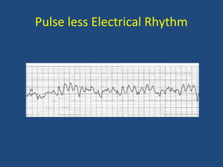
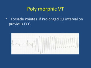
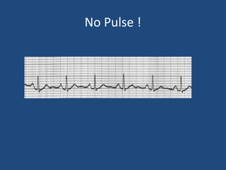
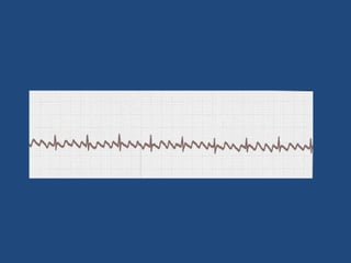
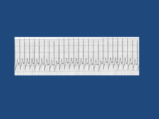
Arrhythmia : What you need to
know for ACLS?
Syed Raza
Introduction
• Rhythm recognition is a key skill that one
needs to demonstrate during cardiac arrest
situation.
• This can be life saving.
• Early defibrillation
• Decision making on the right therapy
Lets Keep it Simple!
• Pulseless Rhythms
• Tachyarrhythmias
• Bradyarrhythmias
Pulse less Electrical Rhythm
Ventricular Fibrillation
• Uncoordinated contractions within the
ventricles of heart.
• Due to multiple cardiac cells that function as
pacemakers and discharge electrical impulses
in a chaotic manner.
• Reduced / No cardiac output : No pulse
• Will result in Asystole if not treated.
• Commonest cause : Hypoxia /Ischemia
• Types : Fine and Coarse
Therapy
Immediate Defibrillation
CPR
I/V Amiodarone after 3 shocks
Ventricular Tachycardia
• Broad Complex Tachycardia (QRS > 0.12s)
• Heart rate > 180 beats /mt
• Mono-morphic
• Poly-morphic / Torsade Pointe
• Pulse less vs with pulse
Mono morphic VT
Poly morphic VT
• Torsade Pointes if Prolonged QT interval on
previous ECG
Treatment
• Pulseless : Defibrillation
• With pulse : stable = Amiodarone
Unstable = DC Cardio version
No Pulse !
Pulse Less Electrical Activity (PEA)
• Organized electrical activity but without a
pulse
• Usually has underlying treatable cause
• Hypovolumea and Hypoxia are the
commonest causes.
• If no underlying cause is identified, it will be
treated same as Asystole.
5 Hs and 5 Ts
5 Hs
Hypovolumia
Hypoxia
Hydrogen Ion (Acidosis)
Hyperkalemia
Hypokalemia
Hypoglycemia
5Ts
Toxins
Tension Pneumothorax
Tamponade
Thrombosis : Coronary
Thrombosis : Pulmonary
Trauma
ASYSTOLE
Follow flat line protocol – check leads and gain
Not a true rhythm
State of no electrical activity
Terminal event
Very poor prognosis : ROSC extremely unlikely
Possible underlying cause : 5Hs and 5Ts
Treatment : CPR and Epinephrine
First Degree AV Block
• PR interval is prolonged > 200ms
• No clinical significance if asymptomatic
• May lead to higher degree AV Block
Second Degree AV Block
Mobitz Type 1
• Progressive prolongation of PR interval.
• Atrial impulse (P waves) may not be conducted
through AVN and gets blocked and hence no
QRS.
• No clinical significance unless symptomatic.
Mobitz Type 2
• Non prolongation and fixed PR interval.
• Non conducted p waves
• No ventricular activity -Drop beats / No QRS
Most times Infranodal
Third Degree AV Block (CHB)
P waves with a regular pp interval
QRS complexes with a regular RR interval
QRS complex may be narrow or wide (escape
rhythm)
No relationship between P waves and QRS
complexes.
Treatment
• Trans cutaneous or Trans Venous pacemaker
• Atropine (0.5 mg) may be tried
Epinephrine 0.5 -1 mg /kg bw
Atrial Fbrillation
• No p waves preceding QRS complexes as no
coordinated atrial contractility
• Irregular (variable) RR intervals
Treatment
• Unstable : Synchronized DC Cardio version
• Stable : Rhythm Control vs Rate Control
• Rhythm : Amiodarone, Sotalol, Flecainide
• Rate control : Beta blocker, Calcium channel
blocker, Digoxin.
• Anticoagulant if indicated.
Atrial Flutter
• Atrial rate 250 – 350 /mt
• Saw Tooth Appearance
• Ventricular rate depends on Degree of AV
block
• Electrical foci usually in RA
Treatment
• Rate Control
• Rhythm Control
• Anti coagulant
• DCC if unstable
Supra Ventricular Tachycardia
• Broad term for various supra ventricular
arrhythmia
• Electrical impulses above the ventricular
electrical conducting system.
• Inverted p waves preceding or following qrs
complexes.
• Review old ECG – exclude WPW
Treatment
Vagal maneuver
Adenosine
•Drugs – Chemical Cardio version or Rate
control.
• Anti coagulant.
•If unstable : sync. DCC
ARRHYTHMIA - WHAT YOU NEED TO KNOW FOR ACLS.

ARRHYTHMIA - WHAT YOU NEED TO KNOW FOR ACLS.

  • 1.
    Arrhythmia : Whatyou need to know for ACLS? Syed Raza
  • 2.
    Introduction • Rhythm recognitionis a key skill that one needs to demonstrate during cardiac arrest situation. • This can be life saving. • Early defibrillation • Decision making on the right therapy
  • 3.
    Lets Keep itSimple! • Pulseless Rhythms • Tachyarrhythmias • Bradyarrhythmias
  • 4.
  • 6.
    Ventricular Fibrillation • Uncoordinatedcontractions within the ventricles of heart. • Due to multiple cardiac cells that function as pacemakers and discharge electrical impulses in a chaotic manner. • Reduced / No cardiac output : No pulse • Will result in Asystole if not treated.
  • 7.
    • Commonest cause: Hypoxia /Ischemia • Types : Fine and Coarse
  • 8.
  • 10.
    Ventricular Tachycardia • BroadComplex Tachycardia (QRS > 0.12s) • Heart rate > 180 beats /mt • Mono-morphic • Poly-morphic / Torsade Pointe • Pulse less vs with pulse
  • 11.
  • 12.
    Poly morphic VT •Torsade Pointes if Prolonged QT interval on previous ECG
  • 13.
    Treatment • Pulseless :Defibrillation • With pulse : stable = Amiodarone Unstable = DC Cardio version
  • 14.
  • 15.
    Pulse Less ElectricalActivity (PEA) • Organized electrical activity but without a pulse • Usually has underlying treatable cause • Hypovolumea and Hypoxia are the commonest causes. • If no underlying cause is identified, it will be treated same as Asystole.
  • 16.
    5 Hs and5 Ts 5 Hs Hypovolumia Hypoxia Hydrogen Ion (Acidosis) Hyperkalemia Hypokalemia Hypoglycemia
  • 17.
    5Ts Toxins Tension Pneumothorax Tamponade Thrombosis :Coronary Thrombosis : Pulmonary Trauma
  • 19.
    ASYSTOLE Follow flat lineprotocol – check leads and gain Not a true rhythm State of no electrical activity Terminal event Very poor prognosis : ROSC extremely unlikely Possible underlying cause : 5Hs and 5Ts Treatment : CPR and Epinephrine
  • 20.
  • 21.
    • PR intervalis prolonged > 200ms • No clinical significance if asymptomatic • May lead to higher degree AV Block
  • 22.
  • 23.
    Mobitz Type 1 •Progressive prolongation of PR interval. • Atrial impulse (P waves) may not be conducted through AVN and gets blocked and hence no QRS. • No clinical significance unless symptomatic. Mobitz Type 2 • Non prolongation and fixed PR interval. • Non conducted p waves • No ventricular activity -Drop beats / No QRS Most times Infranodal
  • 24.
    Third Degree AVBlock (CHB)
  • 25.
    P waves witha regular pp interval QRS complexes with a regular RR interval QRS complex may be narrow or wide (escape rhythm) No relationship between P waves and QRS complexes.
  • 26.
    Treatment • Trans cutaneousor Trans Venous pacemaker • Atropine (0.5 mg) may be tried Epinephrine 0.5 -1 mg /kg bw
  • 28.
    Atrial Fbrillation • Nop waves preceding QRS complexes as no coordinated atrial contractility • Irregular (variable) RR intervals
  • 29.
    Treatment • Unstable :Synchronized DC Cardio version • Stable : Rhythm Control vs Rate Control • Rhythm : Amiodarone, Sotalol, Flecainide • Rate control : Beta blocker, Calcium channel blocker, Digoxin. • Anticoagulant if indicated.
  • 31.
    Atrial Flutter • Atrialrate 250 – 350 /mt • Saw Tooth Appearance • Ventricular rate depends on Degree of AV block • Electrical foci usually in RA
  • 32.
    Treatment • Rate Control •Rhythm Control • Anti coagulant • DCC if unstable
  • 34.
    Supra Ventricular Tachycardia •Broad term for various supra ventricular arrhythmia • Electrical impulses above the ventricular electrical conducting system. • Inverted p waves preceding or following qrs complexes. • Review old ECG – exclude WPW
  • 35.
    Treatment Vagal maneuver Adenosine •Drugs –Chemical Cardio version or Rate control. • Anti coagulant. •If unstable : sync. DCC