A complete presentation about all-aspects of the Alzheimer's disease, including Patho Physiology, Treatment, Nursing Management, Prevention, Disease Overview, Clinical Manifestation, etc.
Factory Supply Best Quality Pmk Oil CAS 28578–16–7 PMK Powder in Stockrebeccabio
Factory Supply Best Quality Pmk Oil CAS 28578–16–7 PMK Powder in Stock
Telegram: bmksupplier
signal: +85264872720
threema: TUD4A6YC
You can contact me on Telegram or Threema
Communicate promptly and reply
Free of customs clearance, Double Clearance 100% pass delivery to USA, Canada, Spain, Germany, Netherland, Poland, Italy, Sweden, UK, Czech Republic, Australia, Mexico, Russia, Ukraine, Kazakhstan.Door to door service
Hot Selling Organic intermediates
ARTIFICIAL INTELLIGENCE IN HEALTHCARE.pdfAnujkumaranit
Artificial intelligence (AI) refers to the simulation of human intelligence processes by machines, especially computer systems. It encompasses tasks such as learning, reasoning, problem-solving, perception, and language understanding. AI technologies are revolutionizing various fields, from healthcare to finance, by enabling machines to perform tasks that typically require human intelligence.
Anti ulcer drugs and their Advance pharmacology ||
Anti-ulcer drugs are medications used to prevent and treat ulcers in the stomach and upper part of the small intestine (duodenal ulcers). These ulcers are often caused by an imbalance between stomach acid and the mucosal lining, which protects the stomach lining.
||Scope: Overview of various classes of anti-ulcer drugs, their mechanisms of action, indications, side effects, and clinical considerations.
These lecture slides, by Dr Sidra Arshad, offer a quick overview of physiological basis of a normal electrocardiogram.
Learning objectives:
1. Define an electrocardiogram (ECG) and electrocardiography
2. Describe how dipoles generated by the heart produce the waveforms of the ECG
3. Describe the components of a normal electrocardiogram of a typical bipolar leads (limb II)
4. Differentiate between intervals and segments
5. Enlist some common indications for obtaining an ECG
Study Resources:
1. Chapter 11, Guyton and Hall Textbook of Medical Physiology, 14th edition
2. Chapter 9, Human Physiology - From Cells to Systems, Lauralee Sherwood, 9th edition
3. Chapter 29, Ganong’s Review of Medical Physiology, 26th edition
4. Electrocardiogram, StatPearls - https://www.ncbi.nlm.nih.gov/books/NBK549803/
5. ECG in Medical Practice by ABM Abdullah, 4th edition
6. ECG Basics, http://www.nataliescasebook.com/tag/e-c-g-basics
Lung Cancer: Artificial Intelligence, Synergetics, Complex System Analysis, S...Oleg Kshivets
RESULTS: Overall life span (LS) was 2252.1±1742.5 days and cumulative 5-year survival (5YS) reached 73.2%, 10 years – 64.8%, 20 years – 42.5%. 513 LCP lived more than 5 years (LS=3124.6±1525.6 days), 148 LCP – more than 10 years (LS=5054.4±1504.1 days).199 LCP died because of LC (LS=562.7±374.5 days). 5YS of LCP after bi/lobectomies was significantly superior in comparison with LCP after pneumonectomies (78.1% vs.63.7%, P=0.00001 by log-rank test). AT significantly improved 5YS (66.3% vs. 34.8%) (P=0.00000 by log-rank test) only for LCP with N1-2. Cox modeling displayed that 5YS of LCP significantly depended on: phase transition (PT) early-invasive LC in terms of synergetics, PT N0—N12, cell ratio factors (ratio between cancer cells- CC and blood cells subpopulations), G1-3, histology, glucose, AT, blood cell circuit, prothrombin index, heparin tolerance, recalcification time (P=0.000-0.038). Neural networks, genetic algorithm selection and bootstrap simulation revealed relationships between 5YS and PT early-invasive LC (rank=1), PT N0—N12 (rank=2), thrombocytes/CC (3), erythrocytes/CC (4), eosinophils/CC (5), healthy cells/CC (6), lymphocytes/CC (7), segmented neutrophils/CC (8), stick neutrophils/CC (9), monocytes/CC (10); leucocytes/CC (11). Correct prediction of 5YS was 100% by neural networks computing (area under ROC curve=1.0; error=0.0).
CONCLUSIONS: 5YS of LCP after radical procedures significantly depended on: 1) PT early-invasive cancer; 2) PT N0--N12; 3) cell ratio factors; 4) blood cell circuit; 5) biochemical factors; 6) hemostasis system; 7) AT; 8) LC characteristics; 9) LC cell dynamics; 10) surgery type: lobectomy/pneumonectomy; 11) anthropometric data. Optimal diagnosis and treatment strategies for LC are: 1) screening and early detection of LC; 2) availability of experienced thoracic surgeons because of complexity of radical procedures; 3) aggressive en block surgery and adequate lymph node dissection for completeness; 4) precise prediction; 5) adjuvant chemoimmunoradiotherapy for LCP with unfavorable prognosis.
- Video recording of this lecture in English language: https://youtu.be/lK81BzxMqdo
- Video recording of this lecture in Arabic language: https://youtu.be/Ve4P0COk9OI
- Link to download the book free: https://nephrotube.blogspot.com/p/nephrotube-nephrology-books.html
- Link to NephroTube website: www.NephroTube.com
- Link to NephroTube social media accounts: https://nephrotube.blogspot.com/p/join-nephrotube-on-social-media.html
Prix Galien International 2024 Forum ProgramLevi Shapiro
June 20, 2024, Prix Galien International and Jerusalem Ethics Forum in ROME. Detailed agenda including panels:
- ADVANCES IN CARDIOLOGY: A NEW PARADIGM IS COMING
- WOMEN’S HEALTH: FERTILITY PRESERVATION
- WHAT’S NEW IN THE TREATMENT OF INFECTIOUS,
ONCOLOGICAL AND INFLAMMATORY SKIN DISEASES?
- ARTIFICIAL INTELLIGENCE AND ETHICS
- GENE THERAPY
- BEYOND BORDERS: GLOBAL INITIATIVES FOR DEMOCRATIZING LIFE SCIENCE TECHNOLOGIES AND PROMOTING ACCESS TO HEALTHCARE
- ETHICAL CHALLENGES IN LIFE SCIENCES
- Prix Galien International Awards Ceremony
Flu Vaccine Alert in Bangalore Karnatakaaddon Scans
As flu season approaches, health officials in Bangalore, Karnataka, are urging residents to get their flu vaccinations. The seasonal flu, while common, can lead to severe health complications, particularly for vulnerable populations such as young children, the elderly, and those with underlying health conditions.
Dr. Vidisha Kumari, a leading epidemiologist in Bangalore, emphasizes the importance of getting vaccinated. "The flu vaccine is our best defense against the influenza virus. It not only protects individuals but also helps prevent the spread of the virus in our communities," he says.
This year, the flu season is expected to coincide with a potential increase in other respiratory illnesses. The Karnataka Health Department has launched an awareness campaign highlighting the significance of flu vaccinations. They have set up multiple vaccination centers across Bangalore, making it convenient for residents to receive their shots.
To encourage widespread vaccination, the government is also collaborating with local schools, workplaces, and community centers to facilitate vaccination drives. Special attention is being given to ensuring that the vaccine is accessible to all, including marginalized communities who may have limited access to healthcare.
Residents are reminded that the flu vaccine is safe and effective. Common side effects are mild and may include soreness at the injection site, mild fever, or muscle aches. These side effects are generally short-lived and far less severe than the flu itself.
Healthcare providers are also stressing the importance of continuing COVID-19 precautions. Wearing masks, practicing good hand hygiene, and maintaining social distancing are still crucial, especially in crowded places.
Protect yourself and your loved ones by getting vaccinated. Together, we can help keep Bangalore healthy and safe this flu season. For more information on vaccination centers and schedules, residents can visit the Karnataka Health Department’s official website or follow their social media pages.
Stay informed, stay safe, and get your flu shot today!
3. Rhythm
• Heart rate is the number of times the heart beats in a minute.
• Heart rhythm is the pattern of the heart beat
• It may be referred to as regular, irregular, fast or slow.
• The normal heart rhythm is called sinus rhythm.
• Arrhythmia:
• a variation in the rhythm of the heartbeat
• Dysrhythmia:
• an unusual rhythm, either in electrical impulses or in speaking
• Arrhythmia VS Dysrhythmia:
• Both refer to an abnormal rhythm of the heart beat.
• Dysrhythmias present with irregular heart beat, but still
4. MECHANISMS OF DYSRHYTHMIAS
Normally the main pacemaker of
the heart is the SA node, which
spontaneously discharges 60 to
100 times per minute
• Disorders of impulse formation
can cause dysrythmies.
Secondary pacemakers may
originate from the AV node or
Bundle of His or Purkinje system.
• Secondary pacemakers can
originate when they discharge more
rapidly than the normal pacemaker
of the SA node
• Triggered beats (early or late)
may come from an ectopic focus
(area outside the normal
conduction pathway) in the atria,
AV node, or ventricles.
5. What is the rhythm of an ECG?
• Sinus rhythm refers to the origination of the electrical activity
coming from the sinoatrial node, or SA node
• The first ECG strip shows a P wave with sinus morphology, thus
normal sinus rhythm.
6. Causes of Arrythmias:
• Coronary artery disease.
• Irritable tissue in the heart (due to genetic or acquired causes).
• Hypertension
• Cardiomyopathy
• Valve disorders.
• Electrolyte imbalances in your blood, such as sodium or
potassium imbalances.
• Injury from a heart attack.
• The healing process after heart surgery.
• Other medical conditions.
7. Risk Factors:
• Advancing age: age of 60 are more likely to
develop dysrhythmia.
• Congenital heart defects:
• Family history:
• Previous heart attacks or surgeries:
8. Symptoms:
vary from silent to severe.
• Chest pain or tightness
• Dizziness or lightheadedness
• Fainting /Syncope
• Palpitation – a feeling of skipped
heart beats or fluttering
• Pounding in the chest
• Shortness of breath
• Weakness or fatigue
9. Diagnosis:
• Electrocardiogram (ECG or EKG)
• Holter monitor, is a portable ECG
device
• Event recorder, is a wearable
ECG device
• Echocardiogram
• Implantable loop recorder
11. Types of Arrythmias:
1. Tachyarrhythmia
a. Supraventricular arrhythmias:
• Arrhythmias that begin in the atria (the heart’s upper
chambers). "Supra” means above. “Ventricular” refers to
lower c ventricles.
b. Ventricular arrhythmias:
• Arrhythmias that begin in the ventricles (the heart’s lower
chambers).
2. Bradyarrhythmia:
• Slow heart rhythms that may be caused by disease in the
heart’s conduction system, such as the sinoatrial (SA)
atrioventricular (AV) node or HIS-Purkinje network
12. Supraventricular Arrythmias
Paroxysmal supraventricular
tachycardia (PSVT):
• is a dysrhythmia originating in an
ectopic focus, anywhere above the
bifurcation of the bundle of His.
• PSVT is a rapid rhythm that starts and
stops suddenly.
• The patient may feel palpitations,
• dizziness, lightheadedness, or
anxiety
13. Premature Atrial
Contraction
(PACs)
• are extra heart beats that
start in the upper chambers
of the heart.
• HR 60 to 100bpm,
• ECG showed abnormal shape
P wave
Atrial tachycardia:
• A rapid heart rhythm that
starts in the atria.
• ECG show inverted P
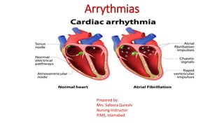
14. Atrial fibrillation (A fib):
• Irregular heart rhythm, with a
total disorganization of atrial
electrical activity due to
multiple ectopic foci resulting
in loss of effective atrial
contraction.
• HR 350 to 600bpm.
• ECG show fibrillatory (fib)
waves.
• May associated with CAD and
RHD, HTN, and cardiac
myopathy.
Fib waves
15. Atrial flutter:
• An atrial tachydysrhythmia,
HR 250 to 350bpm
• Identified by recurring,
regular, sawtooth-shaped
flutter waves (F)waves
• Originate from a single
ectopic focus in the right
atrium.
• May associated with CAD,
HTN, and CLD
16. Sinus Tachycardia
• The conduction pathway is the
same as in normal sinus rhythm.
• May associated Hard exercise,
anxiety, certain drugs, or a fever
• If there is no clear reason,
called inappropriate sinus tachy
• The heart rate might shoot up
with just a little movement as a
result of sympathetic nerve
activation
• HR >100 bpm with regular
rhythm
17. Junctional Dysrhythmias.
• The area of origin is the AV
node, because;
• The SA node has failed to fire or
the signal has been blocked.
• The AV node becomes the
pacemaker of the heart
• HR between 40-140bpm
• Narrow QRS.
• P waves may be hidden,
inverted, retrograde, or
short/upright.
18. Ventricular Arrythmias
Premature ventricular contractions
(PVCs):
• is a contraction originating in an ectopic
focus in the ventricle.
• It is the premature occurrence of QRS
complex, which is wide and distorted in
shape.
• PVCs may sensed as skipped beats.
Because the ventricles are only partially
filled, the PVC frequently does not
generate a pulse.
• Rate: Depends on rate of underlying rhythm
• Rhythm: Irregular whenever a PVC
occurs
• P Waves: None associated with the PVC
• PR Interval: None associated with the
PVC
• QRS: Wide (>0.10 sec), bizarre
appearance
19. Ventricular tachycardia (VT):
• It occurs when an ectopic focus or
foci fire repetitively and the
ventricle takes control as the
pacemaker.
• Rate: 100–250 bpm,
• Rhythm: Regular
• P Waves: None or not associated
with the QRS
• PR Interval: None
• QRS: Wide (>0.10 sec), bizarre
appearance
Monomorphic
VT
Polymorphic VT
20. Ventricular fibrillation (V-fib):
• is characterized by disorganized
firing of impulses from the
ventricles.
• Chaotic electrical activity occurs with no
ventricular depolarization
• Mechanically the ventricle is
quivering and no effective
contraction and consequently NO
CO
• Represents the firing of multiple
ectopic foci in ventricle.
• This is a medical emergency that
must be treated with CPR and
defibrillation .
Ventricular fibrillation
21. Bradyarrhythmia
• Sinus node dysfunction: Slow heart
rhythms due to an abnormal SA node
Sinus bradycardia:
• Has a normal sinus rhythm, but the SA node
fires at a rate less than 60 beats/min & is
referred to as absolute bradycardia.
Heart block:
• Heart block is a type of heart rhythm
disorder (arrhythmia)
• A delay or complete block of the
electrical impulse as it travels from
the sinus node to the ventricles.
• The level of the block or delay may
occur in
• the AV node or HIS-Purkinje system.
• The heartbeat may be irregular
Rate: Slow (<60 bpm)
Rhythm: Regular
P Waves: Normal (upright and
uniform)
PR Interval: Normal (0.12–0.20 sec)
QRS: Normal (0.06–0.10 sec)
Sinus Bradycardia
22. Types / Degree of Heart Block
First-degree (AV ) block.
• This is the least severe type
• AV) block is a condition of
abnormally slow conduction
through the AV node.
• ECG changes include a
• PR interval of greater than
0.20sec without disruption of
atrial to ventricular conduction.
• This condition is asymptomatic
and discovered only on routine
ECG.
• There is no treatment for first-
degree AV block.
23. Second-degree AV block.
• The electrical signals between the atria and
ventricles can intermittently fail to conduct
Types of AV Block:
Mobitz type I
• The electrical signals get slower and slower
between beats.
• Over time the heart drops a beat and then
the process repeats.
Mobitz type II
• A certain number of impulses from the SA
node are not conducted to the ventricles.
• No progressive slowing of the electrical
signal.
• AV block can often progress to third-degree
heart block.
P wave
Missing QRS
24. • Third-degree Block (complete
heart block).
• Occur when there is no connection
or dissociation of electrical activity
from the atria and ventricles
• This is the most severe type, with
complete failure of electrical
conduction.
• The electrical signal from the atria to
the ventricles is completely blocked
• There is a This can result in no
pulse or a very slow pulse if a
backup heart rate is present.
25. Medical Management
Antidysrhythmic Drugs:
• Class I:Sodium channels blockers (decrease conduction velocity ).
Procainamide, lidocaine, Propafenone
• Class II: Beta- blockers (decrease automaticity in SA node, decrease
conduction velocity in AV node). Atenolol, metoprolol
• Class III: Potass ium channels blockers:(Delay repolarization).
Amiodarone, Bretylium
• Class IV: Calcium blockers (Decrease automaticity of SA node, delay
AV node conduction).Verapamil, diltiazem
• Other Antidysrhythmic Drugs. Adenosine, Digoxin,
26. Therapeutic techniques
Vagal maneuvers:
• Certain dysrhythmias, like SVT
supraventricular tachycardia, can be
stopped by using maneuvers that include
holding your breath and straining, dunking
your face in ice water or coughing. These
actions affect the vagus nerves that control
your hear
Medication:
• Adenosine (Adenocard)
Adenosine is the first-line medical
treatment for the termination of paroxysmal
SVT.
27. Defibrillation
Cardioversion / Defibrillation
• For certain dysrhythmias – like
atrial fibrillation – patient may
treated with this procedure, during
which a electric shock is
delivered to your heart through
paddles or patches on your chest.
The current affects the electrical
impulses and can restore a normal
rhythm.
28. Invasive Procedures
Catheter Ablation;
• is a minimally invasive treatment for
fast heartbeats. Catheter ablation is one
type of heart ablation procedure used to
treat abnormal heart rhythms
(arrhythmias). Ablation is a technique
used to strategically destroy abnormal
tissue and restore proper function to the
heart.
• The procedure uses hot or cold energy
to create scars in your heart tissue
where the arrhythmia is occurring. The
scars help block abnormal electrical
impulses and prevent abnormal
rhythms.
29. Device implantation:
pacemaker or
implantable cardioverter-
defibrillator (ICD).
A pacemaker is a small
device placed under the
skin near the collarbone
in a minor surgical
procedure. A wire extends
from the device to the
heart. If a pacemaker
detects an abnormal heart
rate, it emits electrical
impulses that stimulate
the heart to beat at a
normal rate.
30. • Maze procedure: During this
procedure, a surgeon makes a series
of incisions in the upper half of the
heart (atria) to create a pattern (or
maze) of scar tissue to interfere
with stray electrical impulses that
cause some types of dysrhythmia.
The procedure is very effective, but
it is usually reserved for people
who don’t respond to other
treatments or for those who are
having heart surgery for other
reasons.
31. surgery
• Coronary artery bypass
grafting (CABG): This
surgery improves blood
flow to your heart by
creating a bypass around
your narrowed coronary
arteries using arteries or
veins taken from other
parts of your body
33. Pacemaker
• A pacemaker is a small device that's placed (implanted) in the
chest to help control the heartbeat.
• It's used to prevent the heart from beating too slowly.
Implanting a pacemaker in the chest requires a surgical
procedure.
• A pacemaker is also called a cardiac pacing device
Causes:
• Arrhythmias
• Heart blocks
• Heart failure.
• Heart attack
34. Parts of Pacemaker
• Pulse generator. This small metal
container houses a battery and the
electrical circuitry that controls the
rate of electrical pulses sent to the
heart.
• Leads (electrodes). One to three
flexible, insulated wires are each
placed in one or more chambers of
the heart and deliver the electrical
pulses to adjust the heart rate.
However, some newer pacemakers
don't require leads. These devices,
called leadless pacemakers, are
implanted directly into the heart
muscle.
35. Types
• Single chamber pacemaker. This type usually carries
electrical impulses to the right ventricle of heart.
• Dual chamber pacemaker. This type carries electrical
impulses to the right ventricle and the right atrium of your
heart to help control the timing of contractions between the two
chambers.
• Biventricular pacemaker. Biventricular pacing, also
called cardiac resynchronization therapy, is for people who
have heart failure and heartbeat problems. This type of
pacemaker stimulates both of the lower heart chambers (the
right and left ventricles) to make the heart beat more efficiently.
36. Risk factors
• Infection near the site in the heart where the device is implanted
• Swelling, bruising or bleeding at the pacemaker site, especially
if you take blood thinners
• Thromboembolism near the pacemaker site
• Damage to blood vessels or nerves near the pacemaker
• Pneumothorax
• Hemothorax
• Movement (shifting) of the device or leads, which could lead to
cardiac perforation (rare)
37. Monitoring of Patient after procedure
• Continue ECG monitoring to evaluate the pacemaker status such as
• Failure to sense ( pacer lead damage)
• Failure to capture (battery failure)
• Check vital signs
• Observe complications such as;
• infection, hematoma formation, pneumothorax
• Perforation
• Prophylactic antibiotics
• Postinsertion x-rays to check lead placement
• Limited arm and shoulder movement to prevent dislodgment of
pacing lead
38. Home Care Teaching
• Follow-up Care to insertion site
• Report sign of infection
• Keep incision side dry and clean and check for infection
• Avoid lifting arm on pacemaker side above the shoulder
• Avoid vigorous exercises for a period of time
• Avoid direct blows to pacemaker side
• Avoid close proximity to high-voltage electric generators, large
magnetic such as MRI
• Microwaves and cell phones are save but keep your cell 6inch away
• Security alert system may interfere with pacemaker function
• Wear Medic alert ID or pacemaker information card all the time