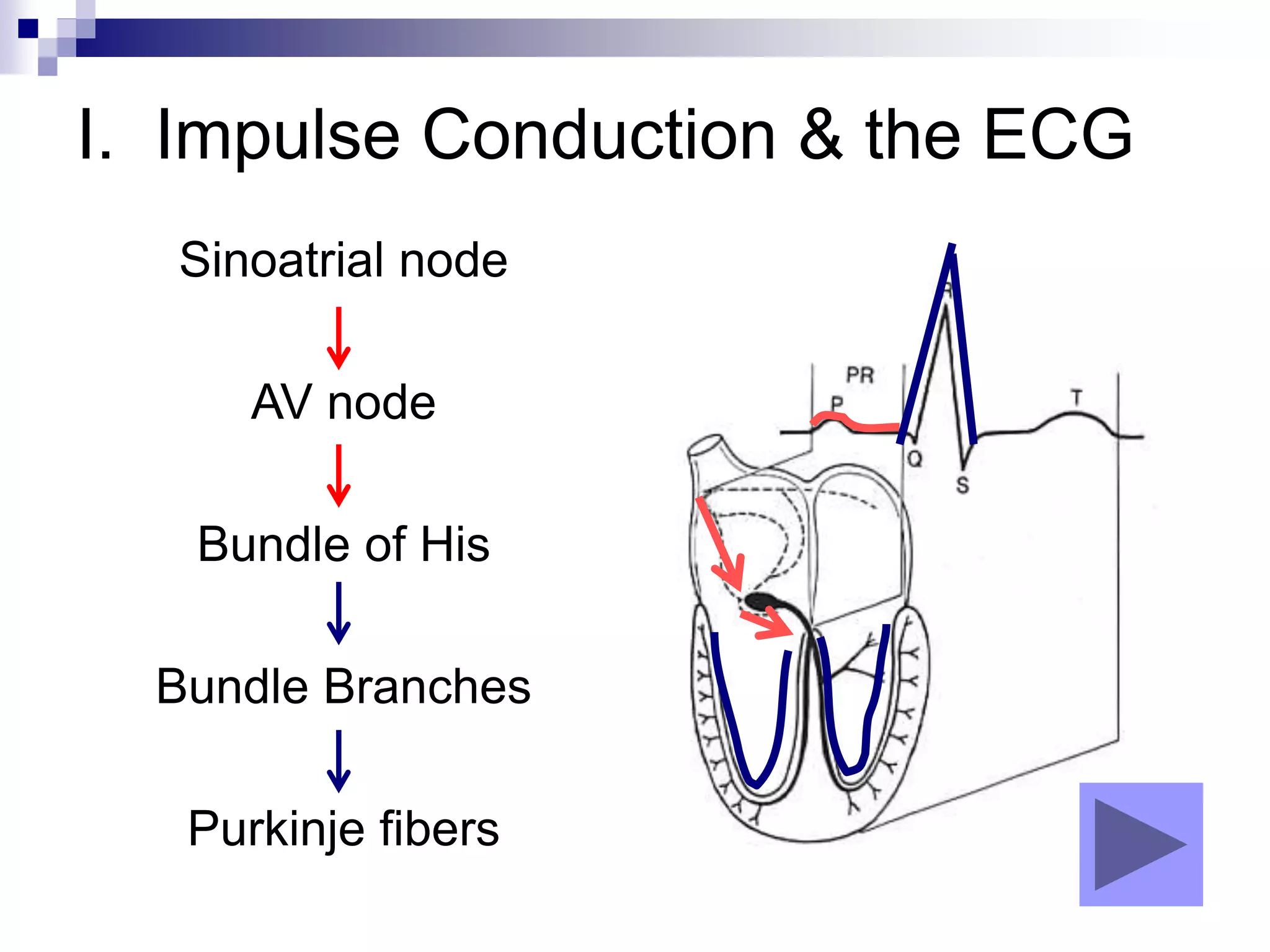
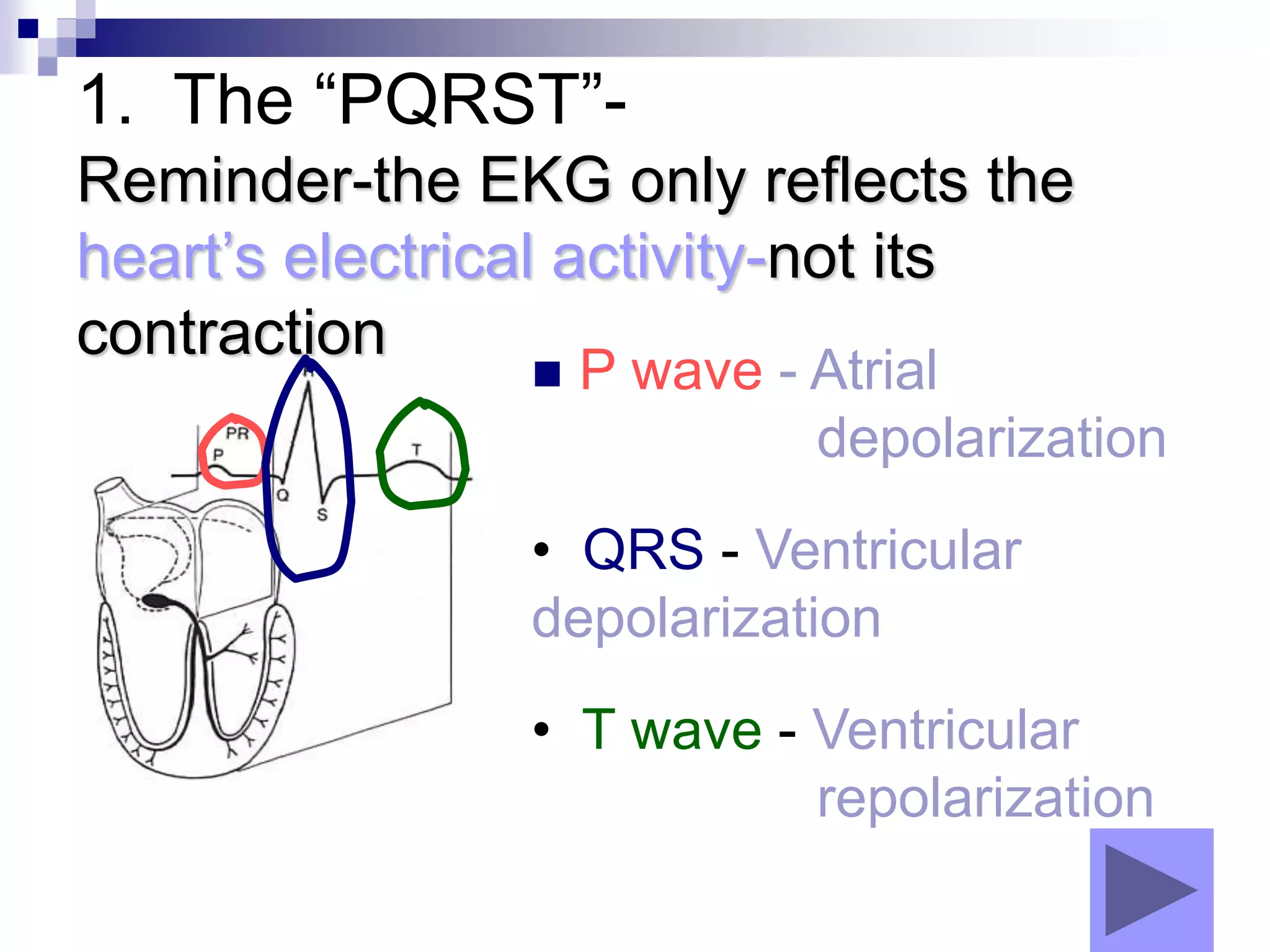
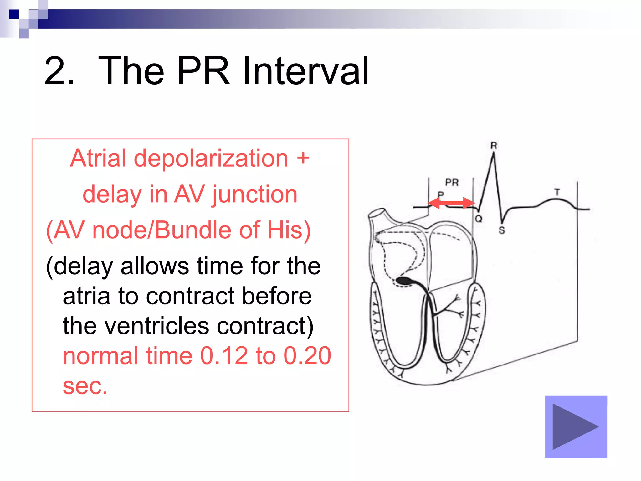
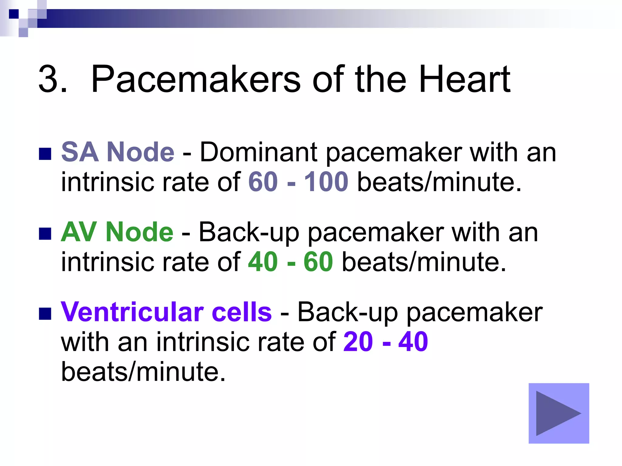
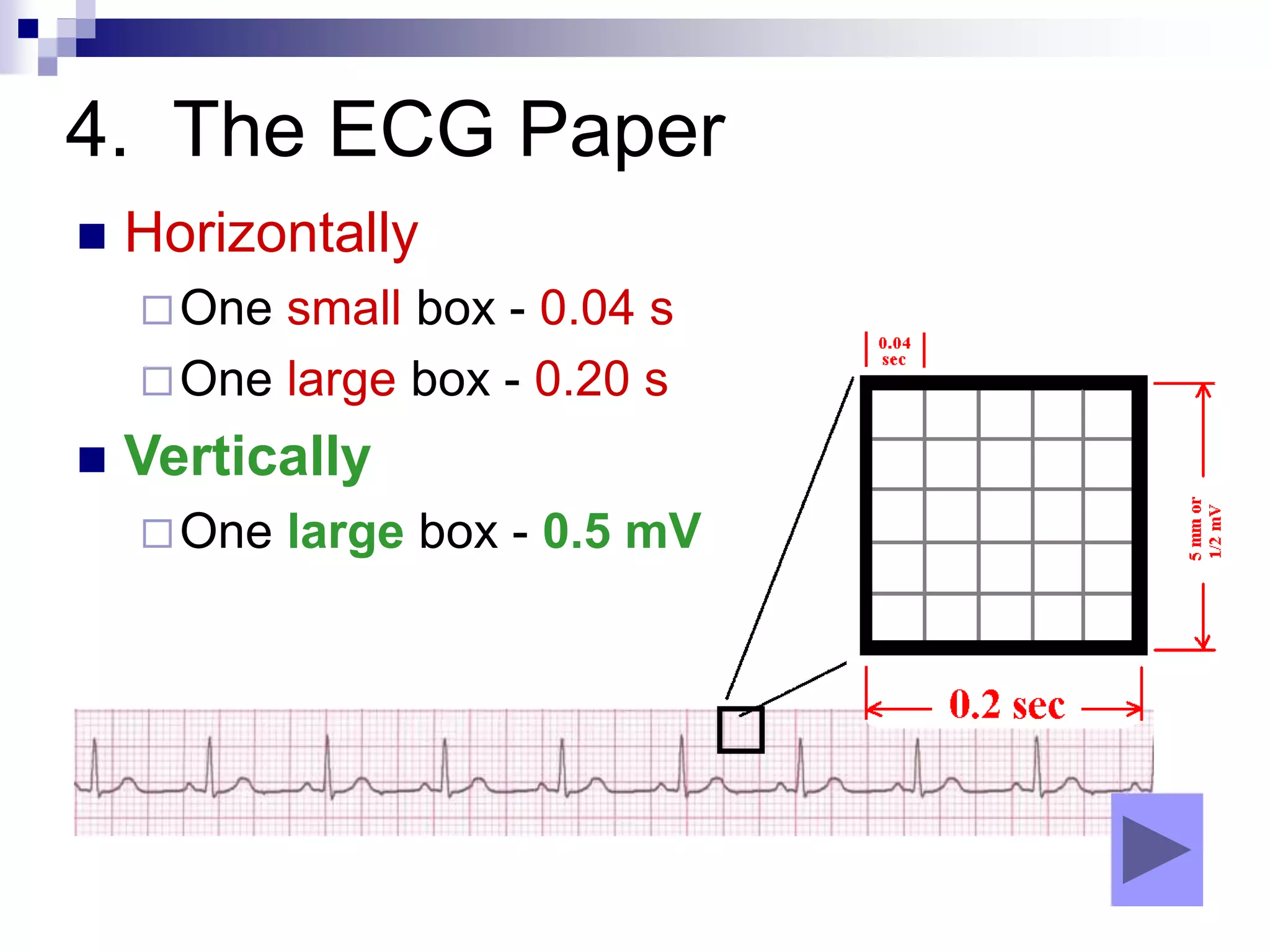
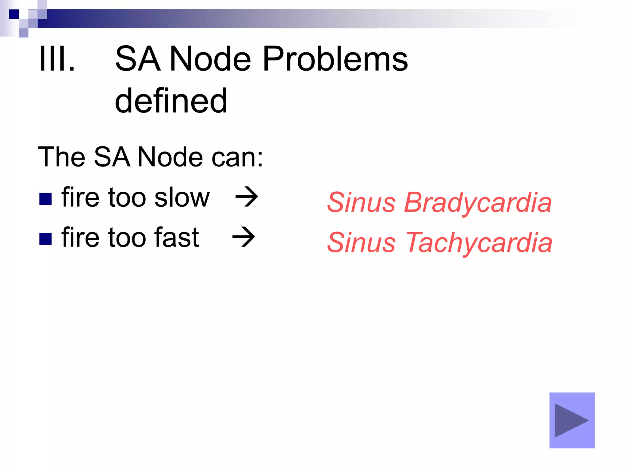
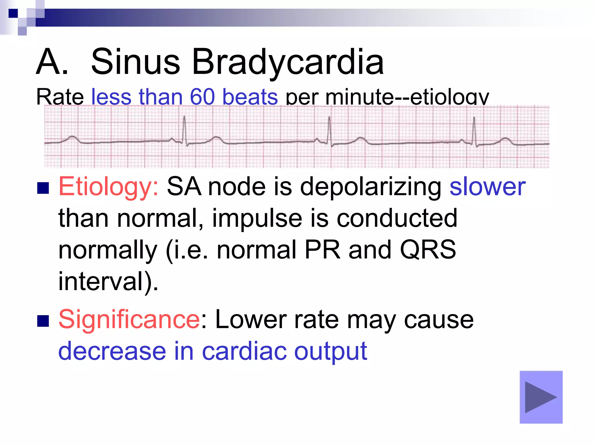
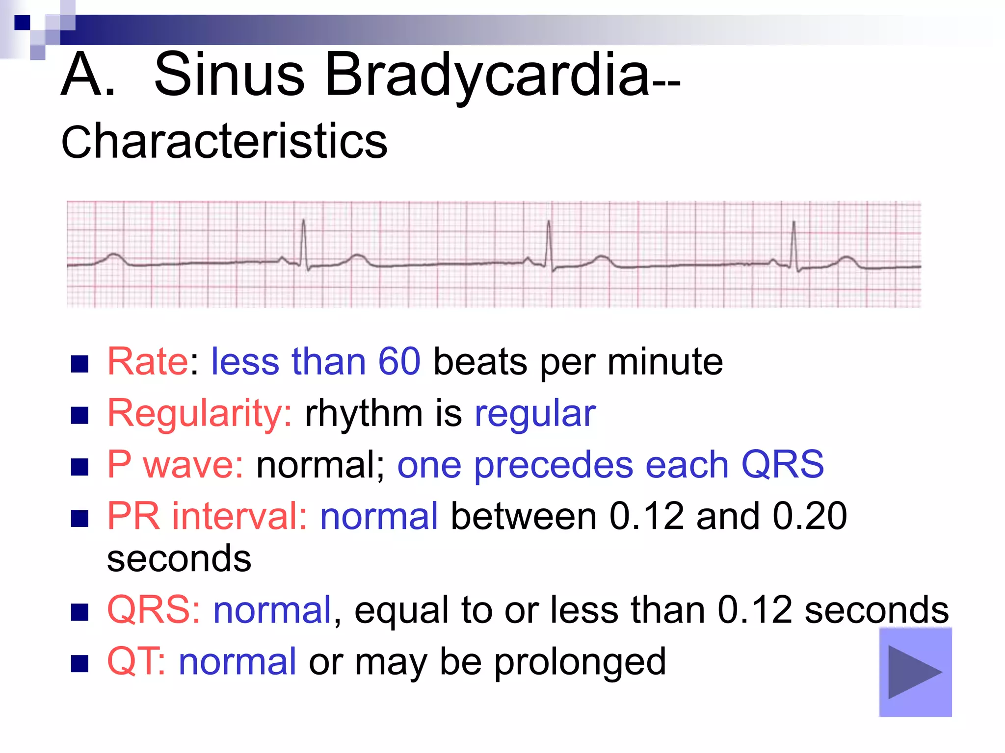
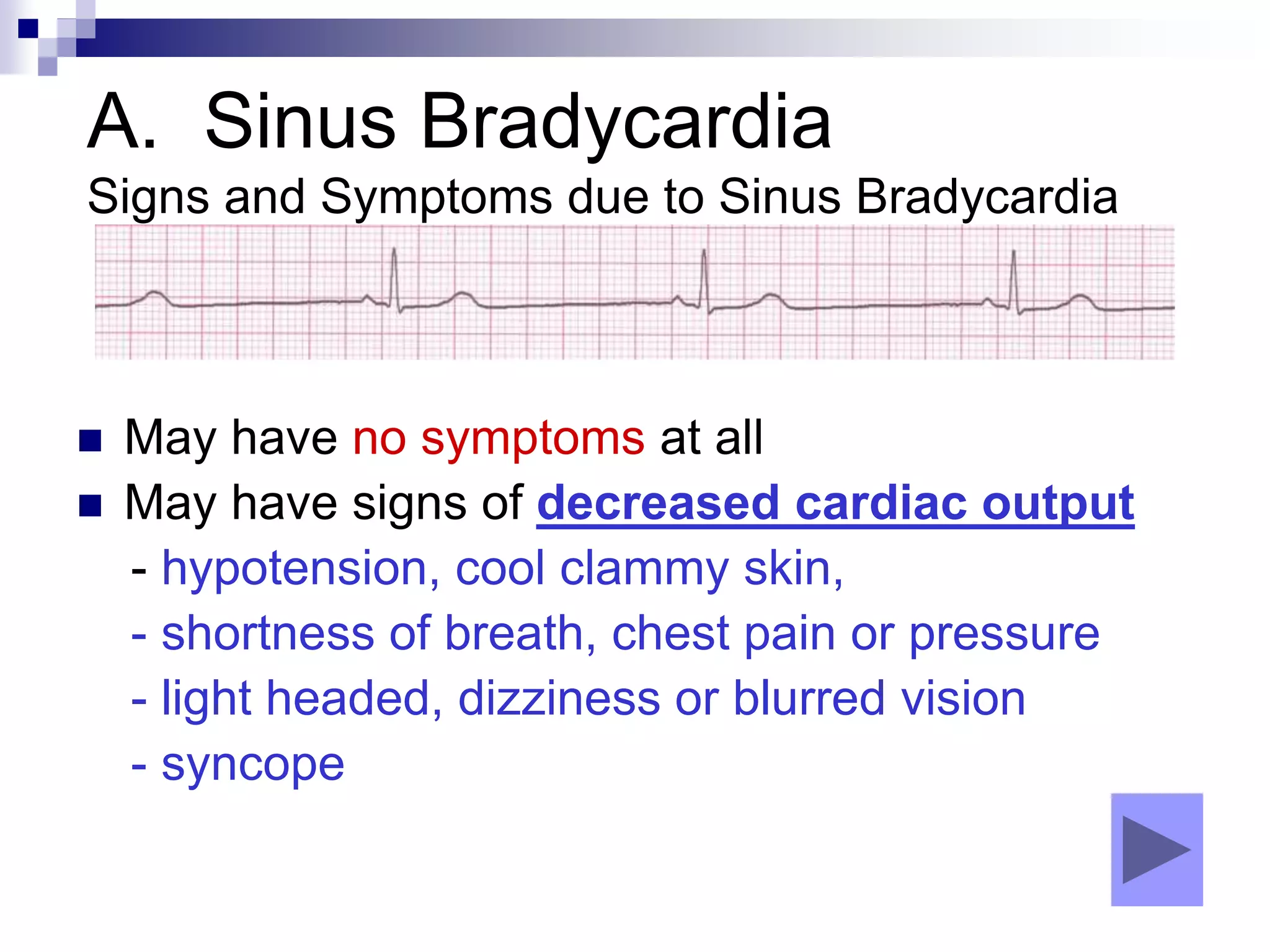
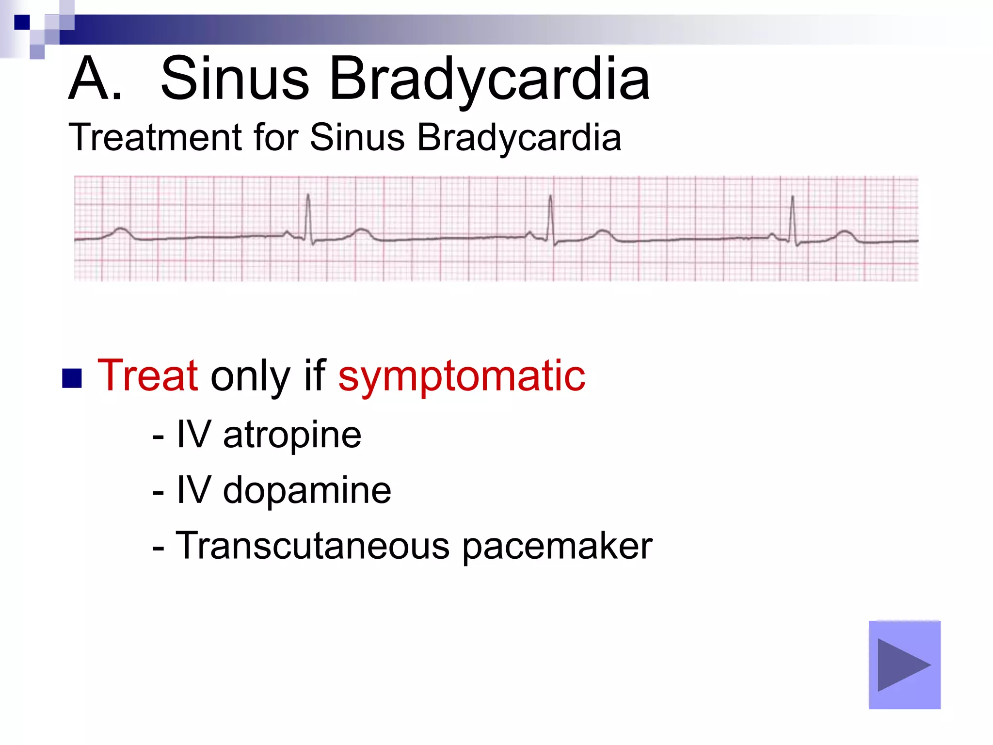
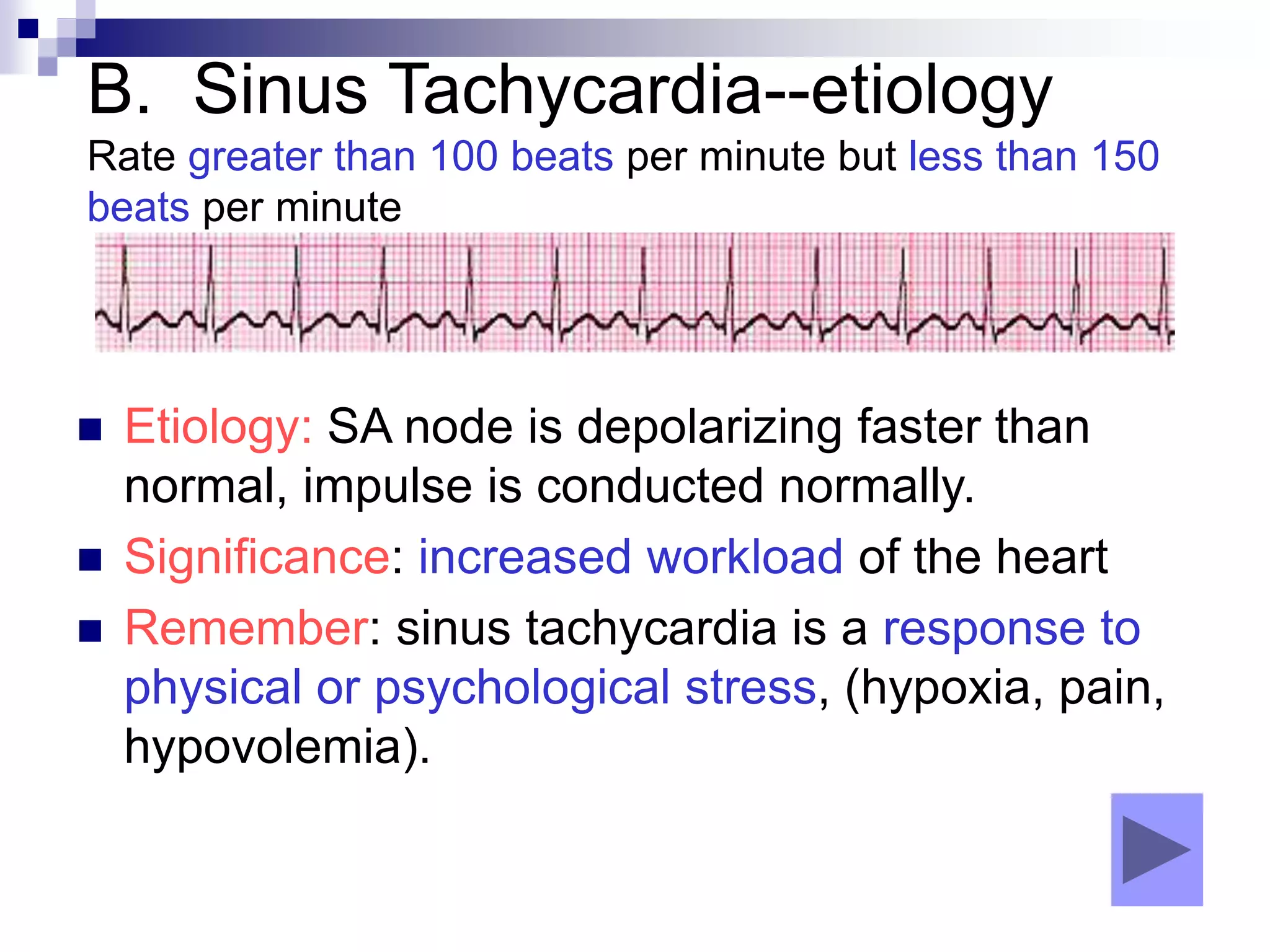
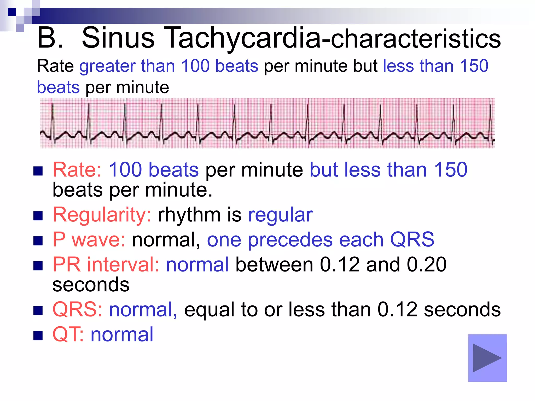
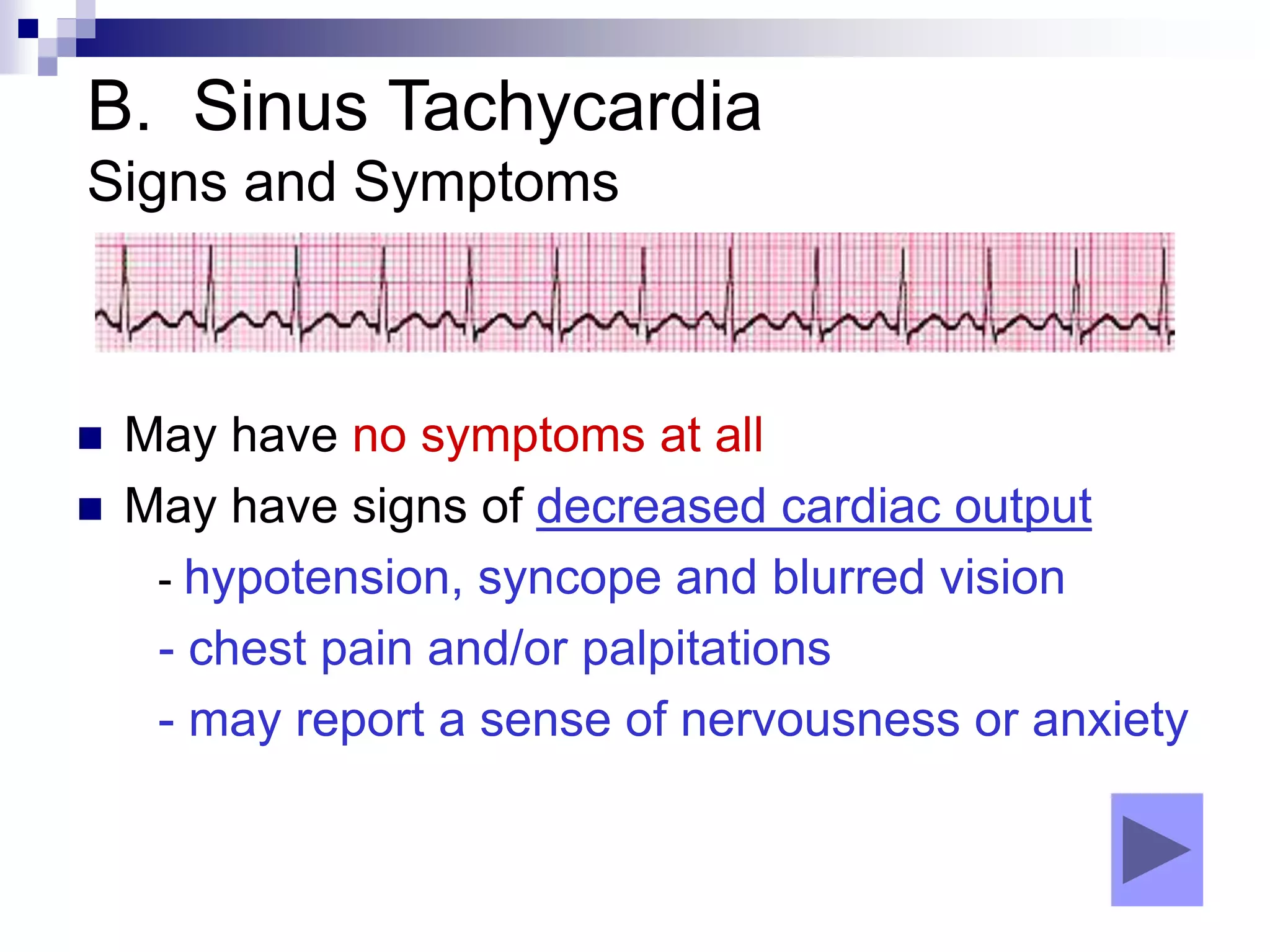
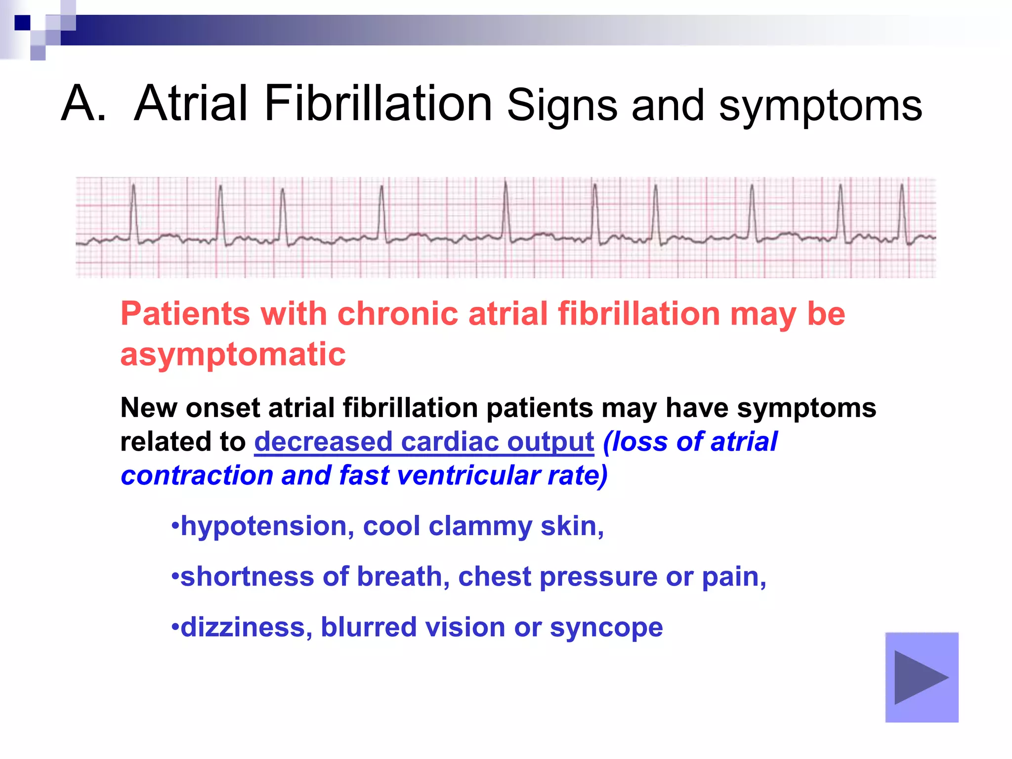
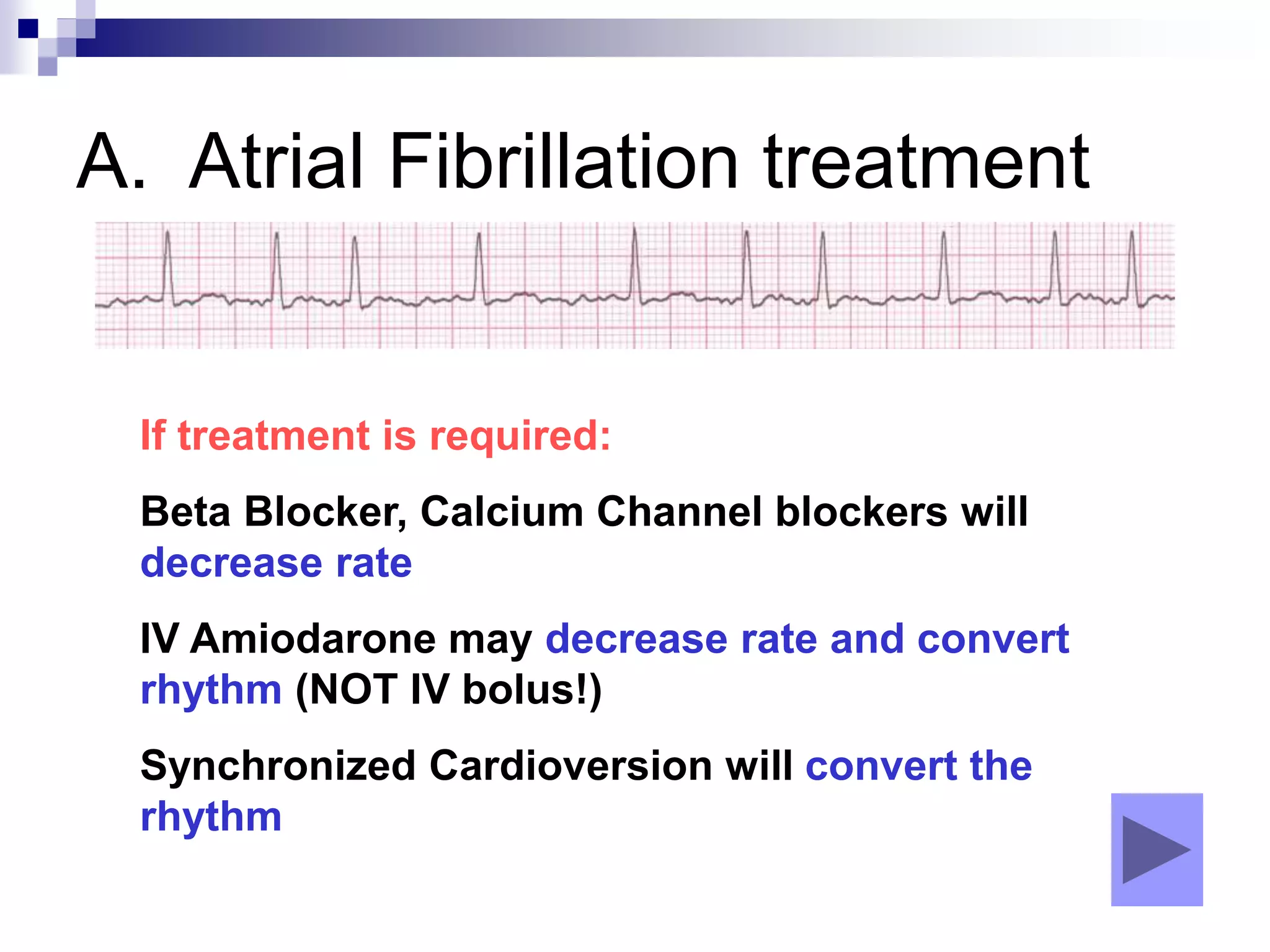
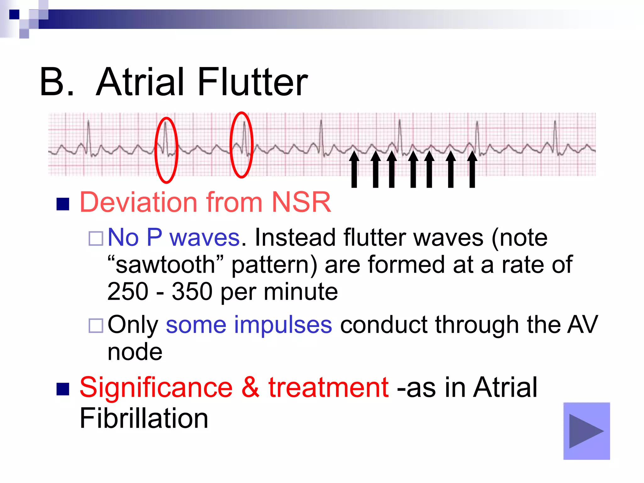
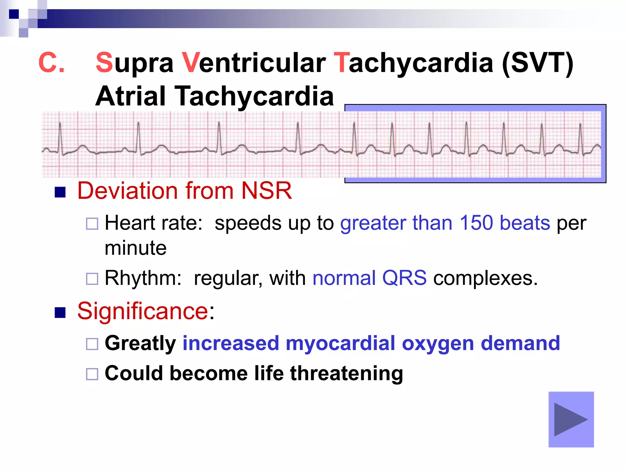
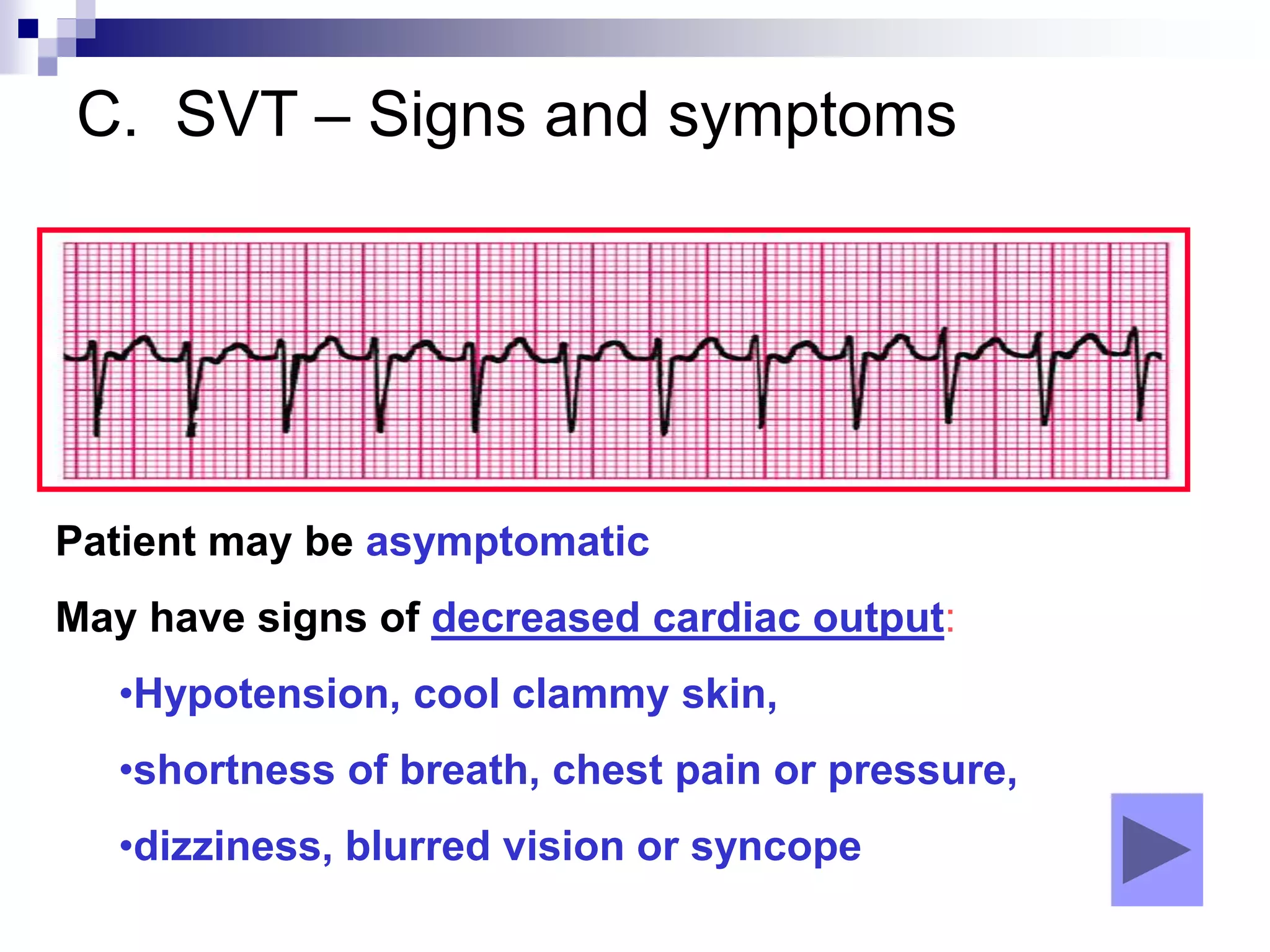
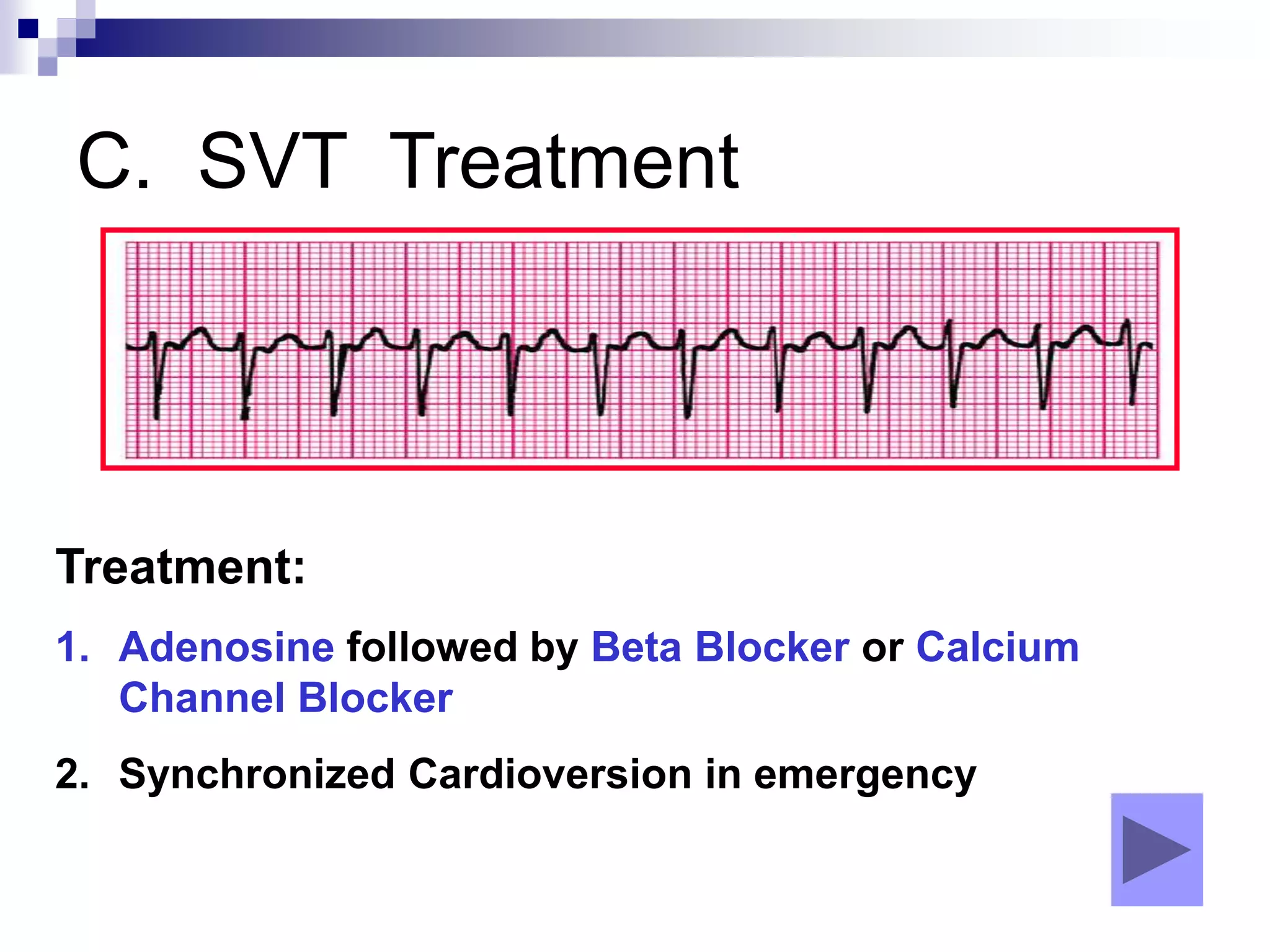
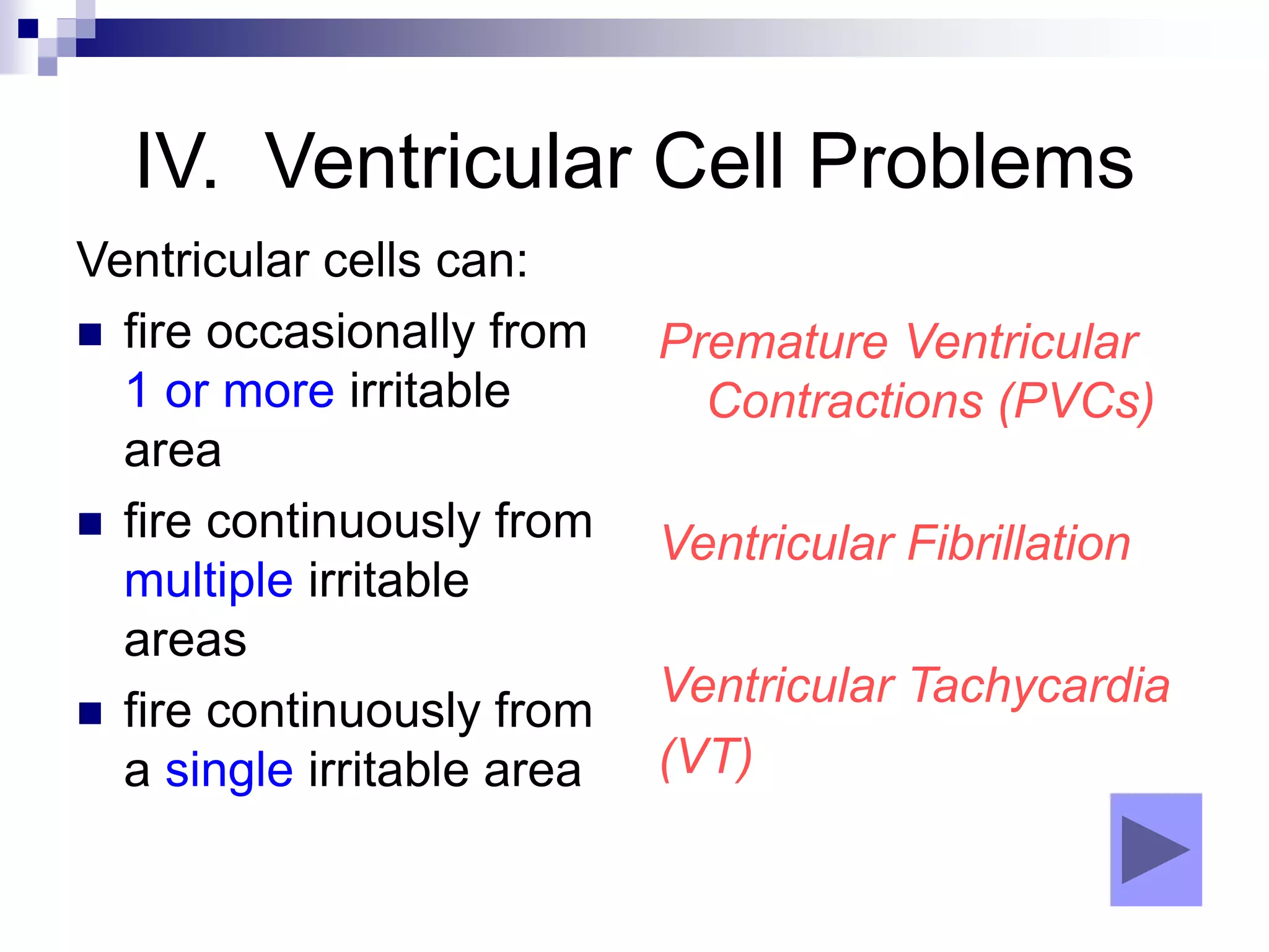
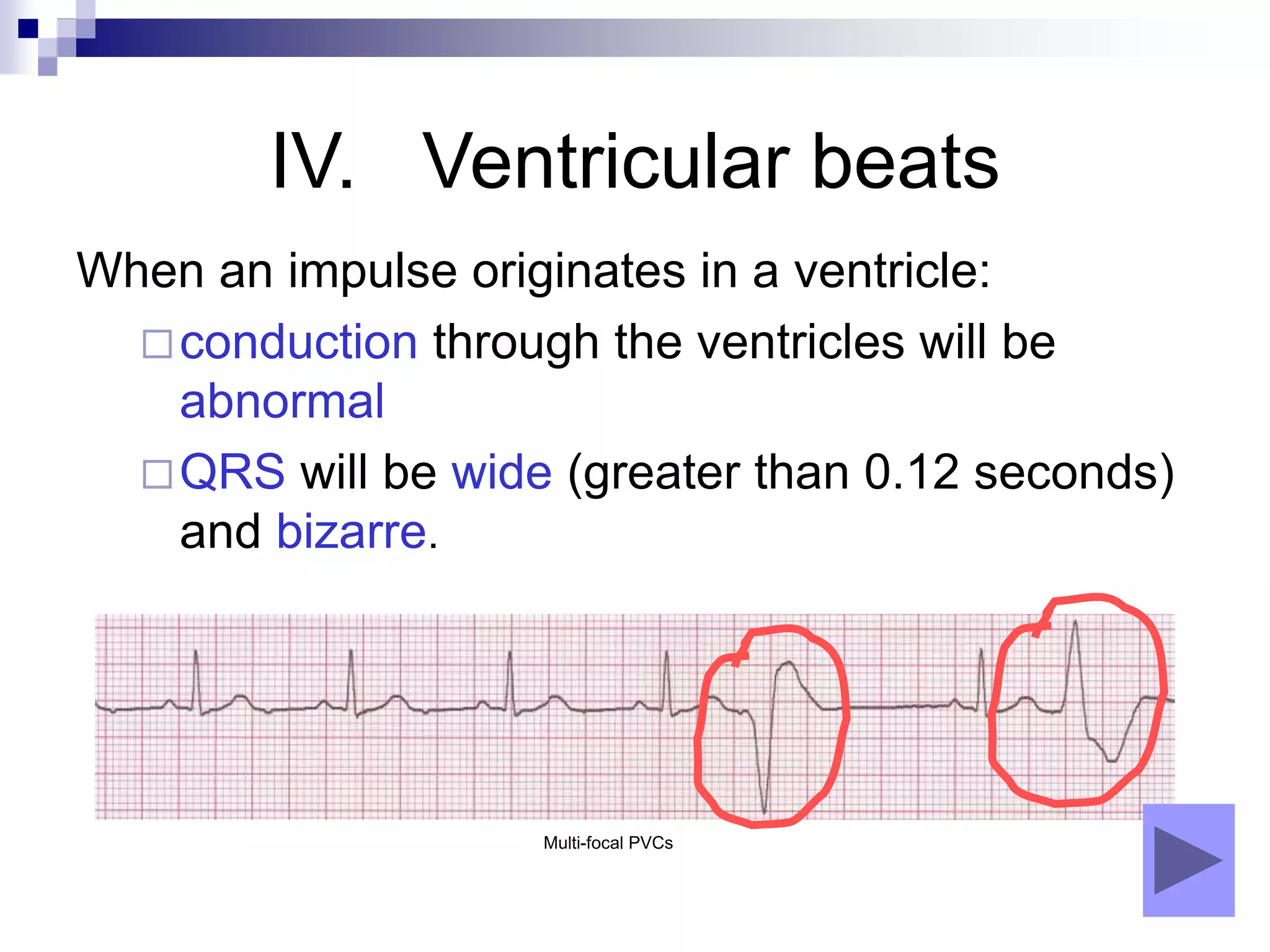
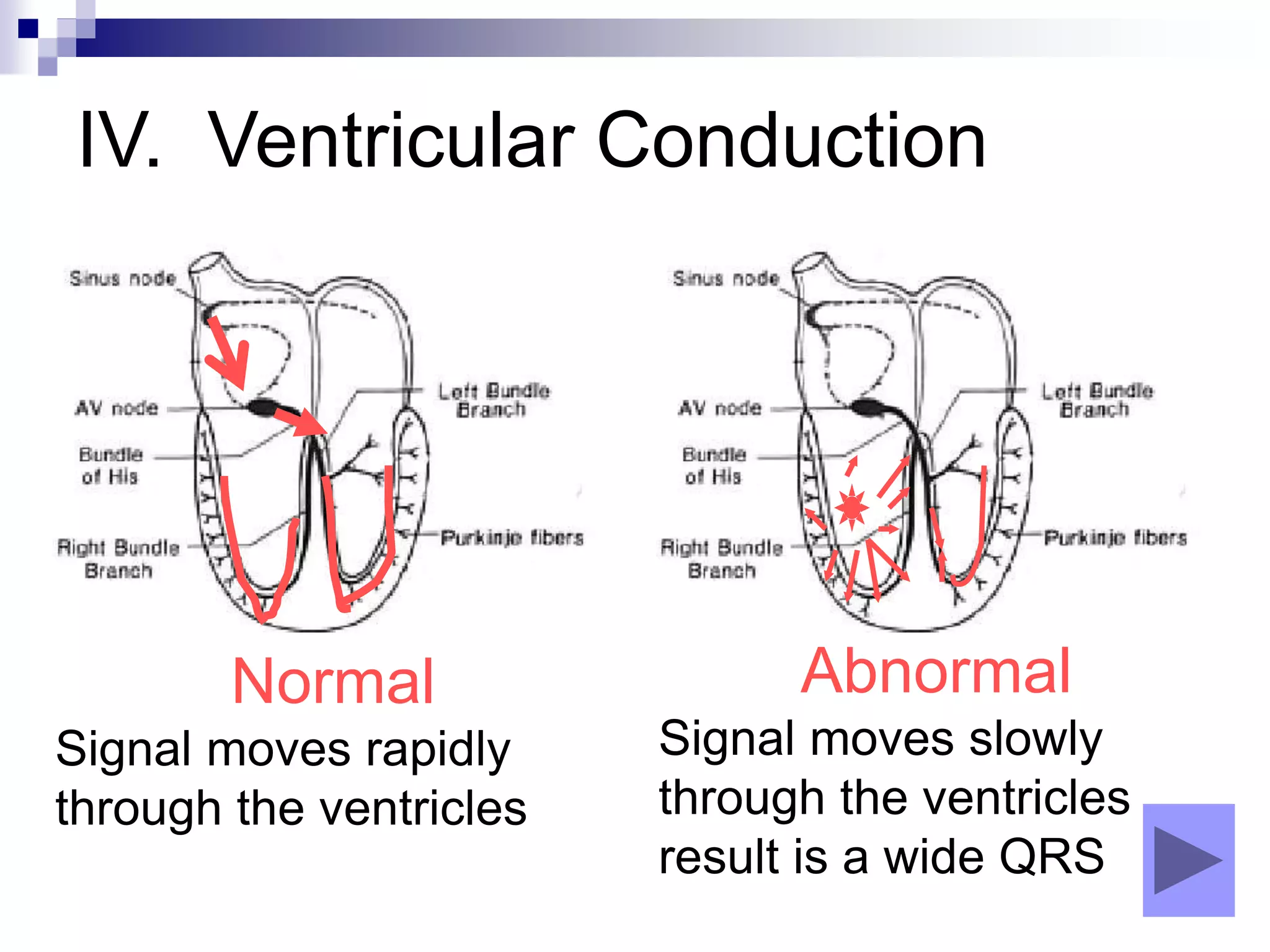
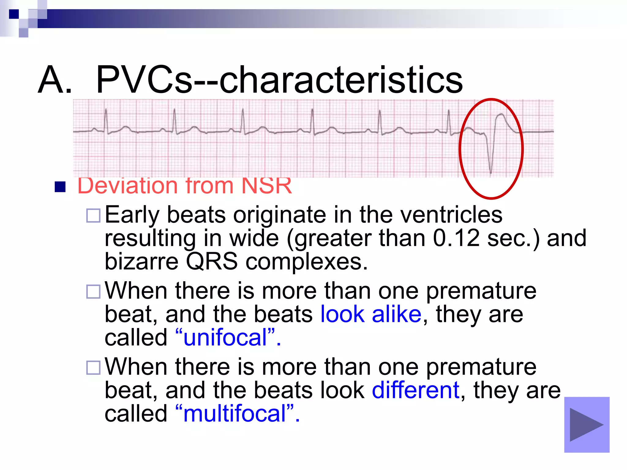
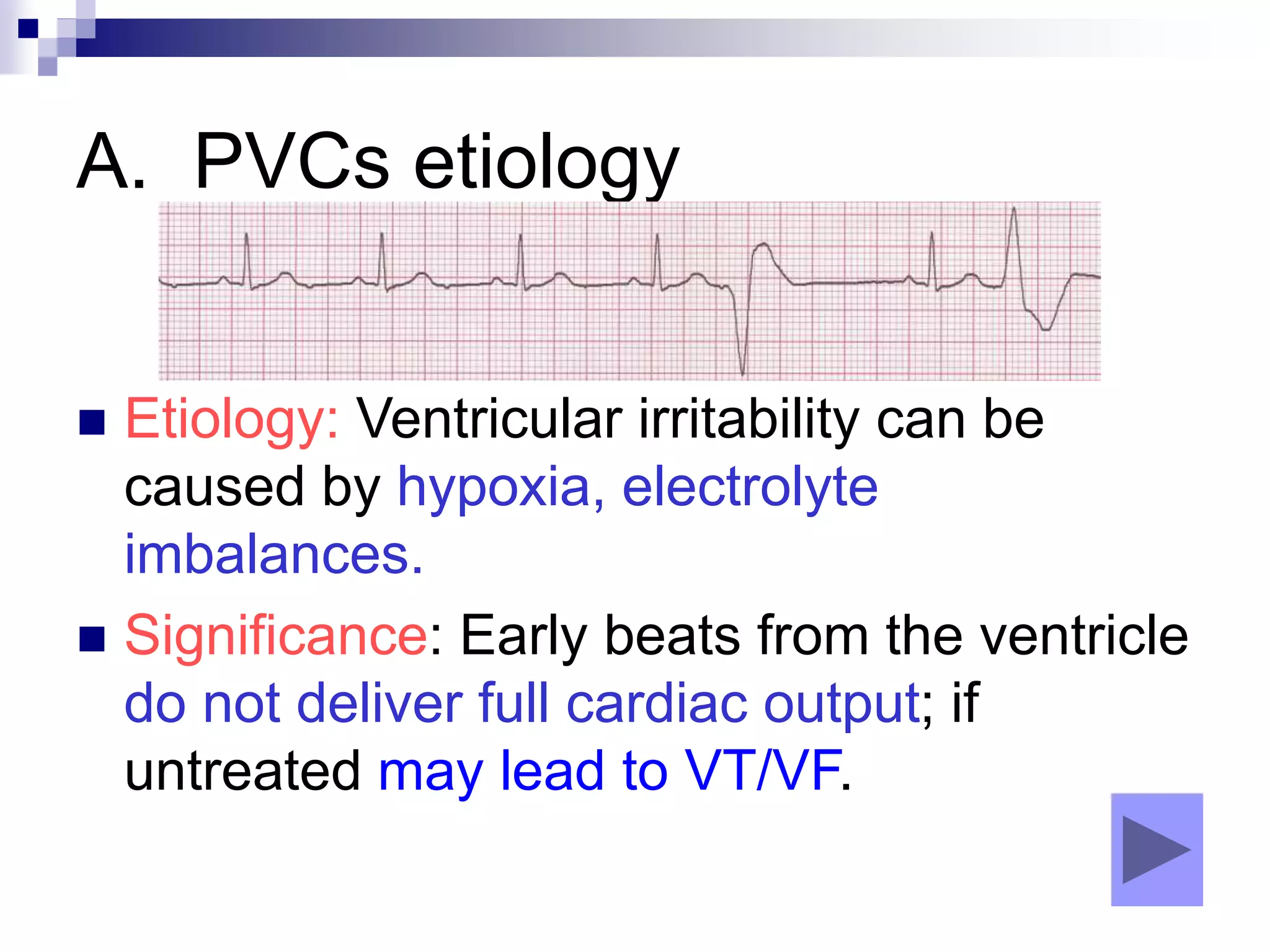
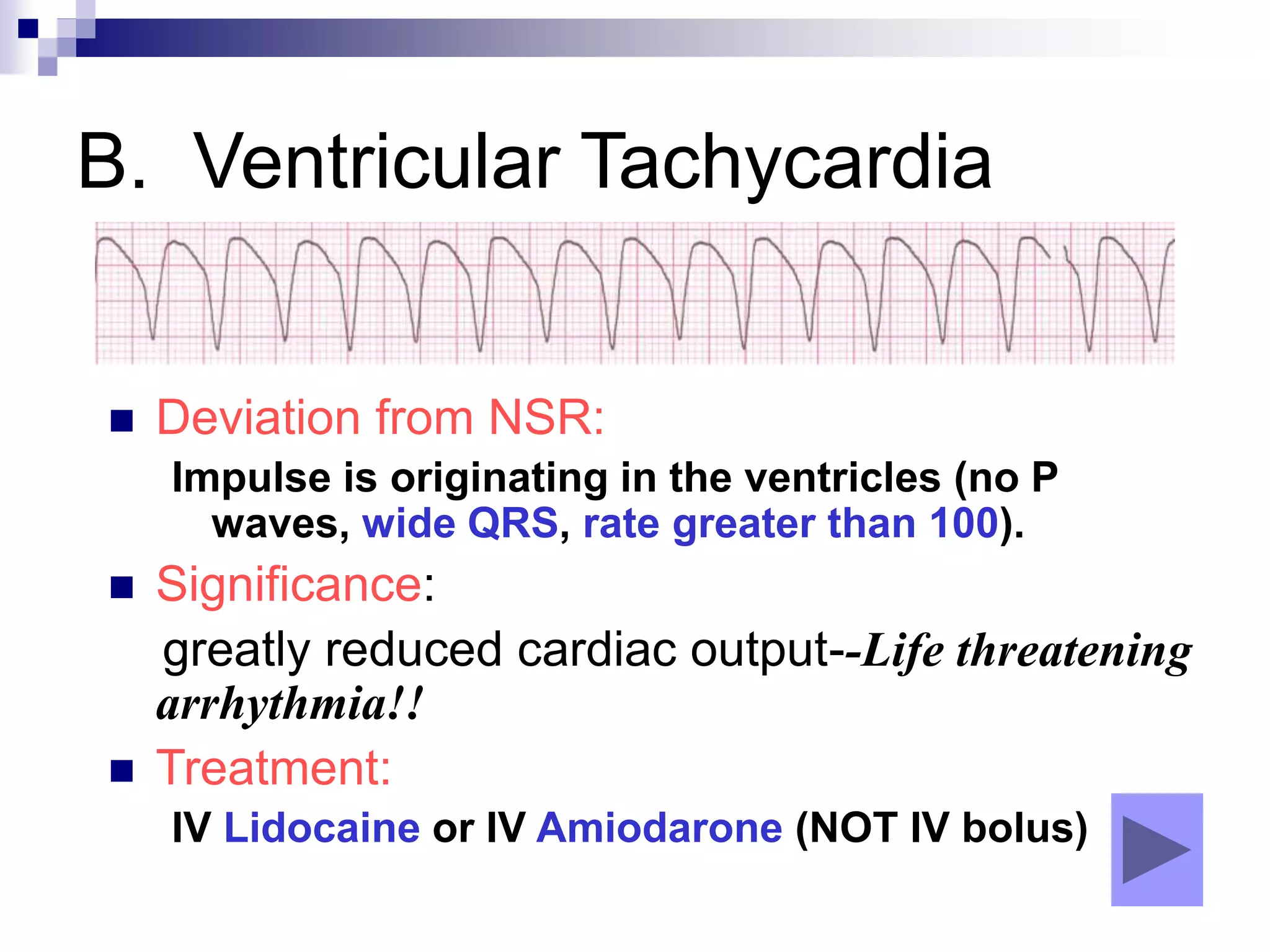
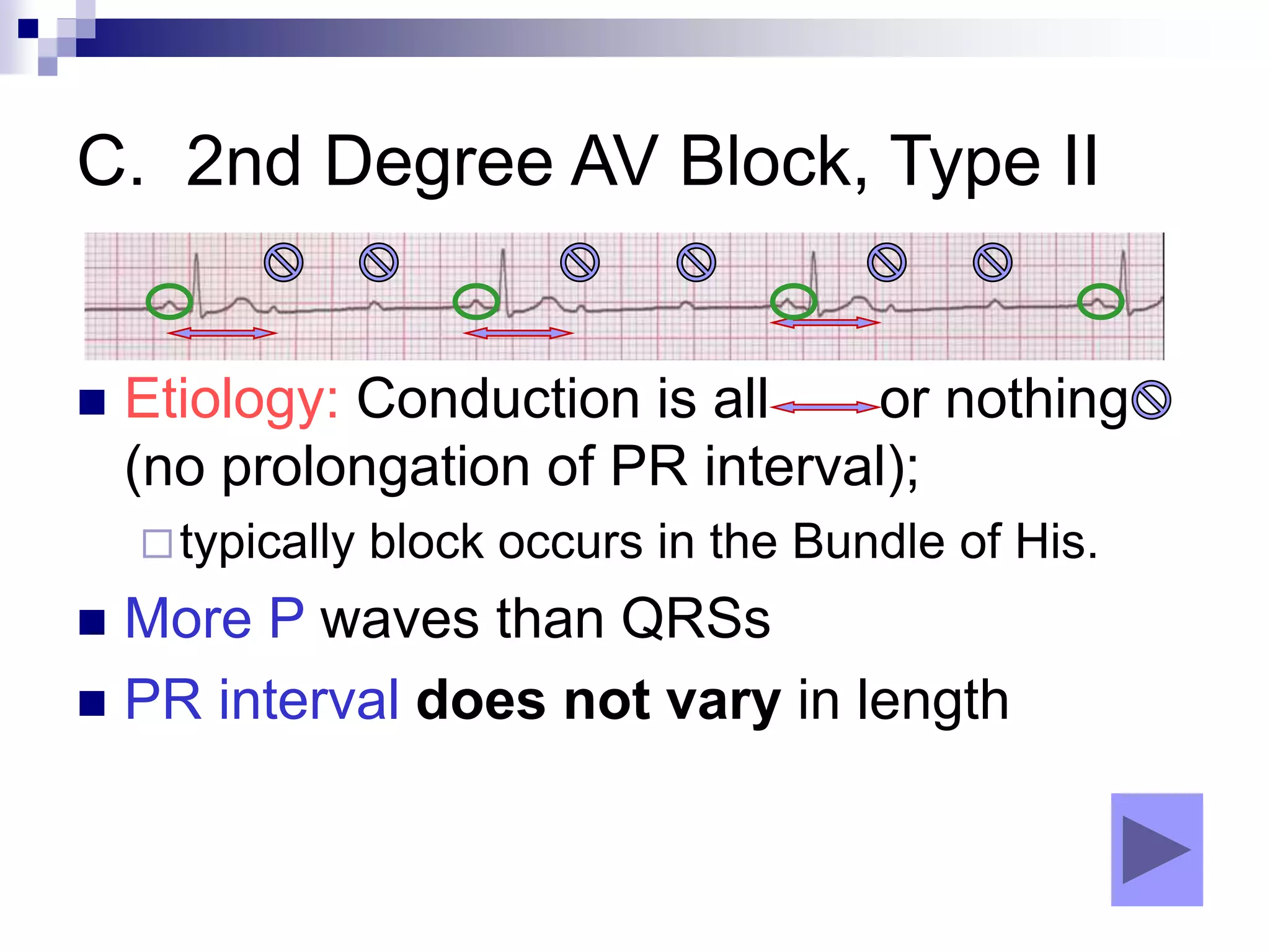
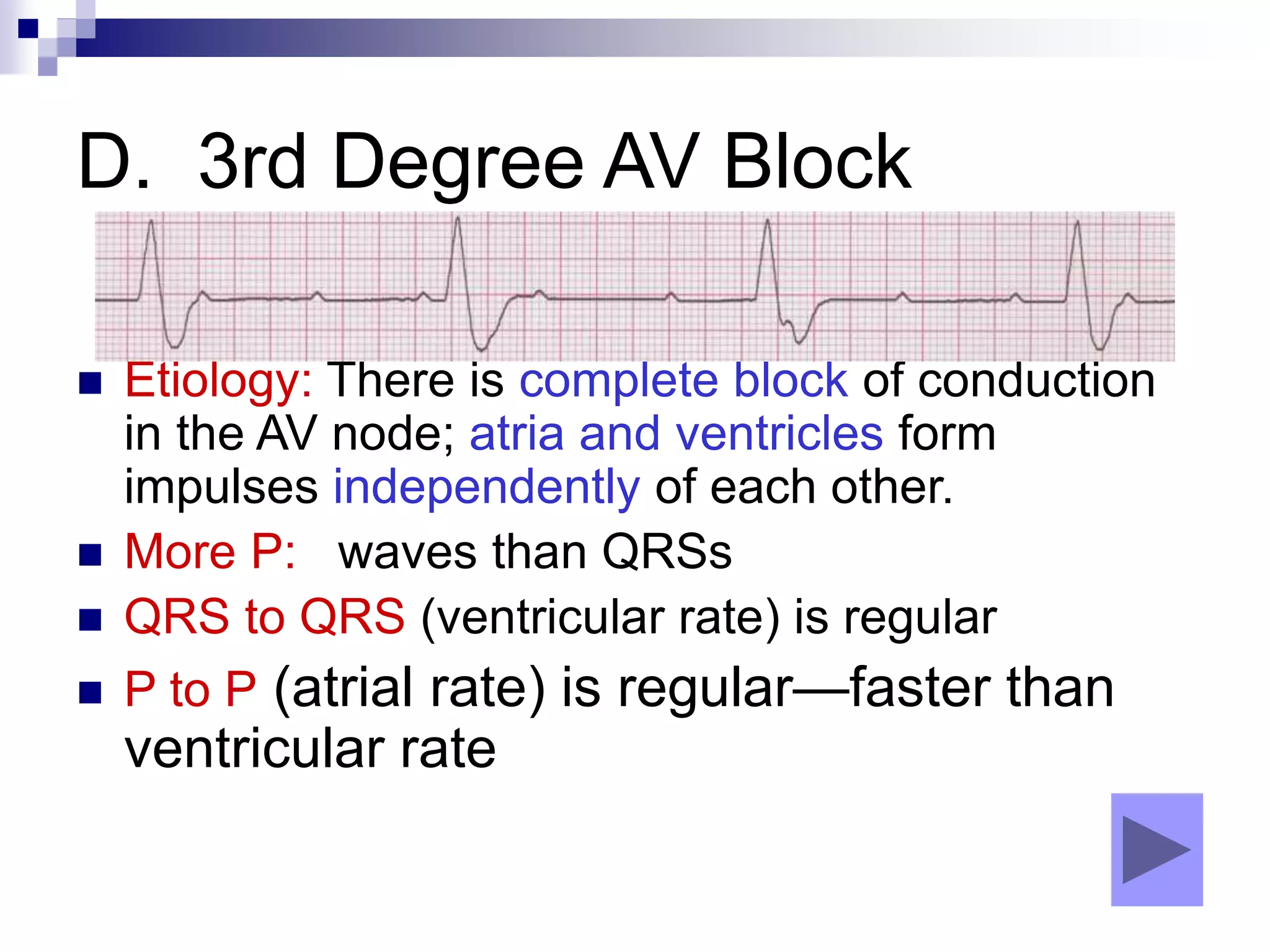
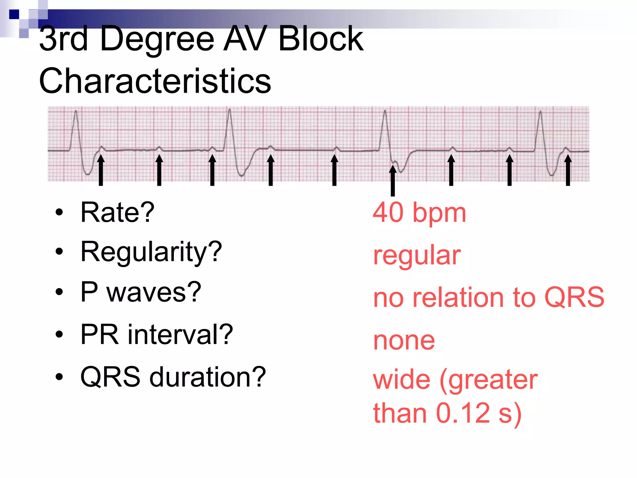
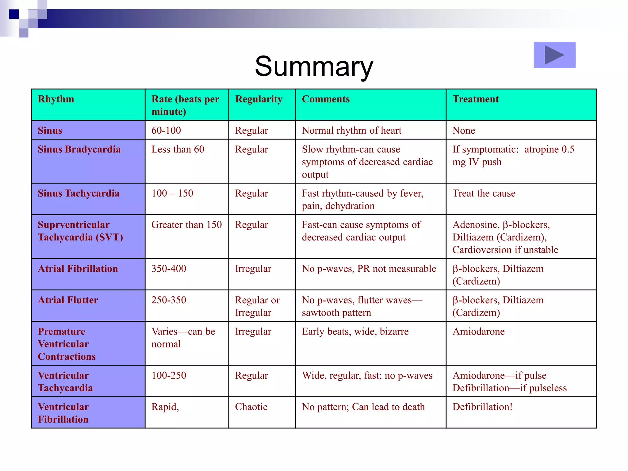
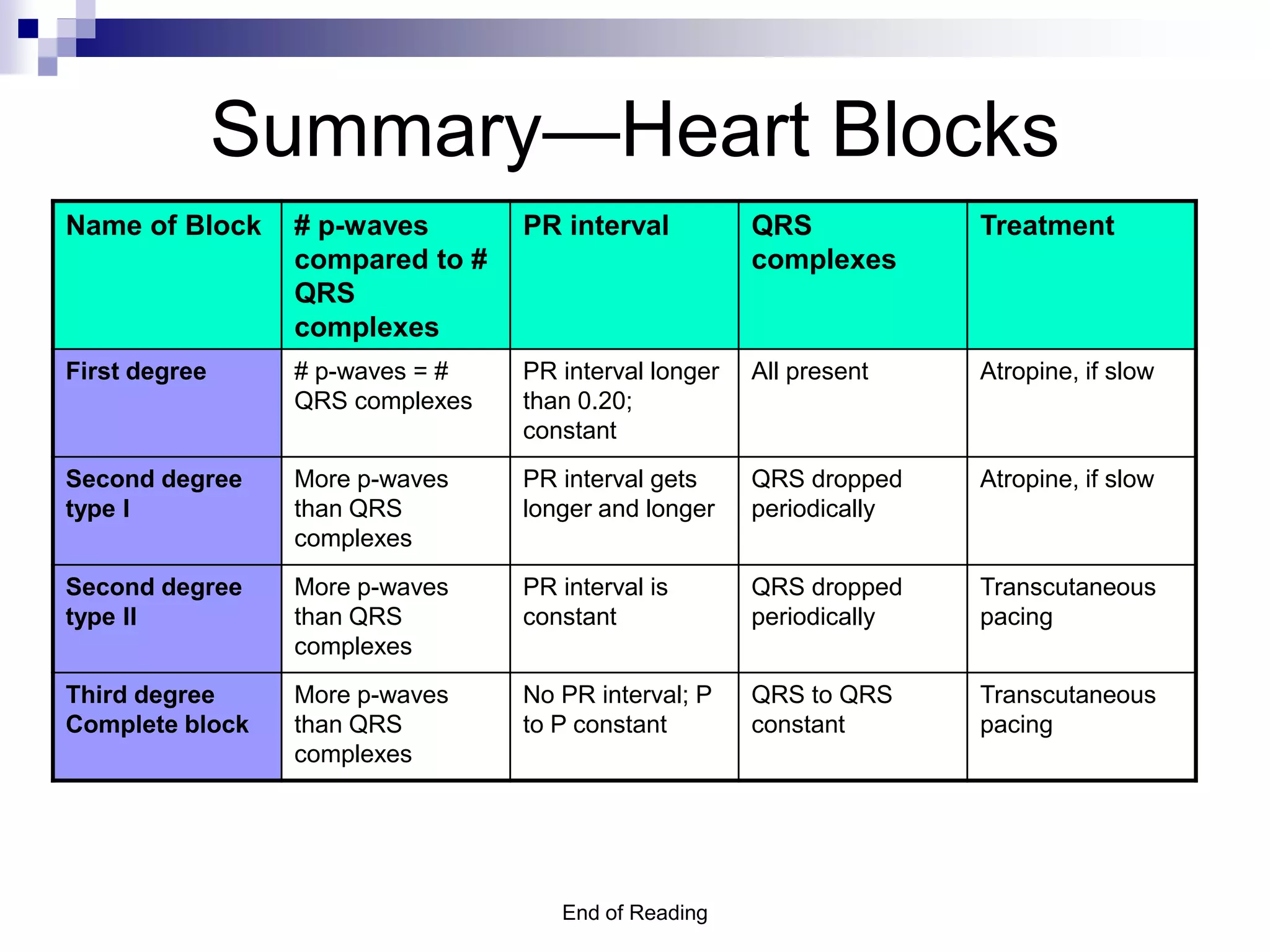
Basic Overview
ECG Rhythm
Interpretation
Objectives
 To recognize the normal rhythm of the
heart - “Normal Sinus Rhythm.”
 To recognize the most common rhythm
disturbances.
 To identify emergency interventions for life
threatening arrhythmias
I. Impulse Conduction & the ECG
Sinoatrial node
AV node
Bundle of His
Bundle Branches
Purkinje fibers
1. The “PQRST”-
Reminder-the EKG only reflects the
heart’s electrical activity-not its
contraction
 P wave - Atrial
depolarization
• T wave - Ventricular
repolarization
• QRS - Ventricular
depolarization
2. The PR Interval
Atrial depolarization +
delay in AV junction
(AV node/Bundle of His)
(delay allows time for the
atria to contract before
the ventricles contract)
normal time 0.12 to 0.20
sec.
3. Pacemakers of the Heart
 SA Node - Dominant pacemaker with an
intrinsic rate of 60 - 100 beats/minute.
 AV Node - Back-up pacemaker with an
intrinsic rate of 40 - 60 beats/minute.
 Ventricular cells - Back-up pacemaker
with an intrinsic rate of 20 - 40
beats/minute.
4. The ECG Paper
 Horizontally
One small box - 0.04 s
One large box - 0.20 s
 Vertically
One large box - 0.5 mV
4. The ECG Paper (cont)
 Every 3 seconds (15 large boxes) is
marked by a vertical line (hash mark).
 This helps when calculating the heart rate.
 You need a 6 second strip to calculate
heart rate (30 large boxes)
3 sec 3 sec
4. The ECG Paper (cont)
 Count the number of P waves in a 6 second strip (30
large boxes) and multiply by 10 to determine atrial rate
 (9 x 10 = 90) Atrial rate = 90 beats/min
 Count the number of QRS complexes in a 6 second strip
(30 large boxes) and multiply by 10 to determine
ventricular rate
 (9 x 10 =90) Ventricular rate + 90 beats/min
3 sec 3 sec
II. Normal Sinus Rhythm (NSR)
Normal Rhythm of Heart--etiology
 Etiology: the electrical impulse is formed in
the SA node and conducted normally.
 This is the normal rhythm of the heart;
other rhythms that do not conduct via the
typical pathway are called arrhythmias.
II. Normal Sinus Rhythm (NSR)
Characteristics
 Rate – 60 to 100
 Regularity – rhythm is regular
 P waves – one P wave preceding each QRS
 PR interval – normal between 0.12 and 0.20 seconds and
constant in length
 QRS – all look the same
 QRS duration – equal to or less than 0.12 seconds and constant
in width
 QT interval – equal to or less than 0.44 seconds and constant in
length
III. SA Node Problems
defined
The SA Node can:
 fire too slow 
 fire too fast 
Sinus Bradycardia
Sinus Tachycardia
A. Sinus Bradycardia
Rate less than 60 beats per minute--etiology
 Etiology: SA node is depolarizing slower
than normal, impulse is conducted
normally (i.e. normal PR and QRS
interval).
 Significance: Lower rate may cause
decrease in cardiac output
A. Sinus Bradycardia--
Characteristics
 Rate: less than 60 beats per minute
 Regularity: rhythm is regular
 P wave: normal; one precedes each QRS
 PR interval: normal between 0.12 and 0.20
seconds
 QRS: normal, equal to or less than 0.12 seconds
 QT: normal or may be prolonged
A. Sinus Bradycardia
Signs and Symptoms due to Sinus Bradycardia
 May have no symptoms at all
 May have signs of decreased cardiac output
- hypotension, cool clammy skin,
- shortness of breath, chest pain or pressure
- light headed, dizziness or blurred vision
- syncope
A. Sinus Bradycardia
Treatment for Sinus Bradycardia
 Treat only if symptomatic
- IV atropine
- IV dopamine
- Transcutaneous pacemaker
B. Sinus Tachycardia--etiology
Rate greater than 100 beats per minute but less than 150
beats per minute
 Etiology: SA node is depolarizing faster than
normal, impulse is conducted normally.
 Significance: increased workload of the heart
 Remember: sinus tachycardia is a response to
physical or psychological stress, (hypoxia, pain,
hypovolemia).
B. Sinus Tachycardia-characteristics
Rate greater than 100 beats per minute but less than 150
beats per minute
 Rate: 100 beats per minute but less than 150
beats per minute.
 Regularity: rhythm is regular
 P wave: normal, one precedes each QRS
 PR interval: normal between 0.12 and 0.20
seconds
 QRS: normal, equal to or less than 0.12 seconds
 QT: normal
B. Sinus Tachycardia
Signs and Symptoms
 May have no symptoms at all
 May have signs of decreased cardiac output
- hypotension, syncope and blurred vision
- chest pain and/or palpitations
- may report a sense of nervousness or anxiety
B. Sinus Tachycardia
Treatment
 Look for the cause (ie. pain, anxiety, fever,
hemorrhage) and treat the cause.
 If tachycardia leads to cardiac ischemia
treatment may include medications to slow the
heart rate
 Beta blockers or
 Calcium channel blockers
III. Atrial Arrhythmias
Single or Multiple
irritable areas can
depolarize and “take
over” from the SA
node.
Atrial tissue
A. Atrial Fibrillation etiology
 Etiology: Atrial impulses are formed in a totally
unpredictable fashion-there are no p waves.
 Significance:
 The atria do not contract in a coordinated way
therefore, the cardiac output is decreased.
 The atria do not empty fully, so there is a risk of
atrial clots
 The AV node allows some of the impulses to pass
through at variable intervals (so rhythm is
irregularly irregular). Rates can get dangerously
high
A. Atrial Fibrillation characteristics
Rate:
Atrial rate: not measurable exceeds 400 beats per
minute.
Ventricular rate: 30 to 220 beats per minute
Regularity: grossly irregular
P waves: absent; erratic baseline
PR interval: not measurable
QRS: normal QT: not measurable
A. Atrial Fibrillation Signs and symptoms
Patients with chronic atrial fibrillation may be
asymptomatic
New onset atrial fibrillation patients may have symptoms
related to decreased cardiac output (loss of atrial
contraction and fast ventricular rate)
•hypotension, cool clammy skin,
•shortness of breath, chest pressure or pain,
•dizziness, blurred vision or syncope
A. Atrial Fibrillation treatment
If treatment is required:
Beta Blocker, Calcium Channel blockers will
decrease rate
IV Amiodarone may decrease rate and convert
rhythm (NOT IV bolus!)
Synchronized Cardioversion will convert the
rhythm
B. Atrial Flutter
 Deviation from NSR
No P waves. Instead flutter waves (note
“sawtooth” pattern) are formed at a rate of
250 - 350 per minute
Only some impulses conduct through the AV
node
 Significance & treatment -as in Atrial
Fibrillation
C. Supra Ventricular Tachycardia (SVT)
Atrial Tachycardia
 Deviation from NSR
 Heart rate: speeds up to greater than 150 beats per
minute
 Rhythm: regular, with normal QRS complexes.
 Significance:
 Greatly increased myocardial oxygen demand
 Could become life threatening
C. SVT – Signs and symptoms
Patient may be asymptomatic
May have signs of decreased cardiac output:
•Hypotension, cool clammy skin,
•shortness of breath, chest pain or pressure,
•dizziness, blurred vision or syncope
C. SVT Treatment
Treatment:
1. Adenosine followed by Beta Blocker or Calcium
Channel Blocker
2. Synchronized Cardioversion in emergency
IV. Ventricular Cell Problems
Ventricular cells can:
 fire occasionally from
1 or more irritable
area
 fire continuously from
multiple irritable
areas
 fire continuously from
a single irritable area
Premature Ventricular
Contractions (PVCs)
Ventricular Fibrillation
Ventricular Tachycardia
(VT)
IV. Ventricular beats
When an impulse originates in a ventricle:
conduction through the ventricles will be
abnormal
QRS will be wide (greater than 0.12 seconds)
and bizarre.
Multi-focal PVCs
IV. Ventricular Conduction
Normal
Signal moves rapidly
through the ventricles
Abnormal
Signal moves slowly
through the ventricles
result is a wide QRS
A. PVCs--characteristics
 Deviation from NSR
Early beats originate in the ventricles
resulting in wide (greater than 0.12 sec.) and
bizarre QRS complexes.
When there is more than one premature
beat, and the beats look alike, they are
called “unifocal”.
When there is more than one premature
beat, and the beats look different, they are
called “multifocal”.
A. PVCs etiology
 Etiology: Ventricular irritability can be
caused by hypoxia, electrolyte
imbalances.
 Significance: Early beats from the ventricle
do not deliver full cardiac output; if
untreated may lead to VT/VF.
B. Ventricular Tachycardia
 Deviation from NSR:
Impulse is originating in the ventricles (no P
waves, wide QRS, rate greater than 100).
 Significance:
greatly reduced cardiac output--Life threatening
arrhythmia!!
 Treatment:
IV Lidocaine or IV Amiodarone (NOT IV bolus)
C. Ventricular Fibrillation
 Etiology: Hundreds of ventricular cells are
excitable and depolarizing randomly
 Significance: no cardiac output; no pulse
 Death producing arrhythmia
 Treatment: CODE BLUE
1. Defibrillation
2. IV Epinephrine
3. IV Amiodarone
V. Heart Blocks
(Bradycardia)
 Etiology: weakness, fatigue or damage to
the AV node
 Significance: Bradycardia may result in
decreased cardiac output
 Treatment (same as for bradycardia):
 IV Atropine
 IV Dopamine
 Transcutaneous Pacing
A. 1st Degree AV Block
 Etiology: Prolonged conduction delay in
the AV node or Bundle of His.
 PR interval: greater than 0.20 seconds
and is constant
B. 2nd Degree AV Block, Type I
 Etiology: Each successive atrial impulse
encounters a longer and longer delay in the AV
node until one impulse (usually the 3rd or 4th)
fails to make it through the AV node.
 P waves: More than QRSs
 P-R interval: gets progressively longer
C. 2nd Degree AV Block, Type II
 Etiology: Conduction is all or nothing
(no prolongation of PR interval);
typically block occurs in the Bundle of His.
 More P waves than QRSs
 PR interval does not vary in length
D. 3rd Degree AV Block
 Etiology: There is complete block of conduction
in the AV node; atria and ventricles form
impulses independently of each other.
 More P: waves than QRSs
 QRS to QRS (ventricular rate) is regular
 P to P (atrial rate) is regular—faster than
ventricular rate
3rd Degree AV Block
Characteristics
40 bpm
• Rate?
• Regularity? regular
no relation to QRS
wide (greater
than 0.12 s)
• P waves?
• PR interval? none
• QRS duration?
Summary
Rhythm Rate (beats per
minute)
Regularity Comments Treatment
Sinus 60-100 Regular Normal rhythm of heart None
Sinus Bradycardia Less than 60 Regular Slow rhythm-can cause
symptoms of decreased cardiac
output
If symptomatic: atropine 0.5
mg IV push
Sinus Tachycardia 100 – 150 Regular Fast rhythm-caused by fever,
pain, dehydration
Treat the cause
Suprventricular
Tachycardia (SVT)
Greater than 150 Regular Fast-can cause symptoms of
decreased cardiac output
Adenosine, -blockers,
Diltiazem (Cardizem),
Cardioversion if unstable
Atrial Fibrillation 350-400 Irregular No p-waves, PR not measurable -blockers, Diltiazem
(Cardizem)
Atrial Flutter 250-350 Regular or
Irregular
No p-waves, flutter waves—
sawtooth pattern
-blockers, Diltiazem
(Cardizem)
Premature
Ventricular
Contractions
Varies—can be
normal
Irregular Early beats, wide, bizarre Amiodarone
Ventricular
Tachycardia
100-250 Regular Wide, regular, fast; no p-waves Amiodarone—if pulse
Defibrillation—if pulseless
Ventricular
Fibrillation
Rapid, Chaotic No pattern; Can lead to death Defibrillation!
End of Reading
Summary—Heart Blocks
Name of Block # p-waves
compared to #
QRS
complexes
PR interval QRS
complexes
Treatment
First degree # p-waves = #
QRS complexes
PR interval longer
than 0.20;
constant
All present Atropine, if slow
Second degree
type I
More p-waves
than QRS
complexes
PR interval gets
longer and longer
QRS dropped
periodically
Atropine, if slow
Second degree
type II
More p-waves
than QRS
complexes
PR interval is
constant
QRS dropped
periodically
Transcutaneous
pacing
Third degree
Complete block
More p-waves
than QRS
complexes
No PR interval; P
to P constant
QRS to QRS
constant
Transcutaneous
pacing

ECG basics ppt

  • 1.
  • 2.
    Objectives  To recognizethe normal rhythm of the heart - “Normal Sinus Rhythm.”  To recognize the most common rhythm disturbances.  To identify emergency interventions for life threatening arrhythmias
  • 3.
    I. Impulse Conduction& the ECG Sinoatrial node AV node Bundle of His Bundle Branches Purkinje fibers
  • 4.
    1. The “PQRST”- Reminder-theEKG only reflects the heart’s electrical activity-not its contraction  P wave - Atrial depolarization • T wave - Ventricular repolarization • QRS - Ventricular depolarization
  • 5.
    2. The PRInterval Atrial depolarization + delay in AV junction (AV node/Bundle of His) (delay allows time for the atria to contract before the ventricles contract) normal time 0.12 to 0.20 sec.
  • 6.
    3. Pacemakers ofthe Heart  SA Node - Dominant pacemaker with an intrinsic rate of 60 - 100 beats/minute.  AV Node - Back-up pacemaker with an intrinsic rate of 40 - 60 beats/minute.  Ventricular cells - Back-up pacemaker with an intrinsic rate of 20 - 40 beats/minute.
  • 7.
    4. The ECGPaper  Horizontally One small box - 0.04 s One large box - 0.20 s  Vertically One large box - 0.5 mV
  • 8.
    4. The ECGPaper (cont)  Every 3 seconds (15 large boxes) is marked by a vertical line (hash mark).  This helps when calculating the heart rate.  You need a 6 second strip to calculate heart rate (30 large boxes) 3 sec 3 sec
  • 9.
    4. The ECGPaper (cont)  Count the number of P waves in a 6 second strip (30 large boxes) and multiply by 10 to determine atrial rate  (9 x 10 = 90) Atrial rate = 90 beats/min  Count the number of QRS complexes in a 6 second strip (30 large boxes) and multiply by 10 to determine ventricular rate  (9 x 10 =90) Ventricular rate + 90 beats/min 3 sec 3 sec
  • 10.
    II. Normal SinusRhythm (NSR) Normal Rhythm of Heart--etiology  Etiology: the electrical impulse is formed in the SA node and conducted normally.  This is the normal rhythm of the heart; other rhythms that do not conduct via the typical pathway are called arrhythmias.
  • 11.
    II. Normal SinusRhythm (NSR) Characteristics  Rate – 60 to 100  Regularity – rhythm is regular  P waves – one P wave preceding each QRS  PR interval – normal between 0.12 and 0.20 seconds and constant in length  QRS – all look the same  QRS duration – equal to or less than 0.12 seconds and constant in width  QT interval – equal to or less than 0.44 seconds and constant in length
  • 12.
    III. SA NodeProblems defined The SA Node can:  fire too slow   fire too fast  Sinus Bradycardia Sinus Tachycardia
  • 13.
    A. Sinus Bradycardia Rateless than 60 beats per minute--etiology  Etiology: SA node is depolarizing slower than normal, impulse is conducted normally (i.e. normal PR and QRS interval).  Significance: Lower rate may cause decrease in cardiac output
  • 14.
    A. Sinus Bradycardia-- Characteristics Rate: less than 60 beats per minute  Regularity: rhythm is regular  P wave: normal; one precedes each QRS  PR interval: normal between 0.12 and 0.20 seconds  QRS: normal, equal to or less than 0.12 seconds  QT: normal or may be prolonged
  • 15.
    A. Sinus Bradycardia Signsand Symptoms due to Sinus Bradycardia  May have no symptoms at all  May have signs of decreased cardiac output - hypotension, cool clammy skin, - shortness of breath, chest pain or pressure - light headed, dizziness or blurred vision - syncope
  • 16.
    A. Sinus Bradycardia Treatmentfor Sinus Bradycardia  Treat only if symptomatic - IV atropine - IV dopamine - Transcutaneous pacemaker
  • 17.
    B. Sinus Tachycardia--etiology Rategreater than 100 beats per minute but less than 150 beats per minute  Etiology: SA node is depolarizing faster than normal, impulse is conducted normally.  Significance: increased workload of the heart  Remember: sinus tachycardia is a response to physical or psychological stress, (hypoxia, pain, hypovolemia).
  • 18.
    B. Sinus Tachycardia-characteristics Rategreater than 100 beats per minute but less than 150 beats per minute  Rate: 100 beats per minute but less than 150 beats per minute.  Regularity: rhythm is regular  P wave: normal, one precedes each QRS  PR interval: normal between 0.12 and 0.20 seconds  QRS: normal, equal to or less than 0.12 seconds  QT: normal
  • 19.
    B. Sinus Tachycardia Signsand Symptoms  May have no symptoms at all  May have signs of decreased cardiac output - hypotension, syncope and blurred vision - chest pain and/or palpitations - may report a sense of nervousness or anxiety
  • 20.
    B. Sinus Tachycardia Treatment Look for the cause (ie. pain, anxiety, fever, hemorrhage) and treat the cause.  If tachycardia leads to cardiac ischemia treatment may include medications to slow the heart rate  Beta blockers or  Calcium channel blockers
  • 21.
    III. Atrial Arrhythmias Singleor Multiple irritable areas can depolarize and “take over” from the SA node. Atrial tissue
  • 22.
    A. Atrial Fibrillationetiology  Etiology: Atrial impulses are formed in a totally unpredictable fashion-there are no p waves.  Significance:  The atria do not contract in a coordinated way therefore, the cardiac output is decreased.  The atria do not empty fully, so there is a risk of atrial clots  The AV node allows some of the impulses to pass through at variable intervals (so rhythm is irregularly irregular). Rates can get dangerously high
  • 23.
    A. Atrial Fibrillationcharacteristics Rate: Atrial rate: not measurable exceeds 400 beats per minute. Ventricular rate: 30 to 220 beats per minute Regularity: grossly irregular P waves: absent; erratic baseline PR interval: not measurable QRS: normal QT: not measurable
  • 24.
    A. Atrial FibrillationSigns and symptoms Patients with chronic atrial fibrillation may be asymptomatic New onset atrial fibrillation patients may have symptoms related to decreased cardiac output (loss of atrial contraction and fast ventricular rate) •hypotension, cool clammy skin, •shortness of breath, chest pressure or pain, •dizziness, blurred vision or syncope
  • 25.
    A. Atrial Fibrillationtreatment If treatment is required: Beta Blocker, Calcium Channel blockers will decrease rate IV Amiodarone may decrease rate and convert rhythm (NOT IV bolus!) Synchronized Cardioversion will convert the rhythm
  • 26.
    B. Atrial Flutter Deviation from NSR No P waves. Instead flutter waves (note “sawtooth” pattern) are formed at a rate of 250 - 350 per minute Only some impulses conduct through the AV node  Significance & treatment -as in Atrial Fibrillation
  • 27.
    C. Supra VentricularTachycardia (SVT) Atrial Tachycardia  Deviation from NSR  Heart rate: speeds up to greater than 150 beats per minute  Rhythm: regular, with normal QRS complexes.  Significance:  Greatly increased myocardial oxygen demand  Could become life threatening
  • 28.
    C. SVT –Signs and symptoms Patient may be asymptomatic May have signs of decreased cardiac output: •Hypotension, cool clammy skin, •shortness of breath, chest pain or pressure, •dizziness, blurred vision or syncope
  • 29.
    C. SVT Treatment Treatment: 1.Adenosine followed by Beta Blocker or Calcium Channel Blocker 2. Synchronized Cardioversion in emergency
  • 30.
    IV. Ventricular CellProblems Ventricular cells can:  fire occasionally from 1 or more irritable area  fire continuously from multiple irritable areas  fire continuously from a single irritable area Premature Ventricular Contractions (PVCs) Ventricular Fibrillation Ventricular Tachycardia (VT)
  • 31.
    IV. Ventricular beats Whenan impulse originates in a ventricle: conduction through the ventricles will be abnormal QRS will be wide (greater than 0.12 seconds) and bizarre. Multi-focal PVCs
  • 32.
    IV. Ventricular Conduction Normal Signalmoves rapidly through the ventricles Abnormal Signal moves slowly through the ventricles result is a wide QRS
  • 33.
    A. PVCs--characteristics  Deviationfrom NSR Early beats originate in the ventricles resulting in wide (greater than 0.12 sec.) and bizarre QRS complexes. When there is more than one premature beat, and the beats look alike, they are called “unifocal”. When there is more than one premature beat, and the beats look different, they are called “multifocal”.
  • 34.
    A. PVCs etiology Etiology: Ventricular irritability can be caused by hypoxia, electrolyte imbalances.  Significance: Early beats from the ventricle do not deliver full cardiac output; if untreated may lead to VT/VF.
  • 35.
    B. Ventricular Tachycardia Deviation from NSR: Impulse is originating in the ventricles (no P waves, wide QRS, rate greater than 100).  Significance: greatly reduced cardiac output--Life threatening arrhythmia!!  Treatment: IV Lidocaine or IV Amiodarone (NOT IV bolus)
  • 36.
    C. Ventricular Fibrillation Etiology: Hundreds of ventricular cells are excitable and depolarizing randomly  Significance: no cardiac output; no pulse  Death producing arrhythmia  Treatment: CODE BLUE 1. Defibrillation 2. IV Epinephrine 3. IV Amiodarone
  • 37.
    V. Heart Blocks (Bradycardia) Etiology: weakness, fatigue or damage to the AV node  Significance: Bradycardia may result in decreased cardiac output  Treatment (same as for bradycardia):  IV Atropine  IV Dopamine  Transcutaneous Pacing
  • 38.
    A. 1st DegreeAV Block  Etiology: Prolonged conduction delay in the AV node or Bundle of His.  PR interval: greater than 0.20 seconds and is constant
  • 39.
    B. 2nd DegreeAV Block, Type I  Etiology: Each successive atrial impulse encounters a longer and longer delay in the AV node until one impulse (usually the 3rd or 4th) fails to make it through the AV node.  P waves: More than QRSs  P-R interval: gets progressively longer
  • 40.
    C. 2nd DegreeAV Block, Type II  Etiology: Conduction is all or nothing (no prolongation of PR interval); typically block occurs in the Bundle of His.  More P waves than QRSs  PR interval does not vary in length
  • 41.
    D. 3rd DegreeAV Block  Etiology: There is complete block of conduction in the AV node; atria and ventricles form impulses independently of each other.  More P: waves than QRSs  QRS to QRS (ventricular rate) is regular  P to P (atrial rate) is regular—faster than ventricular rate
  • 42.
    3rd Degree AVBlock Characteristics 40 bpm • Rate? • Regularity? regular no relation to QRS wide (greater than 0.12 s) • P waves? • PR interval? none • QRS duration?
  • 43.
    Summary Rhythm Rate (beatsper minute) Regularity Comments Treatment Sinus 60-100 Regular Normal rhythm of heart None Sinus Bradycardia Less than 60 Regular Slow rhythm-can cause symptoms of decreased cardiac output If symptomatic: atropine 0.5 mg IV push Sinus Tachycardia 100 – 150 Regular Fast rhythm-caused by fever, pain, dehydration Treat the cause Suprventricular Tachycardia (SVT) Greater than 150 Regular Fast-can cause symptoms of decreased cardiac output Adenosine, -blockers, Diltiazem (Cardizem), Cardioversion if unstable Atrial Fibrillation 350-400 Irregular No p-waves, PR not measurable -blockers, Diltiazem (Cardizem) Atrial Flutter 250-350 Regular or Irregular No p-waves, flutter waves— sawtooth pattern -blockers, Diltiazem (Cardizem) Premature Ventricular Contractions Varies—can be normal Irregular Early beats, wide, bizarre Amiodarone Ventricular Tachycardia 100-250 Regular Wide, regular, fast; no p-waves Amiodarone—if pulse Defibrillation—if pulseless Ventricular Fibrillation Rapid, Chaotic No pattern; Can lead to death Defibrillation!
  • 44.
    End of Reading Summary—HeartBlocks Name of Block # p-waves compared to # QRS complexes PR interval QRS complexes Treatment First degree # p-waves = # QRS complexes PR interval longer than 0.20; constant All present Atropine, if slow Second degree type I More p-waves than QRS complexes PR interval gets longer and longer QRS dropped periodically Atropine, if slow Second degree type II More p-waves than QRS complexes PR interval is constant QRS dropped periodically Transcutaneous pacing Third degree Complete block More p-waves than QRS complexes No PR interval; P to P constant QRS to QRS constant Transcutaneous pacing

Editor's Notes

  • #29 Review the key characteristics of PSVT.
  • #30 Review the key characteristics of PSVT.