Ecg interpretation
•Download as PPT, PDF•
32 likes•3,813 views

berisi tentang ECG interpretation

Recommended
More Related Content
What's hot
What's hot (20)
Viewers also liked
Viewers also liked (11)
Similar to Ecg interpretation
Similar to Ecg interpretation (20)
Ecg interpretation
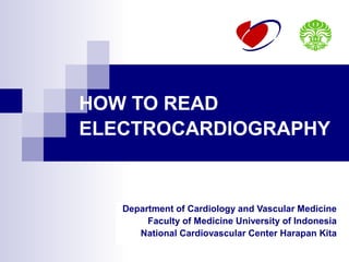
- 1. HOW TO READ ELECTROCARDIOGRAPHY Department of Cardiology and Vascular Medicine Faculty of Medicine University of Indonesia National Cardiovascular Center Harapan Kita
- 7. Unipolar Precodial (Chest) Leads Midclavicular line Anterior axillary line Midaxillary line V6R V6 V5 V5R V4 V4R V3 V3R V2 V1 Mervin J. Goldman, MD. 11th edition Principles of clinical Electrocardiography. Clinical Professor of Medicine University of California School of Medicine San Francisco @1995-1982
- 8. Unipolar Precodial (Chest) Leads Horizontal plane of V 4-6 V7 V8 V9 V 9R V 8R V 7R Mervin J. Goldman, MD. 11th edition Principles of clinical Electrocardiography. Clinical Professor of Medicine University of California School of Medicine San Francisco @1995-1982
- 10. The electrocardiogram (ECG) illustrates conduction of electrical impulses in the heart. The depolarization of the ventricles occurs from the endocardium (inside) to the epicardium (outside) [e] The repolarization of the ventricles occurs in the opposite direction. [g]
- 14. ECG INTERPRETATION 1. RATE 2. RHYTHM 3. AXIS 4. HIPERTROPHIC SIGNS 5. MYOCARDIAL INFARCTION 6. ARRHYTHMIA
- 15. 1. RATE Normal heart rate : 60 – 100 x/minutes • > 100 x/minutes : Sinus Tachycardia • < 60 x/minutes : Sinus Bradicardia Determination heart rate (normal paper speed 25 mm/s): • 300 Count number of large square (bold boxes in one R – R’ interval) • 1500 Count number of small square in one R – R’ intervals • Number of QRS complex in 6 seconds, multiply by 10
- 17. 2. RHYTHM Normal cardiac rhythm : SINUS rhythm Sinus rhythm characteristics : • Rate 60-100 bpm • Constant R – R interval • Negative P wave in aVR and positive di II • P wave is always followed by QRS complex
- 18. 3. AXIS
- 26. 5. MYOCARDIAL INFARCTION Ischemia Injury Necrosis
- 33. ARRHYTHMIA
- 34. AV BLOCK
- 40. WHAT’S WRONG?? Lead Error: V1 and V3 are Transposed! In this normal 12-lead ECG the V1 and V3 chest electrodes are interchanged. Experienced ECG interpreters should be able to spot this lead placement error.
- 57. DISCUSSION
- 59. Sinus arrhythmia
- 62. Subendocardial ischemia. Anterolateral ST-segment depression
- 63. Unstable angina
- 64. acute anterolateral myocardial infarction
- 68. Acute inferoposterior myocardial infarction
- 70. Mobitz I
- 76. Atrial flutter