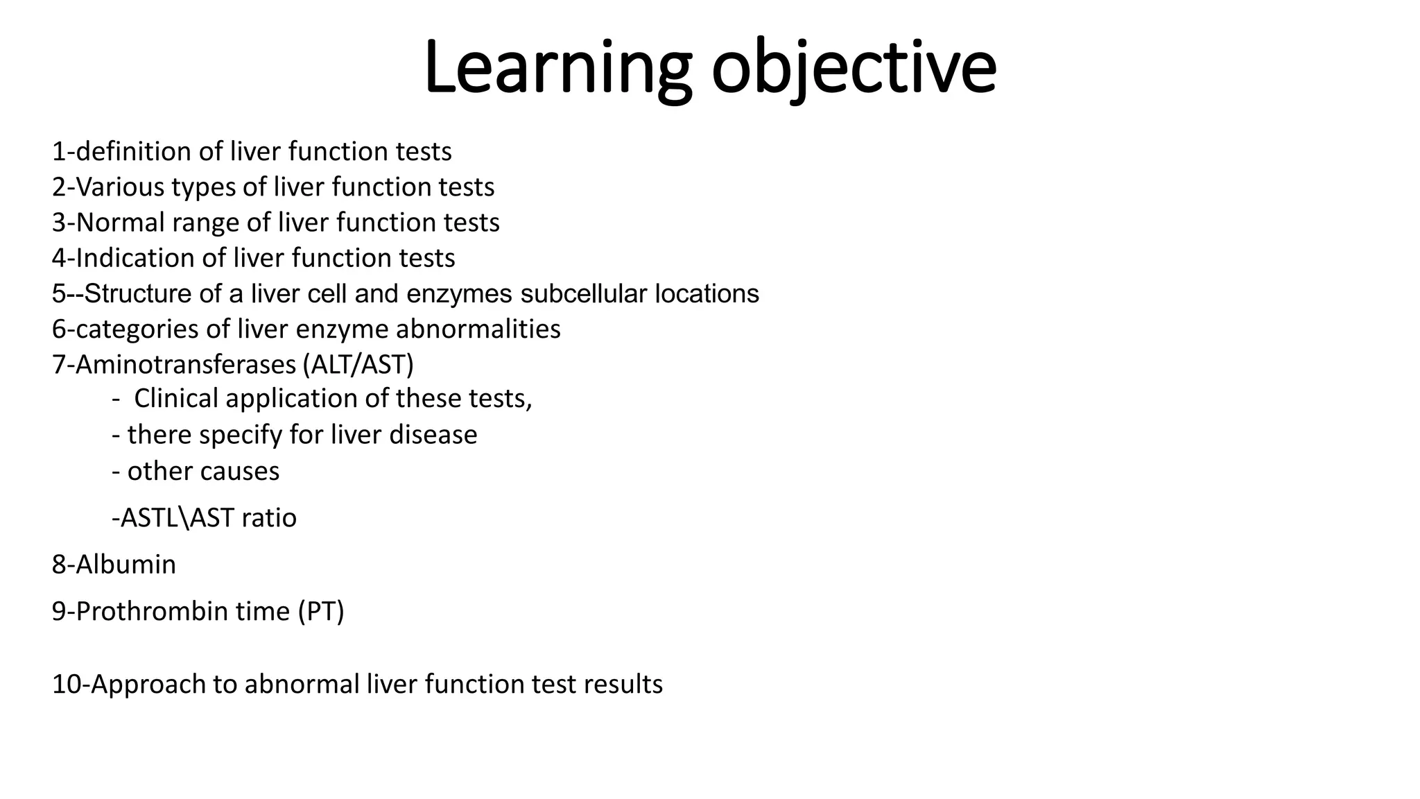
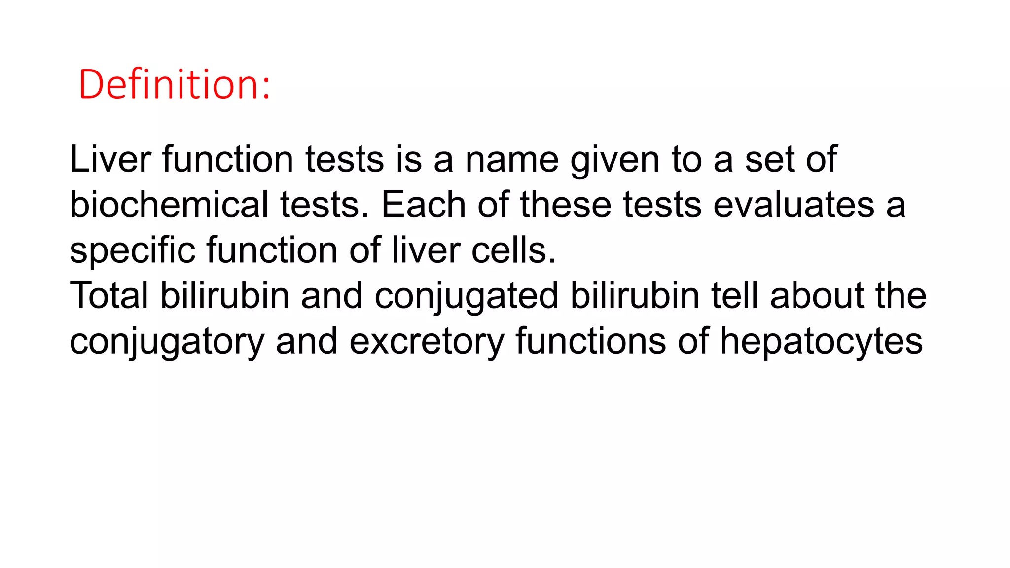
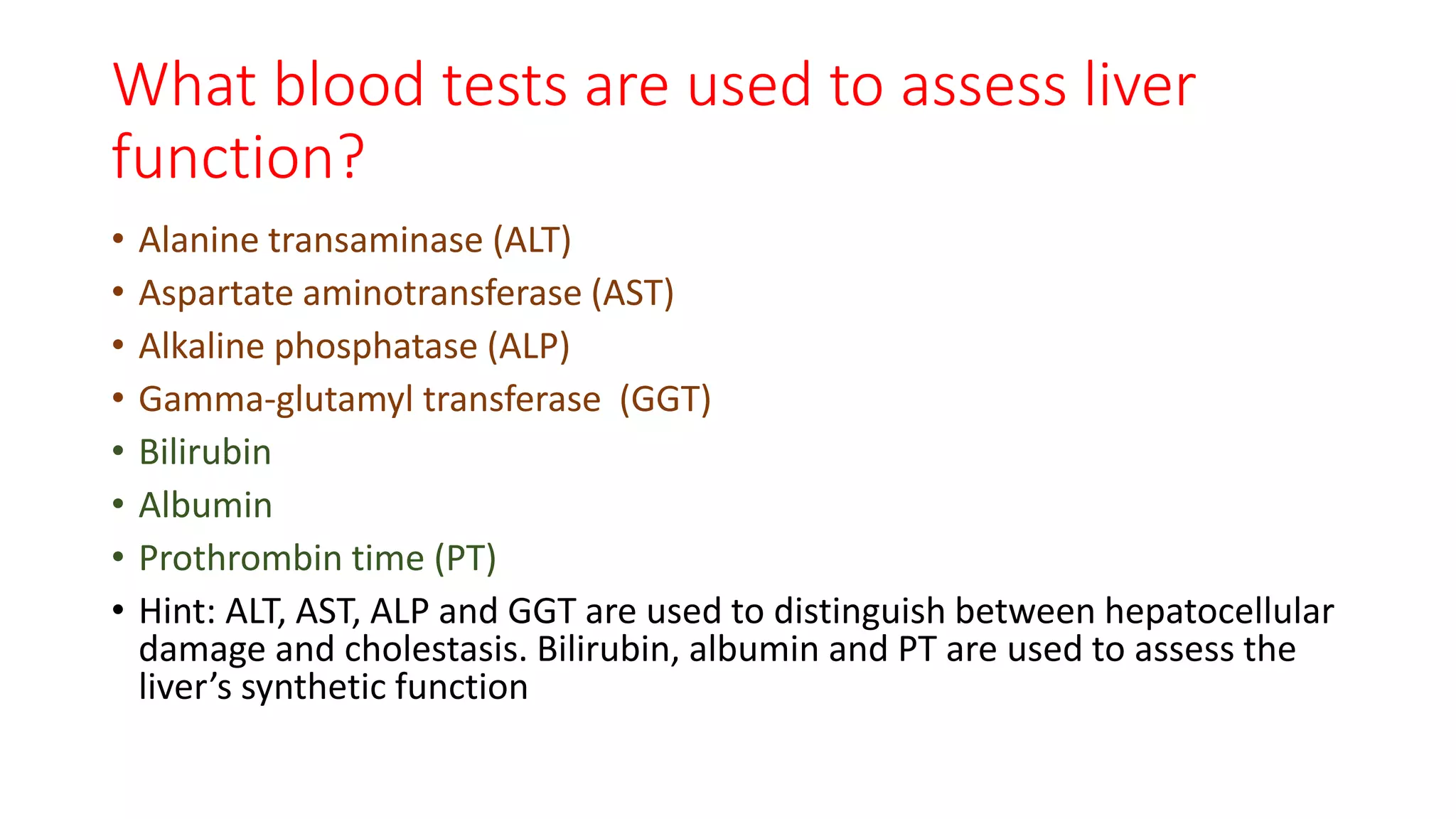
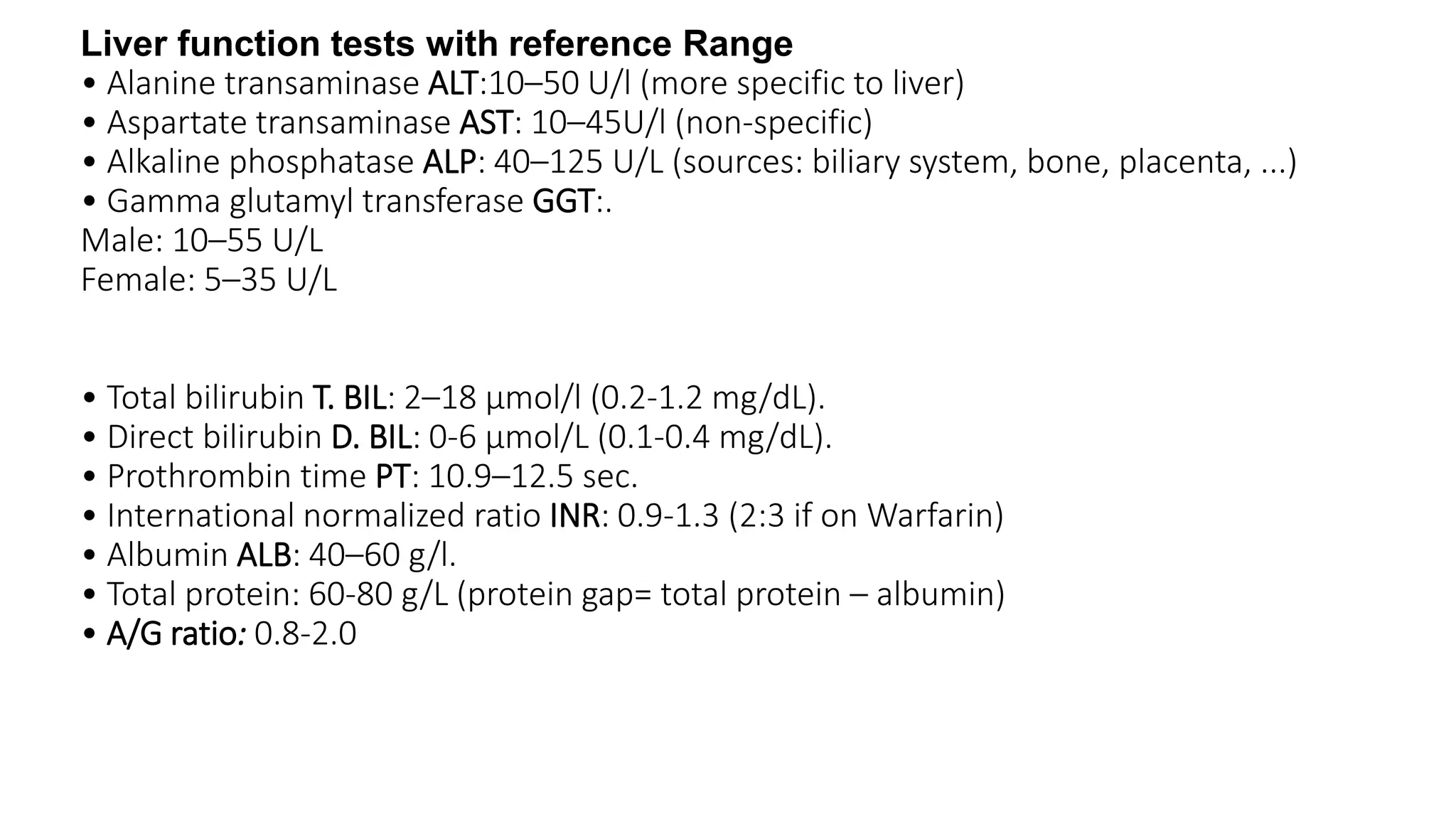
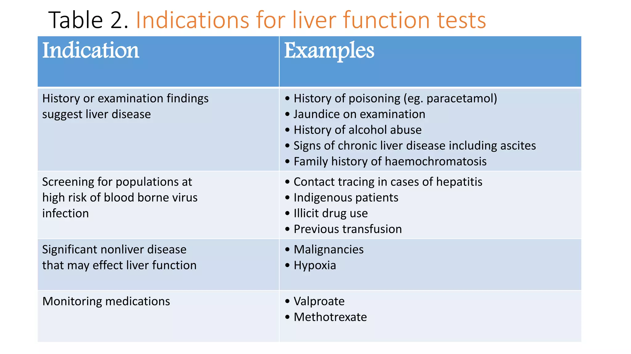
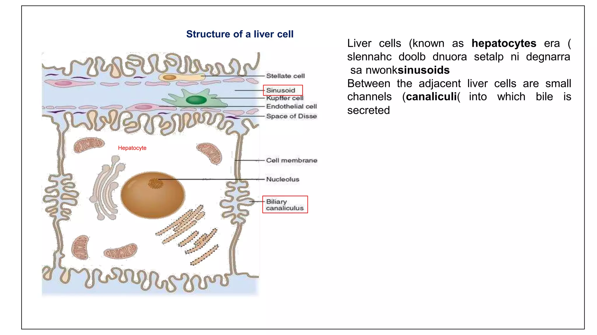
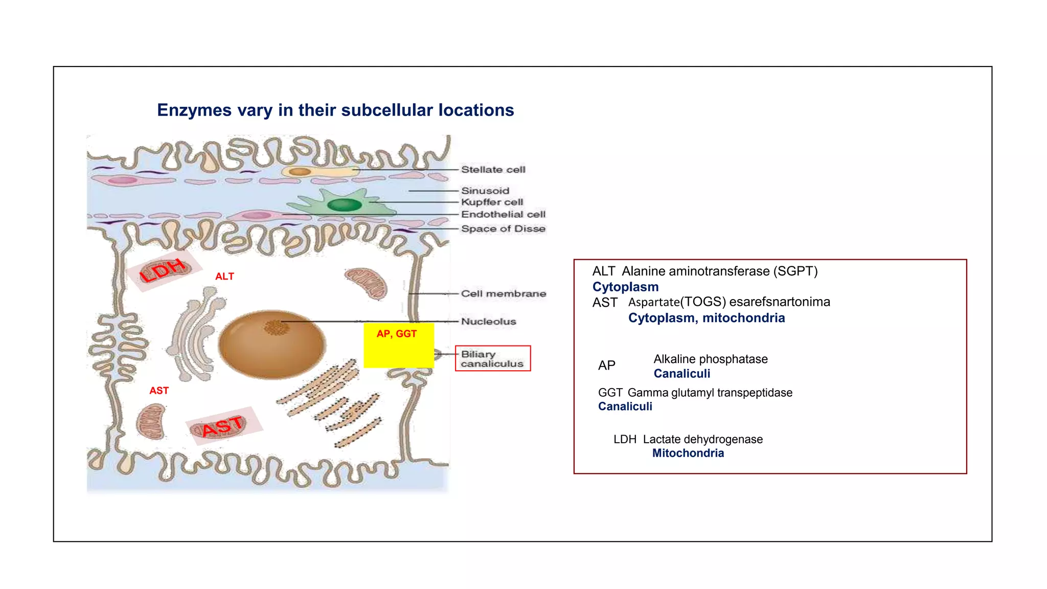
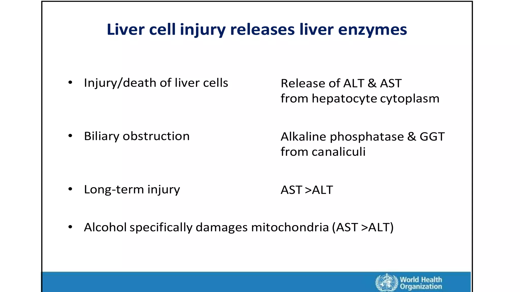
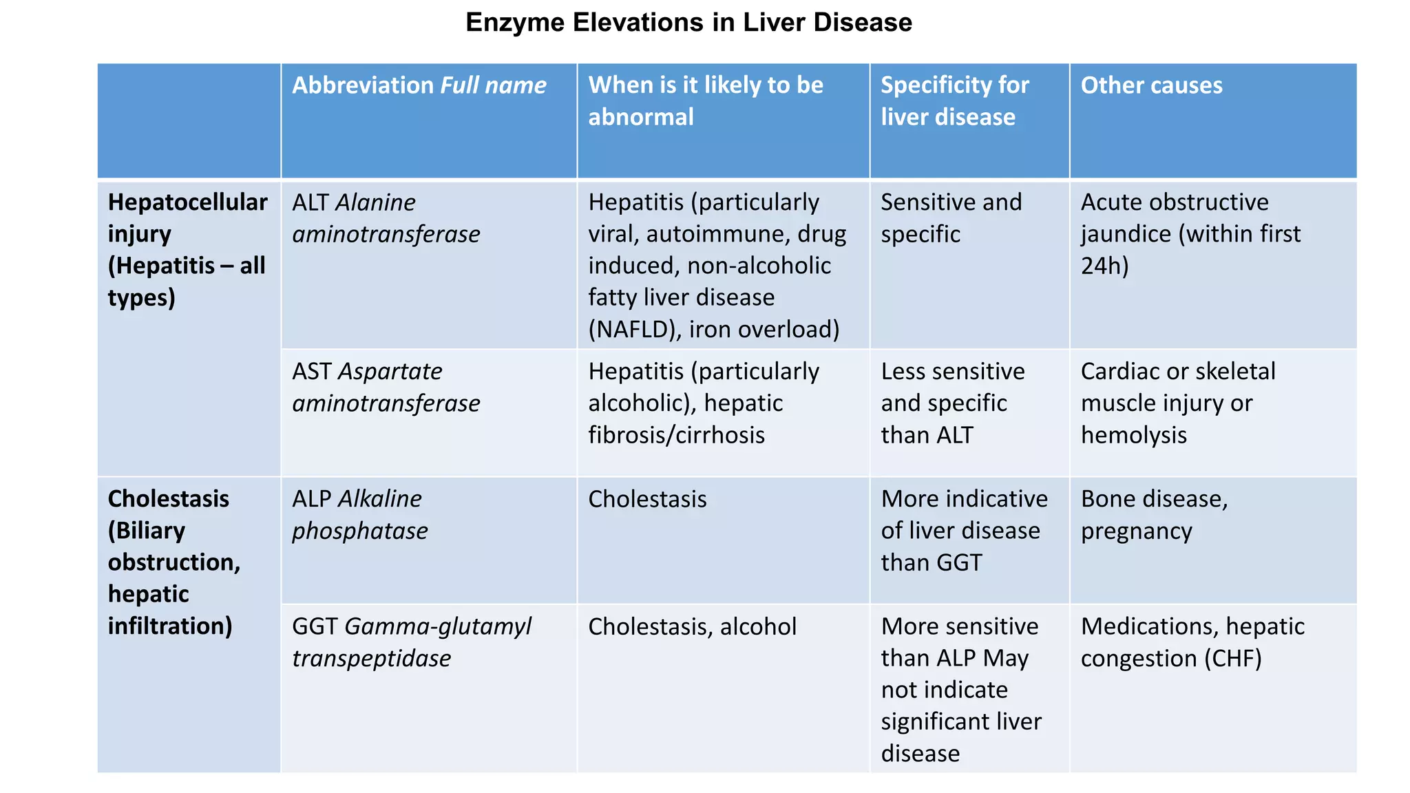
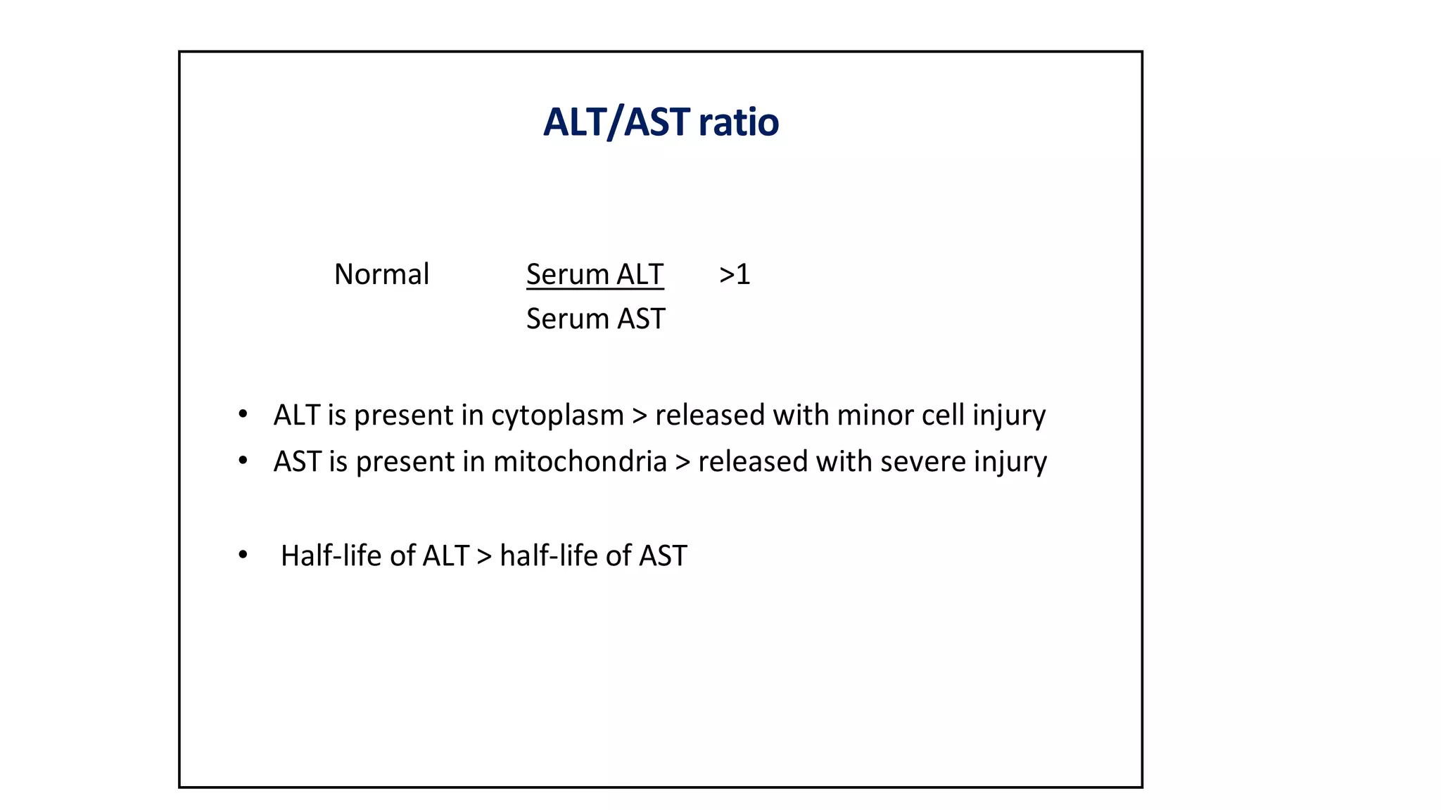
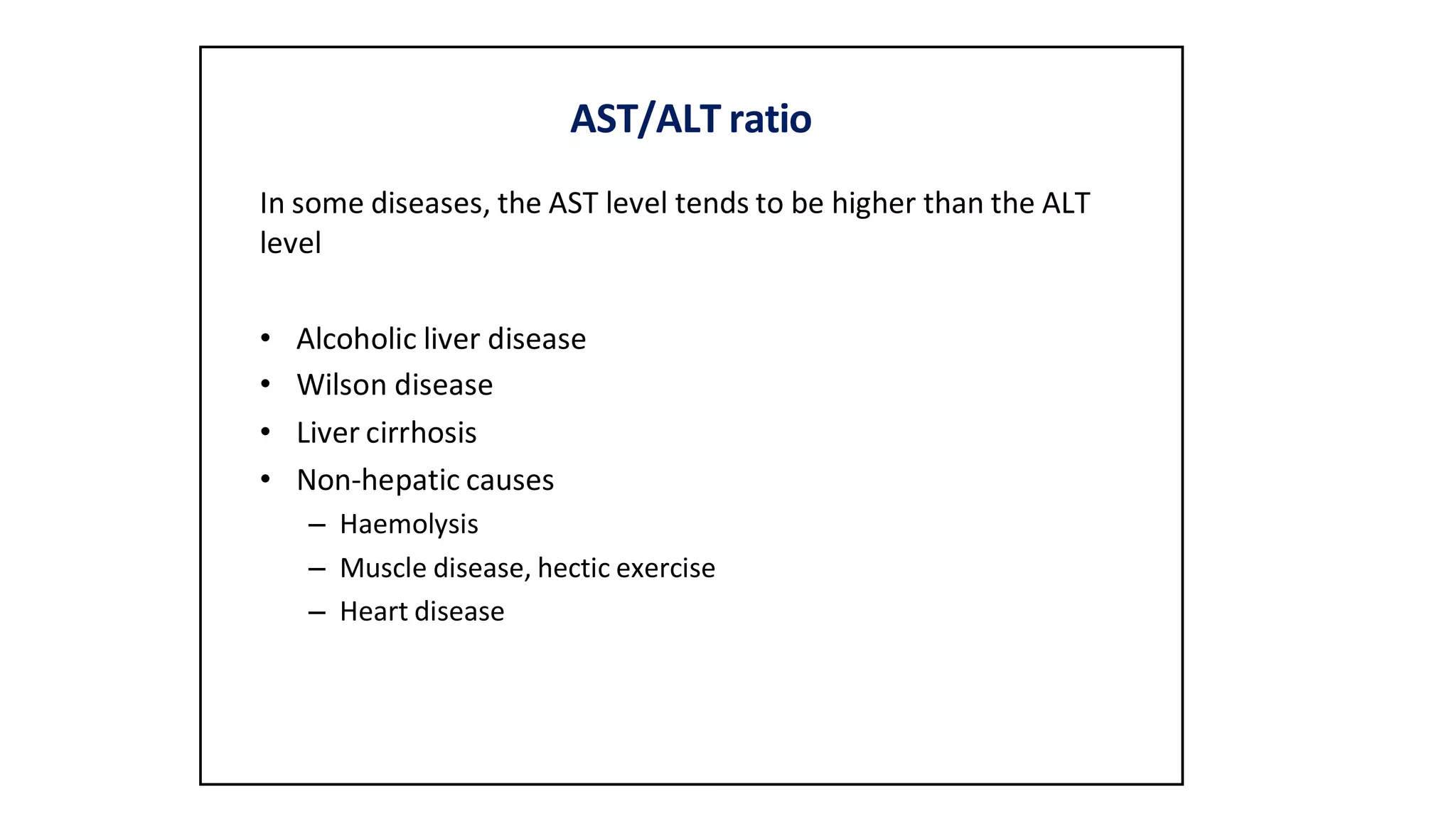
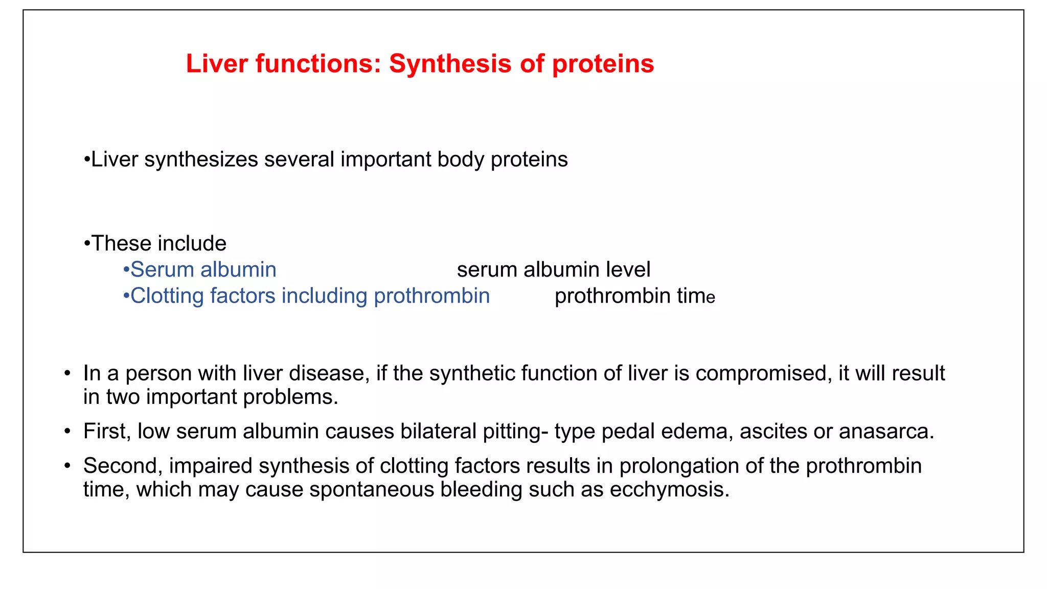
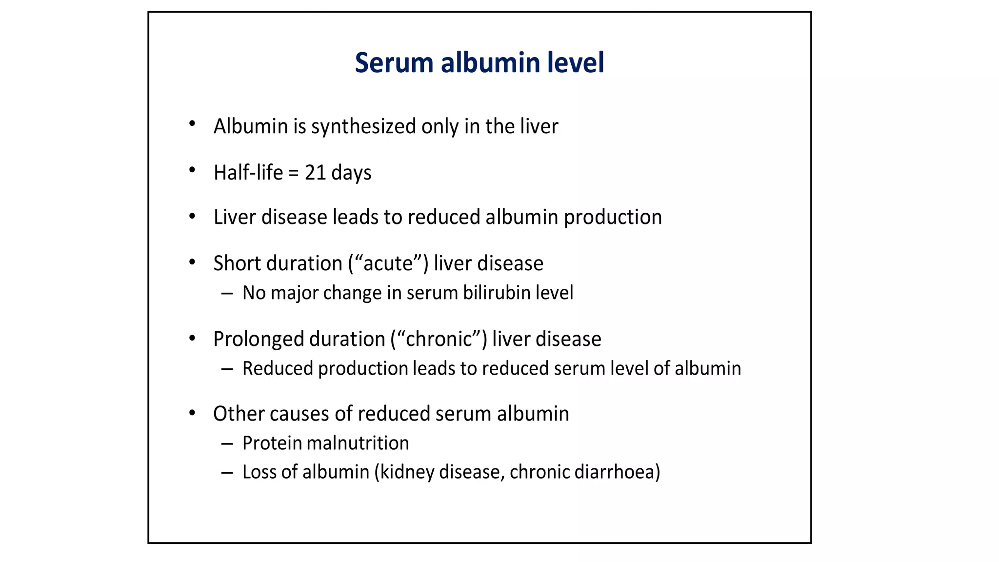
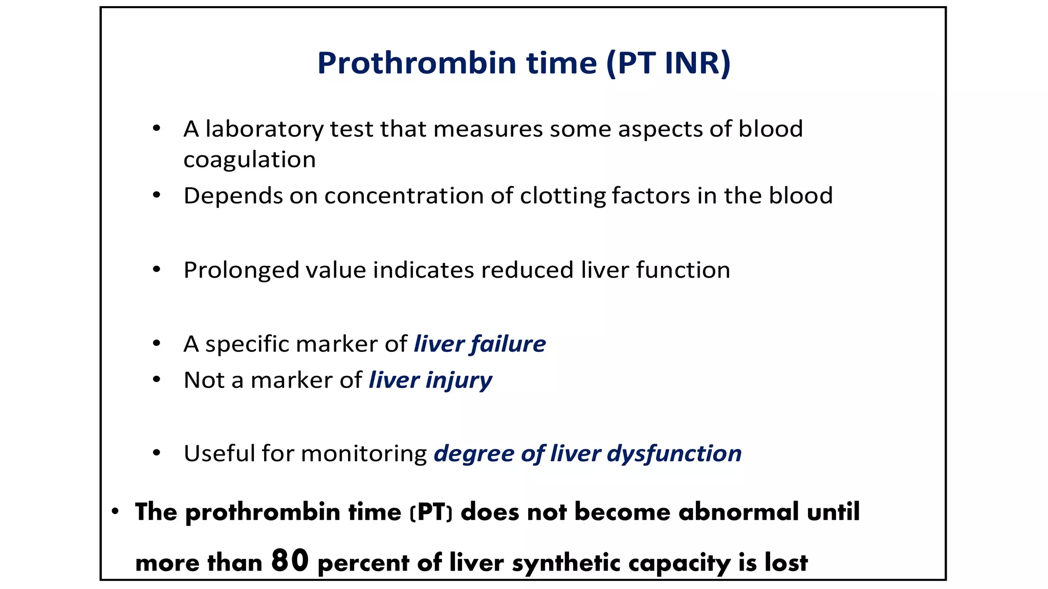
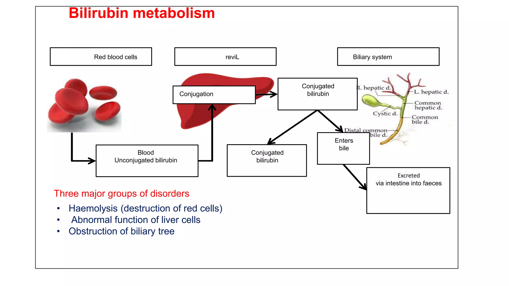
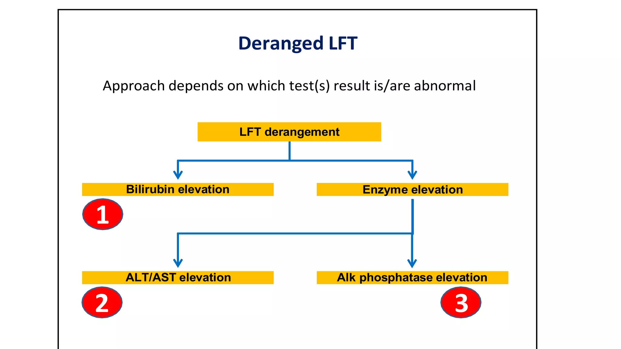
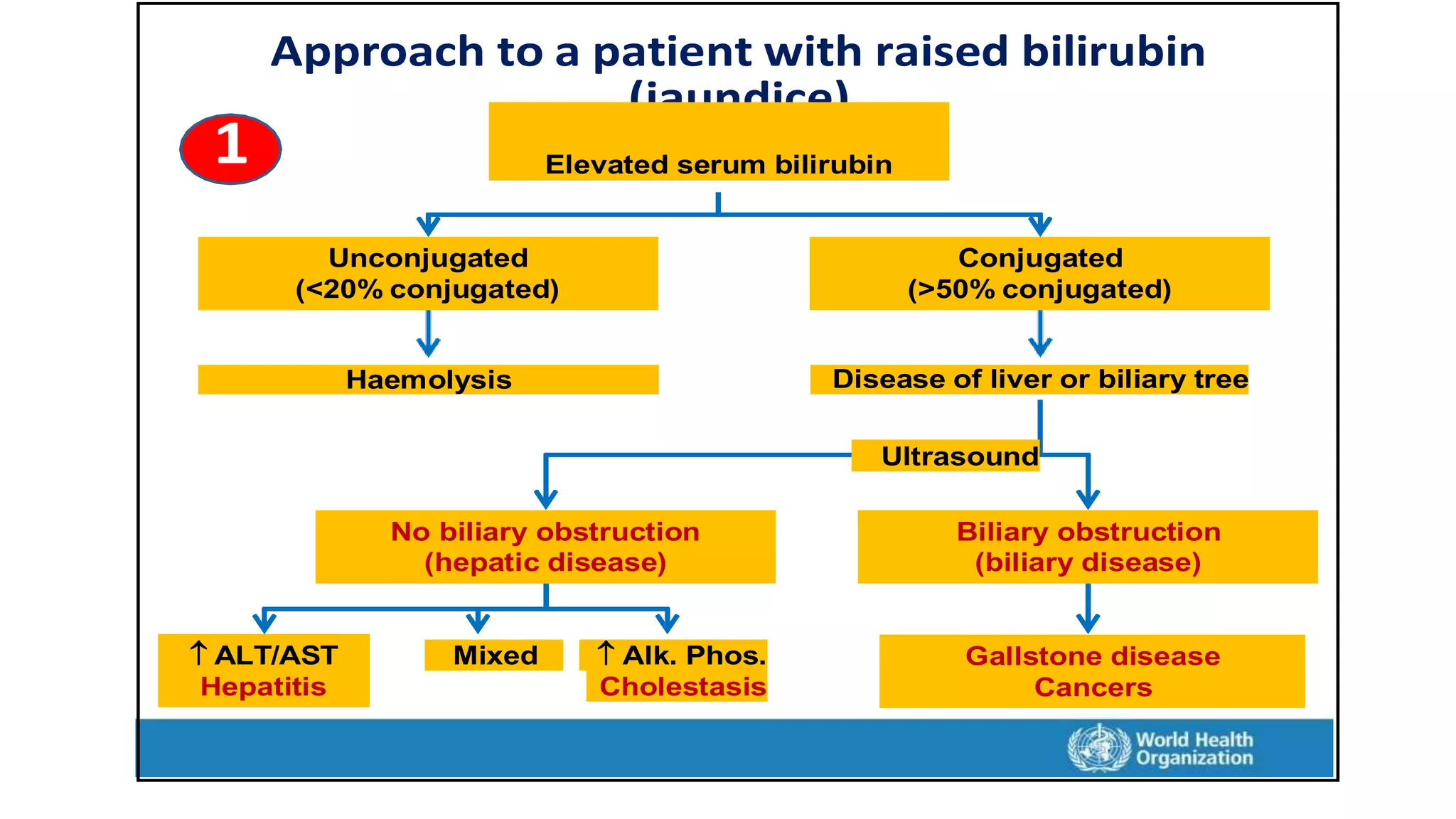
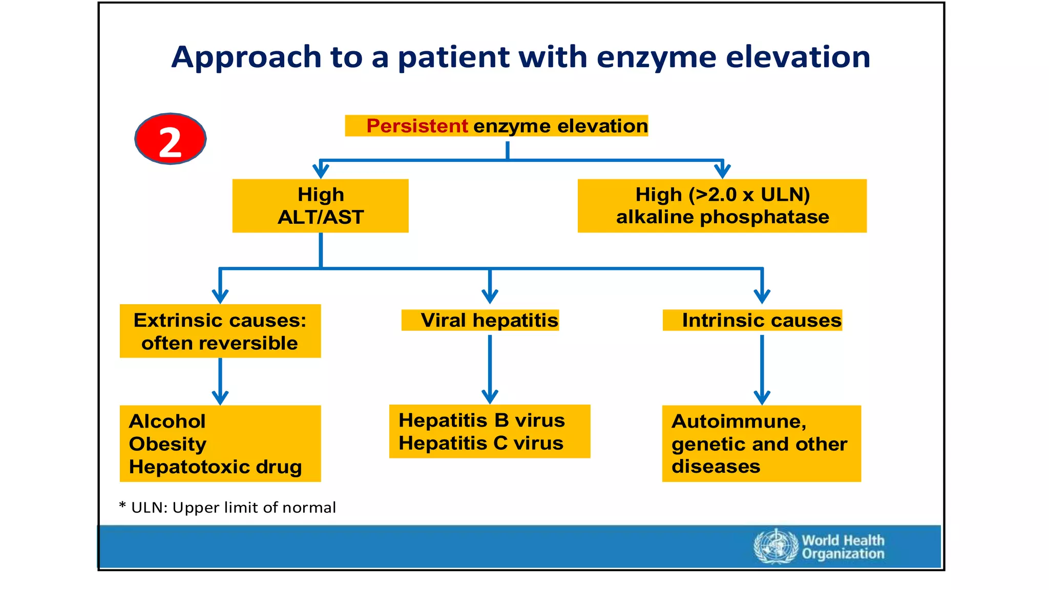
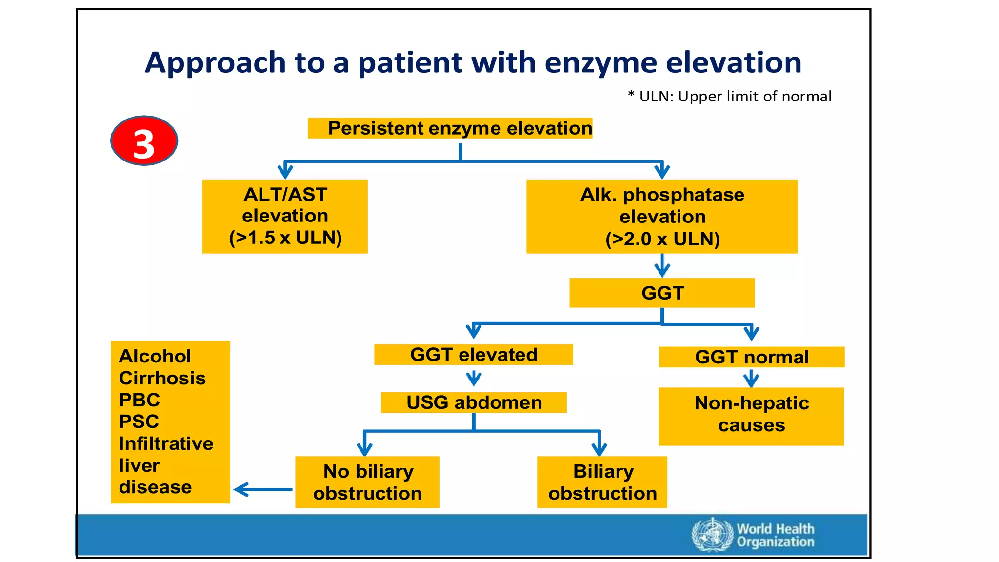
Call Girls Dehradun Just Call 9907093804 Top Class Call Girl Service Available
share.pptx
1. Learning objective
1-definition of liver function tests
2-Various types of liver function tests
3-Normal range of liver function tests
4-Indication of liver function tests
5--Structure of a liver cell and enzymes subcellular locations
6-categories of liver enzyme abnormalities
7-Aminotransferases (ALT/AST)
- Clinical application of these tests,
- there specify for liver disease
- other causes
-ASTLAST ratio
8-Albumin
9-Prothrombin time (PT)
10-Approach to abnormal liver function test results
2. Definition:
Liver function tests is a name given to a set of
biochemical tests. Each of these tests evaluates a
specific function of liver cells.
Total bilirubin and conjugated bilirubin tell about the
conjugatory and excretory functions of hepatocytes
3. What blood tests are used to assess liver
function?
• Alanine transaminase (ALT)
• Aspartate aminotransferase (AST)
• Alkaline phosphatase (ALP)
• Gamma-glutamyl transferase (GGT)
• Bilirubin
• Albumin
• Prothrombin time (PT)
• Hint: ALT, AST, ALP and GGT are used to distinguish between hepatocellular
damage and cholestasis. Bilirubin, albumin and PT are used to assess the
liver’s synthetic function
4. Liver function tests with reference Range
• Alanine transaminase ALT:10–50 U/l (more specific to liver)
• Aspartate transaminase AST: 10–45U/l (non-specific)
• Alkaline phosphatase ALP: 40–125 U/L (sources: biliary system, bone, placenta, ...)
• Gamma glutamyl transferase GGT:.
Male: 10–55 U/L
Female: 5–35 U/L
• Total bilirubin T. BIL: 2–18 μmol/l (0.2-1.2 mg/dL).
• Direct bilirubin D. BIL: 0-6 μmol/L (0.1-0.4 mg/dL).
• Prothrombin time PT: 10.9–12.5 sec.
• International normalized ratio INR: 0.9-1.3 (2:3 if on Warfarin)
• Albumin ALB: 40–60 g/l.
• Total protein: 60-80 g/L (protein gap= total protein – albumin)
• A/G ratio: 0.8-2.0
5. Table 2. Indications for liver function tests
Examples
Indication
• History of poisoning (eg. paracetamol)
• Jaundice on examination
• History of alcohol abuse
• Signs of chronic liver disease including ascites
• Family history of haemochromatosis
History or examination findings
suggest liver disease
• Contact tracing in cases of hepatitis
• Indigenous patients
• Illicit drug use
• Previous transfusion
Screening for populations at
high risk of blood borne virus
infection
• Malignancies
• Hypoxia
Significant nonliver disease
that may effect liver function
• Valproate
• Methotrexate
Monitoring medications
6. Structure of a liver cell
Hepatocyte
Liver cells (known as hepatocytes era )
slennahc doolb dnuora setalp ni degnarra
sa nwonksinusoids
Between the adjacent liver cells are small
channels (canaliculi) into which bile is
secreted
7. Enzymes vary in their subcellular locations
ALT ALT Alanine aminotransferase (SGPT)
Cytoplasm
AST
AP, GGT
Aspartate)TOGS( esarefsnartonima
Cytoplasm, mitochondria
AP
Alkaline phosphatase
Canaliculi
AST GGT Gamma glutamyl transpeptidase
Canaliculi
LDH Lactate dehydrogenase
Mitochondria
8. Liver cell injury releases liver enzymes
• Injury/death of liver cells Release of ALT & AST
from hepatocyte cytoplasm
• Biliary obstruction Alkaline phosphatase & GGT
from canaliculi
• Long-term injury AST >ALT
• Alcohol specifically damages mitochondria (AST >ALT)
9. There are two broad categories of liver enzyme abnormalities:
1. Hepatocellular injury (e.g., hepatitis)
The membranes of liver cells can become permeable when damaged,
allowing for escape of intracellular enzymes into the bloodstream. The
major intracellular enzymes are alanine aminotransferase (ALT) and
aspartate aminotransferase (AST).
10. 2. Cholestasis (e.g., biliary obstruction or hepatic infiltration)
• Obstructed/damaged intra- or extra-hepatic bile ducts cause the
induction of synthesis of alkaline phosphatase (ALP) and gamma-
glutamyl transpeptidase (GGT).
• In acute biliary obstruction, elevation of these enzyme levels often
lags obstruction by approximately 24 hours.
• An isolated minor elevation of GGT is a relatively common finding and
does not necessarily indicate significant liver disease.
11. • Note: Serum bilirubin is not a useful test for distinguishing between
cholestasis and hepatocellular injury because it may be elevated in
both situations.
12. Aminotransferases (ALT/AST)
• ALT is primarily is located in the liver and hence it is a more specific enzyme
for liver injury than AST
• Further, ALT is located in the cytoplasm of the hepatocytes and hence is
released by minor injury. This makes ALT a sensitive marker of liver injury.
• One important aspect is that ALT has a longer half-life than AST. Hence, the
serum ALT level takes a longer time to normalize than the AST after the
injury has subsided.
13. • Serum AST levels are frequently elevated in the presence of heart
disease such as ischaemic heart disease, haemolysis and muscle
injury.
• AST is primarily located inside the mitochondria, and is also found in
the cytoplasm of the hepatocyte in a relatively low concentration. AST
is released from the hepatocyte after more severe injury Hence, AST
is a less sensitive and less specific marker of liver injury than ALT.
• AST is more elevated than ALT in alcohol-induced liver injury.
14. Aminotransferases (ALT/AST)
Several factors can influence the levels of these enzymes
• Some common factors
• Age, gender
• Nutritional status (high body mass index)
• Food intake
• Exercise (muscle)
• Delay in sample processing
15. • Reference range can vary between laboratories and population
groups
• Best expressed as multiples of upper limit of normal (ULN)
• Levels do not correlate well with disease severity or outcome
• Repeated measurements have limited role
• No relation with serum bilirubin level
Aminotransferases considerations: Important
16. • Gamma-Glutamyl Transferase (GGT) test. High levels of GGT in the
blood may be a sign of liver disease or damage to the bile ducts.
• A GGT test can't diagnose the specific cause of liver disease. So it is
usually done along with or after other liver function tests, most often
an alkaline phosphatase (ALP) test.
• Alkaline Phosphatase (ALP)is another type of liver enzyme. It's often
used to help diagnose bone disorders as well as liver disease.
17. Other causes
Specificity for
liver disease
When is it likely to be
abnormal
Abbreviation Full name
Acute obstructive
jaundice (within first
24h)
Sensitive and
specific
Hepatitis (particularly
viral, autoimmune, drug
induced, non-alcoholic
fatty liver disease
(NAFLD), iron overload)
ALT Alanine
aminotransferase
Hepatocellular
injury
(Hepatitis – all
types)
Cardiac or skeletal
muscle injury or
hemolysis
Less sensitive
and specific
than ALT
Hepatitis (particularly
alcoholic), hepatic
fibrosis/cirrhosis
AST Aspartate
aminotransferase
Bone disease,
pregnancy
More indicative
of liver disease
than GGT
Cholestasis
ALP Alkaline
phosphatase
Cholestasis
(Biliary
obstruction,
hepatic
infiltration) Medications, hepatic
congestion (CHF)
More sensitive
than ALP May
not indicate
significant liver
disease
Cholestasis, alcohol
GGT Gamma-glutamyl
transpeptidase
Enzyme Elevations in Liver Disease
18. ALT/AST ratio
Normal Serum ALT >1
Serum AST
• ALT is present in cytoplasm > released with minor cell injury
• AST is present in mitochondria > released with severe injury
• Half-life of ALT > half-life of AST
19. AST/ALT ratio
In some diseases, the AST level tends to be higher than the ALT
level
• Alcoholic liver disease
• Wilson disease
• Liver cirrhosis
• Non-hepatic causes
– Haemolysis
– Muscle disease, hectic exercise
– Heart disease
20. • In a person with liver disease, if the synthetic function of liver is compromised, it will result
in two important problems.
• First, low serum albumin causes bilateral pitting- type pedal edema, ascites or anasarca.
• Second, impaired synthesis of clotting factors results in prolongation of the prothrombin
time, which may cause spontaneous bleeding such as ecchymosis.
Liver functions: Synthesis of proteins
•Liver synthesizes several important body proteins
•These include
•Serum albumin serum albumin level
•Clotting factors including prothrombin prothrombin time
21. Serum albumin level
•
•
•
•
Albumin is synthesized only in the liver
Half-life = 21 days
Liver disease leads to reduced albumin production
Short duration (“acute”) liver disease
– No major change in serum bilirubin level
• Prolonged duration (“chronic”) liver disease
– Reduced production leads to reduced serum level of albumin
• Other causes of reduced serum albumin
– Protein malnutrition
– Loss of albumin (kidney disease, chronic diarrhoea)
22. Prothrombin time (PT INR)
• A laboratory test that measures some aspects of blood
coagulation
• Depends on concentration of clotting factors in the blood
• Prolonged value indicates reduced liver function
• A specific marker of liver failure
• Not a marker of liver injury
• Useful for monitoring degree of liver dysfunction
• The prothrombin time (PT) does not become abnormal until
more than 80 percent of liver synthetic capacity is lost
23. Red blood cells reviL Biliary system
Conjugation
Conjugated
bilirubin
Blood
Unconjugated bilirubin
Conjugated
bilirubin
Enters
bile
Three major groups of disorders
• Haemolysis (destruction of red cells)
• Abnormal function of liver cells
• Obstruction of biliary tree
Excreted
via intestine into faeces
Bilirubin metabolism
24. Jaundice
Jaundice is usually detectable clinically when the plasma bilirubin exceeds
40 μmol/L (∼2.5 mg/dL).
Bilirubin metabolism
Bilirubin in the blood is normally almost all unconjugated and, because it
is not water-soluble, it is bound to albumin and does not enter the urine.
Unconjugated bilirubin is conjugated by glucuronyl transferase into water-soluble
conjugates, which are exported into the bile.
26. Prehepatic jaundice
This is caused either by haemolysis or by congenital hyperbilirubinaemia,
and is characterised by an isolated raised bilirubin level.
In haemolysis, destruction of red blood cells or their marrow precursors
causes increased bilirubin production.
Jaundice because of haemolysis is usually mild because a healthy liver can
excrete a bilirubin load six times greater than normal before unconjugated
bilirubin accumulates in the plasma.
This does not apply to newborns, who have less capacity to metabolise bilirubin.
27. The only common form of nonhaemolytic hyperbilirubinaemia is Gilbert’s
syndrome, an inherited condition causing decreased bilirubin
conjugation, with accumulation of unconjugated bilirubin in the blood. It
has an excellent prognosis and needs no treatment. Other inherited
disorders of bilirubin metabolism are very rare
28. Hepatocellular jaundice
Hepatocellular jaundice results from an inability of the liver to
transport bilirubin into the bile, as a consequence of parenchymal
liver disease.
In hepatocellular jaundice, the concentrations of both unconjugated
and conjugated bilirubin in the blood increase, perhaps because of
the variable way in which bilirubin transport is disturbed.
29. • Parenchymal disease causing jaundice usually also elevates
transaminase levels. Acute jaundice with an alanine aminotransferase
(ALT) greater than 1000 U/L suggests hepatitis A or B, drug toxicity
(e.g. paracetamol) or hepatic ischaemia. Imaging and biopsy are
frequently needed for precise diagnosis.
30. Obstructive (cholestatic) jaundice
Cholestatic jaundice may be caused by:
• Failure of hepatocytes to initiate bile flow.
• Obstruction of the bile ducts or portal tracts.
• Obstruction of bile flow in the extrahepatic bile ducts between the
porta hepatis and the ampulla of Vater
31. Without treatment, cholestatic jaundice tends to progress
because conjugated bilirubin is unable to enter the bile canaliculi
and passes back into the blood.
And also because there is a failure of clearance of unconjugated
bilirubin arriving at the liver cells. Cholestatic jaundice causes an
obstructive pattern of liver function tests.
32. Deranged LFT
Approach depends on which test(s) result is/are abnormal
LFT derangement
Bilirubin elevation
1
Enzyme elevation
ALT/AST elevation
2
Alk phosphatase elevation
3
33. Approach to a patient with raised bilirubin
(jaundice)
1 Elevated serum bilirubin
Unconjugated
(<20% conjugated)
Conjugated
(>50% conjugated)
Haemolysis Disease of liver or biliary tree
Ultrasound
No biliary obstruction
(hepatic disease)
Biliary obstruction
(biliary disease)
ALT/AST
Hepatitis
Mixed Alk. Phos.
Cholestasis
Gallstone disease
Cancers
34. Approach to a patient with enzyme elevation
2
Persistent enzyme elevation
High
ALT/AST
High (>2.0 x ULN)
alkaline phosphatase
Extrinsic causes:
often reversible
Viral hepatitis Intrinsic causes
Alcohol
Obesity
Hepatotoxic drug
Hepatitis B virus
Hepatitis C virus
Autoimmune,
genetic and other
diseases
* ULN: Upper limit of normal
35. Approach to a patient with enzyme elevation
2
>5 <2 <2
Hepatocellular
injury Mixed pattern
Cholestatic
injury
Viral hepatitis
* ULN: Upper limit of normal
36. Non-hepatic
causes
Approach to a patient with enzyme elevation
* ULN: Upper limit of normal
3
Persistent enzyme elevation
ALT/AST
elevation
(>1.5 x ULN)
Alk. phosphatase
elevation
(>2.0 x ULN)
GGT
Alcohol
Cirrhosis
PBC
PSC
Infiltrative
liver
disease
GGT elevated GGT normal
USG abdomen Non-hepatic
causes
No biliary
obstruction
Biliary
obstruction
37. Reference
• AAFP Home | American Academy of Family Physicians
• https://www.aafp.org/home.html
• https://cdn.who.int
• American Gastroenterological Association posi- tion
statement:evaluation of liver chemistry tests. Gastroenterology
2002;123:1364–6.