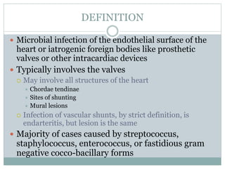
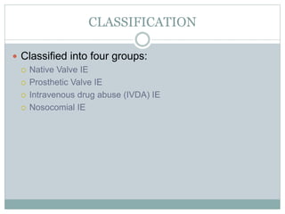
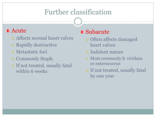
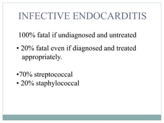
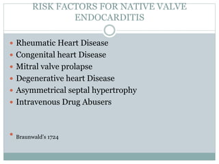
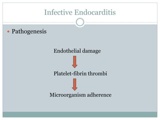
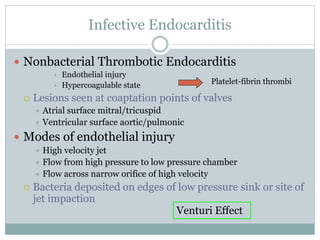
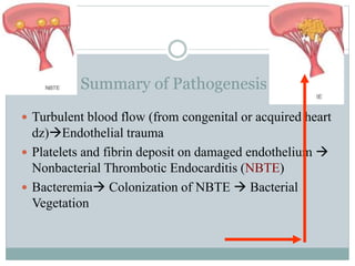
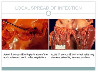
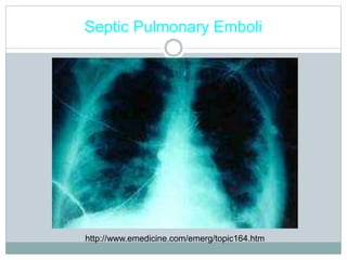
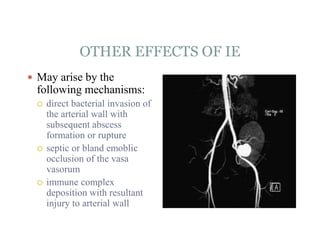
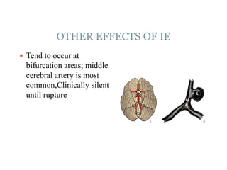
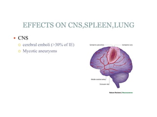
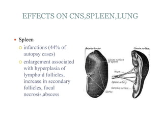
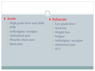
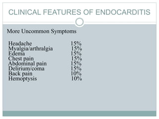
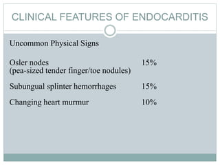
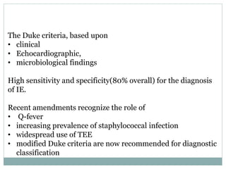
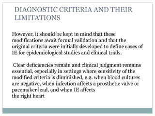
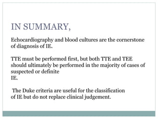
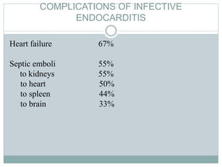
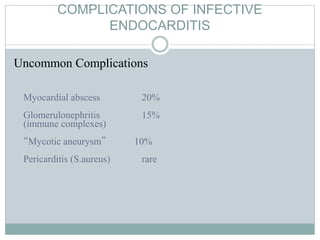
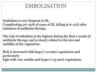
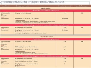
M O D E R A T O R : D R . S A N T O S H K U M A R
D R . A N U P A M A . V . H E G D E
P R E S E N T E R : D R . A B H I N A V K U M A R
INFECTIVE ENDOCARDITIS
DEFINITION
 Microbial infection of the endothelial surface of the
heart or iatrogenic foreign bodies like prosthetic
valves or other intracardiac devices
 Typically involves the valves
 May involve all structures of the heart
 Chordae tendinae
 Sites of shunting
 Mural lesions
 Infection of vascular shunts, by strict definition, is
endarteritis, but lesion is the same
 Majority of cases caused by streptococcus,
staphylococcus, enterococcus, or fastidious gram
negative cocco-bacillary forms
 Gram negative organisms
 P. aeruginosa most common
 HACEK - slow growing, fastidious organisms that may need 3
weeks to grow out of culture
 Haemophilus sp.
 Actinobacillus
 Cardiobacterium
 Eikenella
 Kingella
THE PROTOTYPE LESION
The vegetation
• Variable in size
• Amorphous mass of
fibrin & platelets
• Abundant organisms
• Moderate
inflammatory cells
EPIDEMIOLOGY
 Incidence of IE remained relatively stable from 1950-
2000 at 3.6-7 cases per 100,000 patients year.
 Higher incidence in urban population
 Elderly 4-6 x risk
 Median age-increased to 57.9 years
 (interquartile range 43.2-71.8 years)
 Male:Female ratio of about 2:1
 Upto 75% of pateints with native valve involvement
Have identifiable risk factors
Braunwald’s 1723
CLASSIFICATION
 Classified into four groups:
 Native Valve IE
 Prosthetic Valve IE
 Intravenous drug abuse (IVDA) IE
 Nosocomial IE
Further classification
Acute
 Affects normal heart valves
 Rapidly destructive
 Metastatic foci
 Commonly Staph.
 If not treated, usually fatal
within 6 weeks
Subacute
 Often affects damaged
heart valves
 Indolent nature
 Most commonly S. viridans
or enterococcus
 If not treated, usually fatal
by one year
INFECTIVE ENDOCARDITIS
100% fatal if undiagnosed and untreated
• 20% fatal even if diagnosed and treated
appropriately.
•70% streptococcal
• 20% staphylococcal
RISK FACTORS FOR NATIVE VALVE
ENDOCARDITIS
 Rheumatic Heart Disease
 Congenital heart Disease
 Mitral valve prolapse
 Degenerative heart Disease
 Asymmetrical septal hypertrophy
 Intravenous Drug Abusers
 Braunwald’s 1724
MITRAL VALVE PROLAPSE
 High Prevalence
 2-4 % in general healthy population
 20% of young women
 7-30% of Native valve endocarditis without IVD
 Relative risk of 3.5-8.2 for endocarditis
 MVP+systolic murmurIE is 52/100,000 person yr
 MVP-systolic murmurIE is 4.6/100,000 person yr
 Braunwald,1724
RHEUMATIC HEART DISEASE
 Declining in incidence
 20-25% cases of endocarditis cases in 1970’s
 7-18% cases of endocarditis cases in 1980’s
 Commonly involves;
 Mitral valve in women
 Aortic valve in men
Braunwald,1724
CONGENITAL HEART DISEASES
 Accounts for
 10-20% of endocarditis cases in young adults
 8 % of cases in older adults
 Common lesions:
 patent ductus arteriosus
 ventricular septal defect
 bicuspid aortic valve
Braunwald’s 8 edition1724
INTRAVENOUS DRUG ABUSERS
 Risk of IE 2-5/100 patients year
 Higher risk than rheumatic disease or prosthetic
valve
 65-80% of IVD endocarditis population is male
 Average age 27-37 years
 Commonly involves—Tricuspid valves-46-78%
 Mitral valves-24-32%
 aortic valves-8%-19%
 S.Aureus involved in >50% cases
Braunwald’s 1724
PROSTHETIC VALVES
 Accounts for 10-30% of all endocarditis cases
 Risk is greater in first 6 months
 -EARLY endocarditis occurs in first 60 days
 - LATE endocarditis with onset thereafter
 Incidence about 5% at 5 years
 Risks declines over time
 Mechanical valve has higher risk than bio prosthesis
initially
 After 1 year bioprosthesis is more risky than
mechanical valve
Braunwald’s 1725-26
ETIOLOGY
 Native valve Endocarditis- Streptococcus
viridans, Staphylococci, HACEK
 Oral cavity, skin, upper respiratory tract
 Streptococcus bovis (GIT), Enterococci
 Intravascular catheters,UTI, Nosocomial wound
infections, HD- transient bacteremia
 Prosthetic Valve Endocarditis- Coagulase
negative Staphylococci, S.Aureus, GNB, diptheroids,
fungi
 Within 2 months- intra-operative contamination
 IV Drug abuse endocarditis:- Tricuspid valve
 MRSA
 More varies Etiology if left sided heart valves are involved
 Polymicrobial
 Unusual organisms- Pseudomonas Aeruginosa, Candida,
Bacillus, Lactobacillus, Corynebacterium
 Nosocomial- Transvenous pacemaker lead- and/or
implanted defibrillator associated endocarditis
 5-15% may be culture negative (?prior antibiotic exposure)
 Or fastidious organisms- Coxiella burnetti in Europe, Brucella in
the Middle East
 Tropheryma whipplei- indolent afebrile culture negative
endocarditis
MICROBIOLOGY IN A NUTSHELL
 Any pathogen can cause endocarditis
 Common Organisms:
 strep viridians-28%
 staph aureus-28%
 other strep species-23%
 coag negative staph-7%
 gram negative staph-4%
 other-5%
 Drug Resistance seen more common in IV drug use
Cabell,150
Infective Endocarditis
 Pathogenesis
Endothelial damage
Platelet-fibrin thrombi
Microorganism adherence
Infective Endocarditis
 Nonbacterial Thrombotic Endocarditis
• Endothelial injury
• Hypercoagulable state
 Lesions seen at coaptation points of valves
 Atrial surface mitral/tricuspid
 Ventricular surface aortic/pulmonic
 Modes of endothelial injury
 High velocity jet
 Flow from high pressure to low pressure chamber
 Flow across narrow orifice of high velocity
 Bacteria deposited on edges of low pressure sink or site of
jet impaction
Venturi Effect
Platelet-fibrin thrombi
Venturi Effect
PATHOGENESIS OF INFECTIVE
ENDOCARDITIS
 In systemic lupus erythematosus the vegetations may
form on the undersurface of valve towards ventricular side
called as libman sacks syndrome.
PATHOGENESIS OF INFECTIVE
ENDOCARDITIS
 The adherence of the organism to NBTE is a crucial
step.
1. FimA is a surface adhesin of S.viridans that serves as an
important colonization factor. Homologues of fimA genes
were found in many S.viridans strains and enterococci.
2. Fibronectin is implicated as the host receptor within NBTE.
PATHOGENESIS OF INFECTIVE
ENDOCARDITIS
Adherence of some streptococci to blood clot is facilitated
by dextran (a cell wall component) (especially of
Streptococcus mutans, a viridans group.
Further Some strains of bacteria are stimulators of platelet
aggregation
PATHOGENESIS OF INFECTIVE
ENDOCARDITIS
 Thrombotic vegetations are laden with microbial
organisms they become
large even upto 3cms,
friable and
easily detachable
 colour of vegetationstan grey red or brown and situated
along the line of closure of valve.
PATHOGENESIS OF INFECTIVE
ENDOCARDITIS
Microscopic Pathology
Fibrin, platelets, masses of organisms,
+/- necrosis, +/- neutrophils
Later: +/-lymphocytes, +/- macrophages,
+/- fibroblasts, +/- fibrosis
Summary of Pathogenesis BE
 Turbulent blood flow (from congenital or acquired heart
dz)Endothelial trauma
 Platelets and fibrin deposit on damaged endothelium 
Nonbacterial Thrombotic Endocarditis (NBTE)
 Bacteremia Colonization of NBTE  Bacterial
Vegetation
Conversion of NBTE to IE
 Frequency & magnitude of bacteremia
 Density of colonizing bacteria
• Oral > GU > GI
 Disease state of surface
• Infected surface > colonized surface
 Extent of trauma
 Resistance of organism to host defenses
 Most aerobic gram negatives susceptible to complement-mediated
bactericidal effect of serum
 Tendency to adhere to endothelium
• Dextran producing strep
• Fibronectin receptors on staph, enterococcus, strep, Candida
 Marantic Endocarditis- NBTE due to
hypercoagulable state- malignancy, chronic diseases,
SLE, APLA
Cardiac Manifestations
 New regurgitant murmurs- 30-35% then 85%
 CHF- 30 to 40%- valvular damage (aortic),
myocarditis, intracardiac fistula
 Perivalvular abscess
 Fistulae (Root of aorta to chambers/ between
cardiac chambers)
 Pericarditis
 Heart block/ MI due to embolic phenomena
 Embolic phenomena include systemic, cerebral and
pulmonary emboli are common in >50 % cases.
LOCAL SPREAD OF INFECTION
Acute S. aureus IE with perforation of the
aortic valve and aortic valve vegetations.
Acute S. aureus IE with mitral valve ring
abscess extending into myocardium.
LOCAL EFFECTS OF INFECTIVE
ENDOCARDITIS
 Infection may extends beyond valve cusp may erode &
perforate valve, & may erode into underlying myocardium
to produce an abscess (ring abscess) or Paravalvular
abscess
 Septal abscesses & adjacent abscess
 Fistulae
 Prosthetic dehiscence
LOCAL EFFECTS OF INFECTIVE
ENDOCARDITIS
 Valvular distortion/destruction chordal rupture.
 Conduction abnormalities
 Purulent pericarditis
 Functional valve obstruction
 With treatment, healing occurs by fibrosis and
occasionally calcification.
Septic Pulmonary Emboli
http://www.emedicine.com/emerg/topic164.htm
DISTANT EFFECTS OF INFECTIVE
ENDOCARDITIS
 Vegetations may become detached and produce embolic
effects.
 Embolic phenomena are common (15-35%). septic
infarcts involving: renal, splenic, coronary, or cerebral
circulation.
 Risk for emboli is increased when vegetation >1cm.
IMMUNOLOGICAL EFFECTS OF IE
 IE cause both humural and cellular response
 Rheumatoid factor:
 titers correlate with the level of hypergammaglobulinemia and
decrease with therapy
 Antinuclear antibodies:
 may contribute to the musculoskeletal manifestations, low-
grade fever, or pleuritic pain
 Circulating immune complexes:
 Connected with long duration of illness, extravascular
manifestations, hypocomplemenemia
 May cause diffuse glomerulonephritis, and some of the
peripheral manifestations such as Osler nodes
EFFECTS OF IE ON KIDNEY
 Pathological processes: abscess, infarction, glomerulonephritis
(focal, segmental), membranoproliferative GN
 May be normal is size or slightly swollen
 10 to 15% of IE exhibit immune complex GN (as in SLE).
Supporting IC rather than emboli:
1. Bacteria rarely seen in lesion
2. GN can occur with right-sided IE
3. GN is rare in acute IE even though large vegetation result in
metastatic abscess formation
4. IF staining reveals IC-typical distribution
5. Antibacterial antibodies eluted from lesions
OTHER EFFECTS OF IE
1. Mycotic aneurysm is a
localized, irreversible
arterial dilatation due
to destruction of the
vessel wall by
infection
 More common with
S.viridans
OTHER EFFECTS OF IE
 May arise by the
following mechanisms:
 direct bacterial invasion of
the arterial wall with
subsequent abscess
formation or rupture
 septic or bland emoblic
occlusion of the vasa
vasorum
 immune complex
deposition with resultant
injury to arterial wall
OTHER EFFECTS OF IE
 Tend to occur at
bifurcation areas; middle
cerebral artery is most
common,Clinically silent
until rupture
EFFECTS ON CNS,SPLEEN,LUNG
 CNS
 cerebral emboli (>30% of IE)
 Mycotic aneurysms
EFFECTS ON CNS,SPLEEN,LUNG
 Spleen
 infarctions (44% of
autopsy cases)
 enlargement associated
with hyperplasia of
lymphoid follicles,
increase in secondary
follicles, focal
necrosis,abscess
EFFECTS ON CNS,SPLEEN,LUNG
 Lung
 associated with right-sided
IE
 pulmonary embolism,
acute pneumonia, pleural
effusion, or empyema
PETECHIAE
1. Nonspecific may result from local vasculitis or emboli
2.Often located on extremities or mucous membranes
3.Petechiae are red because they contain red blood that
has leaked from the capillaries
Splinter Hemorrhages
1. Nonspecific,Nonblanching
2. Usually 2-3mm long in long axis of nail
3. Initially blue-purple in colour,change to brown of black in 1-2 days
4. Move distal with nail growth
5. Trauma most common;20% of population have them
Osler’s Nodes- immune
American College of Rheumatology
webrheum.bham.ac.uk/.../ default/pages/3b5.htm
1. More specific
2. Painful and
erythematous nodules
3. Located on pulp of
fingers and toes
4. More common in
subacute IE
5. Due to infective emboli
or immune complex
deposits
6. 4 P’s:
 Pink
 Painful
 Pea-sized
 Pulp of the
fingers/toes
Janeway Lesions
1. More specific
2. Erythematous, blanching macules
3. Nonpainful
4. Located on palms and soles
5. Micro abscesses of the dermis with marked necrosis and
inflammatory infiltrate not involving the epidermis, which is due to
the deposition of circulating immune complexes in small blood vessels.
Janeway Lesions
Roth Spots
Eye
• Roth's spots are retinal
hemorrhages with white
or pale centers
composed of coagulated
fibrin.
• observed via fundoscopy
(using an
ophthalmoscope to view
inside the eye) or slit
lamp exam
EFFECTS ON SKIN&EYE
 Eye
 Roth spots
 Caused by immune complex
mediated vasculitis often
resulting from bacterial
endocarditis. Roth's spots may
be observed in
 leukemia,
 diabetes,
 subacute bacterial
endocarditis,
 pernicious anemia,
 ischemic events, and
 in HIV retinopathy.
EFFECTS ON SKIN&EYE
 Infective endocarditis
also can give rise to
conjunctival
haemorrhages
EFFECTS ON SKIN&EYE
 Clubbing is also
known to occur in
infective endocarditis.
THINGS TO REMEMBER IN INFECTIVE
ENDOCARDITIS
 Infective endocarditis affects
Left-sided valves 75%
Right-sided valves 15%
Both 5%
Other 5%
THINGS TO REMEMBER IN INFECTIVE
ENDOCARDITIS
Mitral valve alone 35%
Aortic valve alone 20%
Mitral plus aortic 20%
Tricuspid 14%
Pulmonic 1%
With changing murmurs in character pitch
duration etc.fungal vegetations are large
vegetations.
THINGS TO REMEMBER IN INFECTIVE
ENDOCARDITIS
 Infective endocarditis may be culture negative either due
to prior antibiotic treatment or due to atypical microbial
organisms or due to fungus etc.then called as non bacterial
endocarditis.
Acute
 High grade fever and chills
 SOB
 Arthralgias/ myalgias
 Abdominal pain
 Pleuritic chest pain
 Back pain
Subacute
 Low grade fever
 Anorexia
 Weight loss
 Fatigue
 Arthralgias/ myalgias
 Abdominal pain
 N/V
Clinical Features
 Interval between index bacteremia & onset of sx’s
usually < 2 weeks
 May be substantially longer in early PVE
 Fever most common sign
 May be absent in elderly/debilitated pt.
 Murmur present in 80 – 85%
 Generally indication of underlying lesion
 Frequently absent in tricuspid IE
 Changing murmur
CLINICAL FEATURES OF ENDOCARDITIS
Uncommon Symptoms
Haematuria 25%
Cough 25%
Sweats 25%
Anorexia 25%
Weight loss 25%
Malaise 25%
Skin lesions 20%
Nausea/vomiting 20%
Stroke 20%
CLINICAL FEATURES OF ENDOCARDITIS
More Uncommon Symptoms
Headache 15%
Myalgia/arthralgia 15%
Edema 15%
Chest pain 15%
Abdominal pain 15%
Delirium/coma 15%
Back pain 10%
Hemoptysis 10%
CLINICAL FEATURES OF ENDOCARDITIS
Uncommon Physical Signs
Osler nodes 15%
(pea-sized tender finger/toe nodules)
Subungual splinter hemorrhages 15%
Changing heart murmur 10%
LABORATORY FINDINGS
Laboratory Findings
Elevated ESR (mean 57 mm/hr) 95%
(erythrocyte sedimentation rate)
Circulating immune complexes 90%
Anemia 80%
Proteinuria 60%
LABORATORY FINDINGS
Laboratory Findings
Rheumatoid factor 50%
(anti-IgG antibodies)
Hematuria 40%
Leukocytosis 25%
Hypergammaglobulinemia 25%
Elevated creatinine 10%
Leukopenia 10%
Thrombocytopenia 10%
Clinical Features
 Systemic emboli
 Incidence decreases with effective anti-microbial Rx
 Neurological sequelae
 Embolic stroke 15 – 20% of patients
 Mycotic aneurysm
 Cerebritis
 CHF
 Due to mechanical disruption
 High mortality without surgical intervention
 Renal insufficiency
 Immune complex mediated
 Impaired hemodynamics/drug toxicity
Blood Cultures in IE
 3 separate venepunctures during one hour period
at least 10 ml blood before giving antibiotics.
 Adherence to above practice yields positive
culture in 90 %
 But at least 30 % are prescribed antibiotics before
taking culture.
 Sterile culture ---western =2.5 to 31%
---Indian = 48 to 54 %
Blood Cultures
 MULTIPLE BLOOD CULTURES BEFORE
EMPIRIC THERAPY
 If not critically ill
 3 blood cultures over 12-24 hour period
 ? Delay therapy until diagnosis confirmed
 If critically ill
 3 blood cultures over one hour
 No more than 2 from same venepuncture
 Relatively constant bacteremia
“Culture Negative” IE
 Infective endocarditis may be culture negative either due
to prior antibiotic treatment or due to atypical microbial
organisms or due to fungus etcnon bacterial
endocarditis.
 Less common with improved blood culture methods
 Special media required
 Brucella, Mycoplasma, Chlamydia, Histoplasma,
Legionella, Bartonella
 Longer incubation may be required
 HACEK
 Coxiella burnetii (Q Fever), Trophyrema whipplei
will not grow in cell-free media
IMAGING
Chest x-ray
 Non Specific findings
 Cardiomegaly
 Nodular infilterates-Tricuspid valve endocarditis causing
septic emboli
EKG
 Rarely diagnostic
 Look for evidence of ischemia, conduction delay, and
arrhythmias
 CT & MRI
 still mostly experimental
 Primarily evaluate brain for complications
 isolated CT case reports
 -large aortic root abscess and AV fistula
 MRI potentially diagnose complications of aortic
root aneurysms or abscesses
Sachdeva,192-193
NUCLEAR IMAGING
 Tagged WBC scans have been used
 -can identify vegetation's
 -non specific
 -high false negative
 Case reports suggest that positive scans can be used
to detect local complications of endocarditis
 Useful to detect metastatic septic embolism
Sachdeva,193
Indications for Echocardiography
Transthoracic echocardiography (TTE)
 First line if suspected IE
 Native valves
Transesophageal echocardiography (TEE)
 Prosthetic valves
 High risk patients
 Intracardiac complications
 Inadequate TTE
 Fungal or S. aureus or bacteremia
ECHOCARDIOGRAPHY
 Major duke criteria
 Diagnose and management of infective endocarditis
 Vegetations-detectes in 67% of “definite” cases by
duke criteria
 -Irregular shape
 -Occur on low pressure side of turbulent jet
 *Atrial side in mitral and Tricuspid regurgitation
 *Ventricular side in aortic and pulmonic
regurgitation
 May occur on other nonvalvular locations
Sachdeva,187-188
VEGETATION CHARACTERSTICS
 Large vegetation(>10mm) has 3 times risk of
embolization compared to small ones1
 Prolapsing vegetation's or extravalvular involvement
carries higher risk of heart failure, brain
embolization, need for valve replacement2
 However, poor interobserver reproducibility of these
characteristics.
VALVULAR LOCATION
 Small series show 26% mortality of aortic location vs 16%
with mitral location
 Aortic valve endocarditis more resistant to antibiotic
therapy,more likely to need surgery
 Mitral valve endocarditis,especially anterior leaflet,has
highest incidence of embolization
 Sachdeva 189-190
Typical echo features
 Oscillating intracardiac mass on a valve or
supporting structure or device or in the path of a
regurgitant stream
 Abscess
 New partial dehiscence of prosthetic valve
 New valvular regurgitation
 Echo is useful in predicting complications based on
the size of the vegetation, mobility , extent,&
consistency, either embolisation or destruction.
 Vegetations greater than 10 mm often embolise
 The absence of vegetations on echocardiogram
does not exclude the diagnosis of endocarditis
TTE v/s TEE
 TTE – Initial echo. Sensitive in VSD and aortic valve
repair. vegetation above AV or suture site
 TEE-
- Can detect structure upto 1 mm
 Pulmonic and prosthetic valve lesion aare better visualised
 Sensitivity 81- 100%
 Specificity – 91 to 100%
Use of Echo in Diagnosis of IE
 TEE:
 Prosthetic valves
 Poor visualization on TTE and high suspicion
 Detection of associated complications
 Preoperative
 Reevaluation in complex IE
An approach to the diagnostic use of echocardiography (echo). *High-risk echocardiographic
features include large and/or mobile vegetations, valvular insufficiency, suggestion of
perivalvular extension, or secondary ventricular dysfunction (see text). †For ...
Baddour L M et al. Circulation 2005;111:e394-e434
Copyright © American Heart Association
Duke’s Criteria For Diagnosis of Infective
Endocarditis
Duke Criteria – Simplified
 Requires 2 major,
 or 1 major + 3 minor
 or 5 minor criteria
SEVERAL REASONS TO UPDATE THE
PREVIOUS GUIDELINES PUBLISHED IN
2004.
• IE is clearly an evolving disease,with changes in its
microbiological profile,
• A higher incidence of health care-associated cases,
elderly patients, and patients with intracardiac devices or
prostheses.
• Conversely, cases related to rheumatic disease have
become
less frequent in industrialized nations.
The Duke criteria, based upon
• clinical
• Echocardiographic,
• microbiological findings
High sensitivity and specificity(80% overall) for the diagnosis
of IE.
Recent amendments recognize the role of
• Q-fever
• increasing prevalence of staphylococcal infection
• widespread use of TEE
• modified Duke criteria are now recommended for diagnostic
classification
However, it should be kept in mind that these
modifications await formal validation and that the
original criteria were initially developed to define cases of
IE for epidemiological studies and clinical trials.
Clear deficiencies remain and clinical judgment remains
essential, especially in settings where sensitivity of the
modified criteria is diminished, e.g. when blood cultures
are negative, when infection affects a prosthetic valve or
pacemaker lead, and when IE affects
the right heart
DIAGNOSTIC CRITERIA AND THEIR
LIMITATIONS
IN SUMMARY,
Echocardiography and blood cultures are the cornerstone
of diagnosis of IE.
TTE must be performed first, but both TTE and TEE
should ultimately be performed in the majority of cases of
suspected or definite
IE.
The Duke criteria are useful for the classification
of IE but do not replace clinical judgement.
COMPLICATIONS OF INFECTIVE
ENDOCARDITIS
Heart failure 67%
Septic emboli 55%
to kidneys 55%
to heart 50%
to spleen 44%
to brain 33%
COMPLICATIONS OF INFECTIVE
ENDOCARDITIS
Uncommon Complications
Myocardial abscess 20%
Glomerulonephritis 15%
(immune complexes)
“Mycotic aneurysm” 10%
Pericarditis (S.aureus) rare
Embolism is very frequent in IE,
Complicating 20–50% of cases of IE, falling to 6–21% after
initiation of antibiotic therapy.
The risk of embolism is the highest during the first 2 weeks of
antibiotic therapy and is clearly related to the size and
mobility of the vegetation.
Risk is increased with large (>10 mm) vegetations and
particularly
high with very mobile and larger (>15 mm) vegetations.
EMBOLISATION
ACUTE RENAL FAILURE
Acute renal failure is a common complication of IE which
occurs in
30% of patients and predicts poor prognosis.Causes are often
multifactorial
• Immune complex and vasculitic glomerulonephritis
• Renal infarction
• Haemodynamic impairment in cases with HF or severe sepsis,
or after cardiac surgery
• Antibiotic toxicity
•Nephrotoxicity of contrast agents used for imaging purposes
INFECTIVE ENDOCARDITIS
DURING PREGNANCY
A challenge for the physician during pregnancy in the cardiac
patient is the changing cardiovascular physiology which can
mimic cardiac disease and confuse the clinical picture.
The incidence of IE during pregnancy0.006%.382
IE in pregnancy is extremely rare, and is
either a complication of a pre-existing cardiac lesion
TREATMENT OF IE GENERAL COMMENTS
 Complete eradication takes weeks, relapses may occur
though rare. This is due to:
 1. The infection exists in an area of impaired host
defense and is tightly encased in a fibrin meshwork
 2. The bacteria reach very high population densities,
such that the organism may exist in a state of reduced
metabolic activity and cell division
TREATMENT OF IE GENERAL COMMENTS
 Effective antimicrobial
treatment should lead to
defervescence within 7 – 10
days
 Persistent fever in IE may
be due to staph,
pseudomonas, culture
negative IE or with
microvascular
complications/major emboli
or due to drug reaction.
 OOPS! You didn’t
premedicate patient and you
encounter unexpected
bleeding!Don’t Panic
 Stop procedure, administer
antibiotics, and resume
working
 Antibiotics administered up
to 2 hours following a
procedure may still
protective
INDICATIONS FOR PROPHYLAXIS
Prophylaxis is indicated for HIGH RISK GROUP
 Prosthetic heart valves
 Dental procedures –PERIAPICAL REGION
 Congenital heart disease with manifestations
 Acquired heart disease with manifestations
 Hypertrophic cardiomyopathy
 Mitral valve prolapse with regurgitation
 Previous history of endocarditis
 Surgery involving GI, respiratory mucosa
Indications for Surgery
 (When removal of an infected valve is necessary).
 Refractory CHF
 Severe valvular dysfunction
 Uncontrolled infection
 Valve perforation
 Dehiscence
 Fistula
 Abscess
Indications for Surgery
 Embolic event with persistent large vegetation
 or >1 episode of embolization
 Prosthetic valve infection
 Fungal IE
 New heart block
 Refractory CHF
 Uncontrolled infection
 Ineffective antimicrobial therapy
Indications for Surgery
 Periannular extension of
infection
 Infected prosthetic
material: less than 1 year
out from original heart
surgery
 Refractory congestive heart
failure (Leading cause of
death)
 Unresponsive infection/
continued infection despite
appropriate antibiotics
ANTITHROMBOTIC THERAPY
There is no indication for the initiation of antithrombotic
drugs
(thrombolytic drugs, anticoagulant or antiplatelet therapy)
during
the active phase of IE.
In patients already taking oral anticoagulants,
There is a risk of intracranial haemorrhage which seems to be
highest in patients with S. aureus
TREATMENT OF IE
ANTIBIOTIC TREATMENT OF IE DUE TO STAPHYLOCOCCUS
TREATMENT OF IE
 If organisms are resistant to this then give
 Vancomycin, 15mg/kg IV 12 hourly daily, plus
Gentamicin 1 to 1.5 mg/kg 8 hourly, both 4 to 6 weeks.
ANTIBIOTIC TREATMENT OF BLOOD CULTURE-NEGATIVE
INFECTIVE ENDOCARDITIS
Persisting infection
Persisting fever is a frequent problem observed during treatment
of IE.
Usually, temperature normalizes within 5–10 days under
specific antibiotic therapy.
Persisting fever may be related to several reasons, including
inadequate antibiotic therapy, resistant organisms, infected lines,
locally uncontrolled infection, embolic complications or extra cardiac
site of infection, and adverse reaction to antibiotics.
Management of persisting fever includes replacement
of intravenous lines, repeat laboratory measurements,
blood cultures and echocardiography, and research for intracardiac
or extracardiac focus of infection.
Perivalvular extension of IE is the most frequent cause of
uncontrolled infection and is associated with poor prognosis
and high likelihood of need for surgery. Perivalvular
complications include abscess formation, pseudoaneurysms,
and fistulae
Perivalvular abscess is more common in aortic
Perivalvular extension in infective
endocarditis
PREDICTORS OF POOR OUTCOME IN IE
THANK YOU
HELP THOSE IN SUFFERING

Infective endocarditis

  • 1.
    M O DE R A T O R : D R . S A N T O S H K U M A R D R . A N U P A M A . V . H E G D E P R E S E N T E R : D R . A B H I N A V K U M A R INFECTIVE ENDOCARDITIS
  • 2.
    DEFINITION  Microbial infectionof the endothelial surface of the heart or iatrogenic foreign bodies like prosthetic valves or other intracardiac devices  Typically involves the valves  May involve all structures of the heart  Chordae tendinae  Sites of shunting  Mural lesions  Infection of vascular shunts, by strict definition, is endarteritis, but lesion is the same  Majority of cases caused by streptococcus, staphylococcus, enterococcus, or fastidious gram negative cocco-bacillary forms
  • 3.
     Gram negativeorganisms  P. aeruginosa most common  HACEK - slow growing, fastidious organisms that may need 3 weeks to grow out of culture  Haemophilus sp.  Actinobacillus  Cardiobacterium  Eikenella  Kingella
  • 4.
    THE PROTOTYPE LESION Thevegetation • Variable in size • Amorphous mass of fibrin & platelets • Abundant organisms • Moderate inflammatory cells
  • 5.
    EPIDEMIOLOGY  Incidence ofIE remained relatively stable from 1950- 2000 at 3.6-7 cases per 100,000 patients year.  Higher incidence in urban population  Elderly 4-6 x risk  Median age-increased to 57.9 years  (interquartile range 43.2-71.8 years)  Male:Female ratio of about 2:1  Upto 75% of pateints with native valve involvement Have identifiable risk factors Braunwald’s 1723
  • 6.
    CLASSIFICATION  Classified intofour groups:  Native Valve IE  Prosthetic Valve IE  Intravenous drug abuse (IVDA) IE  Nosocomial IE
  • 7.
    Further classification Acute  Affectsnormal heart valves  Rapidly destructive  Metastatic foci  Commonly Staph.  If not treated, usually fatal within 6 weeks Subacute  Often affects damaged heart valves  Indolent nature  Most commonly S. viridans or enterococcus  If not treated, usually fatal by one year
  • 8.
    INFECTIVE ENDOCARDITIS 100% fatalif undiagnosed and untreated • 20% fatal even if diagnosed and treated appropriately. •70% streptococcal • 20% staphylococcal
  • 9.
    RISK FACTORS FORNATIVE VALVE ENDOCARDITIS  Rheumatic Heart Disease  Congenital heart Disease  Mitral valve prolapse  Degenerative heart Disease  Asymmetrical septal hypertrophy  Intravenous Drug Abusers  Braunwald’s 1724
  • 10.
    MITRAL VALVE PROLAPSE High Prevalence  2-4 % in general healthy population  20% of young women  7-30% of Native valve endocarditis without IVD  Relative risk of 3.5-8.2 for endocarditis  MVP+systolic murmurIE is 52/100,000 person yr  MVP-systolic murmurIE is 4.6/100,000 person yr  Braunwald,1724
  • 11.
    RHEUMATIC HEART DISEASE Declining in incidence  20-25% cases of endocarditis cases in 1970’s  7-18% cases of endocarditis cases in 1980’s  Commonly involves;  Mitral valve in women  Aortic valve in men Braunwald,1724
  • 12.
    CONGENITAL HEART DISEASES Accounts for  10-20% of endocarditis cases in young adults  8 % of cases in older adults  Common lesions:  patent ductus arteriosus  ventricular septal defect  bicuspid aortic valve Braunwald’s 8 edition1724
  • 13.
    INTRAVENOUS DRUG ABUSERS Risk of IE 2-5/100 patients year  Higher risk than rheumatic disease or prosthetic valve  65-80% of IVD endocarditis population is male  Average age 27-37 years  Commonly involves—Tricuspid valves-46-78%  Mitral valves-24-32%  aortic valves-8%-19%  S.Aureus involved in >50% cases Braunwald’s 1724
  • 14.
    PROSTHETIC VALVES  Accountsfor 10-30% of all endocarditis cases  Risk is greater in first 6 months  -EARLY endocarditis occurs in first 60 days  - LATE endocarditis with onset thereafter  Incidence about 5% at 5 years  Risks declines over time  Mechanical valve has higher risk than bio prosthesis initially  After 1 year bioprosthesis is more risky than mechanical valve Braunwald’s 1725-26
  • 15.
    ETIOLOGY  Native valveEndocarditis- Streptococcus viridans, Staphylococci, HACEK  Oral cavity, skin, upper respiratory tract  Streptococcus bovis (GIT), Enterococci  Intravascular catheters,UTI, Nosocomial wound infections, HD- transient bacteremia  Prosthetic Valve Endocarditis- Coagulase negative Staphylococci, S.Aureus, GNB, diptheroids, fungi  Within 2 months- intra-operative contamination
  • 16.
     IV Drugabuse endocarditis:- Tricuspid valve  MRSA  More varies Etiology if left sided heart valves are involved  Polymicrobial  Unusual organisms- Pseudomonas Aeruginosa, Candida, Bacillus, Lactobacillus, Corynebacterium  Nosocomial- Transvenous pacemaker lead- and/or implanted defibrillator associated endocarditis  5-15% may be culture negative (?prior antibiotic exposure)  Or fastidious organisms- Coxiella burnetti in Europe, Brucella in the Middle East  Tropheryma whipplei- indolent afebrile culture negative endocarditis
  • 17.
    MICROBIOLOGY IN ANUTSHELL  Any pathogen can cause endocarditis  Common Organisms:  strep viridians-28%  staph aureus-28%  other strep species-23%  coag negative staph-7%  gram negative staph-4%  other-5%  Drug Resistance seen more common in IV drug use Cabell,150
  • 18.
    Infective Endocarditis  Pathogenesis Endothelialdamage Platelet-fibrin thrombi Microorganism adherence
  • 19.
    Infective Endocarditis  NonbacterialThrombotic Endocarditis • Endothelial injury • Hypercoagulable state  Lesions seen at coaptation points of valves  Atrial surface mitral/tricuspid  Ventricular surface aortic/pulmonic  Modes of endothelial injury  High velocity jet  Flow from high pressure to low pressure chamber  Flow across narrow orifice of high velocity  Bacteria deposited on edges of low pressure sink or site of jet impaction Venturi Effect Platelet-fibrin thrombi
  • 20.
  • 21.
    PATHOGENESIS OF INFECTIVE ENDOCARDITIS In systemic lupus erythematosus the vegetations may form on the undersurface of valve towards ventricular side called as libman sacks syndrome.
  • 22.
    PATHOGENESIS OF INFECTIVE ENDOCARDITIS The adherence of the organism to NBTE is a crucial step. 1. FimA is a surface adhesin of S.viridans that serves as an important colonization factor. Homologues of fimA genes were found in many S.viridans strains and enterococci. 2. Fibronectin is implicated as the host receptor within NBTE.
  • 23.
    PATHOGENESIS OF INFECTIVE ENDOCARDITIS Adherenceof some streptococci to blood clot is facilitated by dextran (a cell wall component) (especially of Streptococcus mutans, a viridans group. Further Some strains of bacteria are stimulators of platelet aggregation
  • 24.
    PATHOGENESIS OF INFECTIVE ENDOCARDITIS Thrombotic vegetations are laden with microbial organisms they become large even upto 3cms, friable and easily detachable  colour of vegetationstan grey red or brown and situated along the line of closure of valve.
  • 25.
    PATHOGENESIS OF INFECTIVE ENDOCARDITIS MicroscopicPathology Fibrin, platelets, masses of organisms, +/- necrosis, +/- neutrophils Later: +/-lymphocytes, +/- macrophages, +/- fibroblasts, +/- fibrosis
  • 26.
    Summary of PathogenesisBE  Turbulent blood flow (from congenital or acquired heart dz)Endothelial trauma  Platelets and fibrin deposit on damaged endothelium  Nonbacterial Thrombotic Endocarditis (NBTE)  Bacteremia Colonization of NBTE  Bacterial Vegetation
  • 27.
    Conversion of NBTEto IE  Frequency & magnitude of bacteremia  Density of colonizing bacteria • Oral > GU > GI  Disease state of surface • Infected surface > colonized surface  Extent of trauma  Resistance of organism to host defenses  Most aerobic gram negatives susceptible to complement-mediated bactericidal effect of serum  Tendency to adhere to endothelium • Dextran producing strep • Fibronectin receptors on staph, enterococcus, strep, Candida
  • 28.
     Marantic Endocarditis-NBTE due to hypercoagulable state- malignancy, chronic diseases, SLE, APLA
  • 29.
    Cardiac Manifestations  Newregurgitant murmurs- 30-35% then 85%  CHF- 30 to 40%- valvular damage (aortic), myocarditis, intracardiac fistula  Perivalvular abscess  Fistulae (Root of aorta to chambers/ between cardiac chambers)  Pericarditis  Heart block/ MI due to embolic phenomena  Embolic phenomena include systemic, cerebral and pulmonary emboli are common in >50 % cases.
  • 30.
    LOCAL SPREAD OFINFECTION Acute S. aureus IE with perforation of the aortic valve and aortic valve vegetations. Acute S. aureus IE with mitral valve ring abscess extending into myocardium.
  • 31.
    LOCAL EFFECTS OFINFECTIVE ENDOCARDITIS  Infection may extends beyond valve cusp may erode & perforate valve, & may erode into underlying myocardium to produce an abscess (ring abscess) or Paravalvular abscess  Septal abscesses & adjacent abscess  Fistulae  Prosthetic dehiscence
  • 32.
    LOCAL EFFECTS OFINFECTIVE ENDOCARDITIS  Valvular distortion/destruction chordal rupture.  Conduction abnormalities  Purulent pericarditis  Functional valve obstruction  With treatment, healing occurs by fibrosis and occasionally calcification.
  • 33.
  • 34.
    DISTANT EFFECTS OFINFECTIVE ENDOCARDITIS  Vegetations may become detached and produce embolic effects.  Embolic phenomena are common (15-35%). septic infarcts involving: renal, splenic, coronary, or cerebral circulation.  Risk for emboli is increased when vegetation >1cm.
  • 35.
    IMMUNOLOGICAL EFFECTS OFIE  IE cause both humural and cellular response  Rheumatoid factor:  titers correlate with the level of hypergammaglobulinemia and decrease with therapy  Antinuclear antibodies:  may contribute to the musculoskeletal manifestations, low- grade fever, or pleuritic pain  Circulating immune complexes:  Connected with long duration of illness, extravascular manifestations, hypocomplemenemia  May cause diffuse glomerulonephritis, and some of the peripheral manifestations such as Osler nodes
  • 36.
    EFFECTS OF IEON KIDNEY  Pathological processes: abscess, infarction, glomerulonephritis (focal, segmental), membranoproliferative GN  May be normal is size or slightly swollen  10 to 15% of IE exhibit immune complex GN (as in SLE). Supporting IC rather than emboli: 1. Bacteria rarely seen in lesion 2. GN can occur with right-sided IE 3. GN is rare in acute IE even though large vegetation result in metastatic abscess formation 4. IF staining reveals IC-typical distribution 5. Antibacterial antibodies eluted from lesions
  • 37.
    OTHER EFFECTS OFIE 1. Mycotic aneurysm is a localized, irreversible arterial dilatation due to destruction of the vessel wall by infection  More common with S.viridans
  • 38.
    OTHER EFFECTS OFIE  May arise by the following mechanisms:  direct bacterial invasion of the arterial wall with subsequent abscess formation or rupture  septic or bland emoblic occlusion of the vasa vasorum  immune complex deposition with resultant injury to arterial wall
  • 39.
    OTHER EFFECTS OFIE  Tend to occur at bifurcation areas; middle cerebral artery is most common,Clinically silent until rupture
  • 40.
    EFFECTS ON CNS,SPLEEN,LUNG CNS  cerebral emboli (>30% of IE)  Mycotic aneurysms
  • 41.
    EFFECTS ON CNS,SPLEEN,LUNG Spleen  infarctions (44% of autopsy cases)  enlargement associated with hyperplasia of lymphoid follicles, increase in secondary follicles, focal necrosis,abscess
  • 42.
    EFFECTS ON CNS,SPLEEN,LUNG Lung  associated with right-sided IE  pulmonary embolism, acute pneumonia, pleural effusion, or empyema
  • 43.
    PETECHIAE 1. Nonspecific mayresult from local vasculitis or emboli 2.Often located on extremities or mucous membranes 3.Petechiae are red because they contain red blood that has leaked from the capillaries
  • 44.
    Splinter Hemorrhages 1. Nonspecific,Nonblanching 2.Usually 2-3mm long in long axis of nail 3. Initially blue-purple in colour,change to brown of black in 1-2 days 4. Move distal with nail growth 5. Trauma most common;20% of population have them
  • 46.
    Osler’s Nodes- immune AmericanCollege of Rheumatology webrheum.bham.ac.uk/.../ default/pages/3b5.htm 1. More specific 2. Painful and erythematous nodules 3. Located on pulp of fingers and toes 4. More common in subacute IE 5. Due to infective emboli or immune complex deposits 6. 4 P’s:  Pink  Painful  Pea-sized  Pulp of the fingers/toes
  • 48.
    Janeway Lesions 1. Morespecific 2. Erythematous, blanching macules 3. Nonpainful 4. Located on palms and soles 5. Micro abscesses of the dermis with marked necrosis and inflammatory infiltrate not involving the epidermis, which is due to the deposition of circulating immune complexes in small blood vessels.
  • 49.
  • 51.
    Roth Spots Eye • Roth'sspots are retinal hemorrhages with white or pale centers composed of coagulated fibrin. • observed via fundoscopy (using an ophthalmoscope to view inside the eye) or slit lamp exam
  • 52.
    EFFECTS ON SKIN&EYE Eye  Roth spots  Caused by immune complex mediated vasculitis often resulting from bacterial endocarditis. Roth's spots may be observed in  leukemia,  diabetes,  subacute bacterial endocarditis,  pernicious anemia,  ischemic events, and  in HIV retinopathy.
  • 53.
    EFFECTS ON SKIN&EYE Infective endocarditis also can give rise to conjunctival haemorrhages
  • 54.
    EFFECTS ON SKIN&EYE Clubbing is also known to occur in infective endocarditis.
  • 55.
    THINGS TO REMEMBERIN INFECTIVE ENDOCARDITIS  Infective endocarditis affects Left-sided valves 75% Right-sided valves 15% Both 5% Other 5%
  • 56.
    THINGS TO REMEMBERIN INFECTIVE ENDOCARDITIS Mitral valve alone 35% Aortic valve alone 20% Mitral plus aortic 20% Tricuspid 14% Pulmonic 1% With changing murmurs in character pitch duration etc.fungal vegetations are large vegetations.
  • 57.
    THINGS TO REMEMBERIN INFECTIVE ENDOCARDITIS  Infective endocarditis may be culture negative either due to prior antibiotic treatment or due to atypical microbial organisms or due to fungus etc.then called as non bacterial endocarditis.
  • 58.
    Acute  High gradefever and chills  SOB  Arthralgias/ myalgias  Abdominal pain  Pleuritic chest pain  Back pain Subacute  Low grade fever  Anorexia  Weight loss  Fatigue  Arthralgias/ myalgias  Abdominal pain  N/V
  • 59.
    Clinical Features  Intervalbetween index bacteremia & onset of sx’s usually < 2 weeks  May be substantially longer in early PVE  Fever most common sign  May be absent in elderly/debilitated pt.  Murmur present in 80 – 85%  Generally indication of underlying lesion  Frequently absent in tricuspid IE  Changing murmur
  • 60.
    CLINICAL FEATURES OFENDOCARDITIS Uncommon Symptoms Haematuria 25% Cough 25% Sweats 25% Anorexia 25% Weight loss 25% Malaise 25% Skin lesions 20% Nausea/vomiting 20% Stroke 20%
  • 61.
    CLINICAL FEATURES OFENDOCARDITIS More Uncommon Symptoms Headache 15% Myalgia/arthralgia 15% Edema 15% Chest pain 15% Abdominal pain 15% Delirium/coma 15% Back pain 10% Hemoptysis 10%
  • 62.
    CLINICAL FEATURES OFENDOCARDITIS Uncommon Physical Signs Osler nodes 15% (pea-sized tender finger/toe nodules) Subungual splinter hemorrhages 15% Changing heart murmur 10%
  • 63.
    LABORATORY FINDINGS Laboratory Findings ElevatedESR (mean 57 mm/hr) 95% (erythrocyte sedimentation rate) Circulating immune complexes 90% Anemia 80% Proteinuria 60%
  • 64.
    LABORATORY FINDINGS Laboratory Findings Rheumatoidfactor 50% (anti-IgG antibodies) Hematuria 40% Leukocytosis 25% Hypergammaglobulinemia 25% Elevated creatinine 10% Leukopenia 10% Thrombocytopenia 10%
  • 65.
    Clinical Features  Systemicemboli  Incidence decreases with effective anti-microbial Rx  Neurological sequelae  Embolic stroke 15 – 20% of patients  Mycotic aneurysm  Cerebritis  CHF  Due to mechanical disruption  High mortality without surgical intervention  Renal insufficiency  Immune complex mediated  Impaired hemodynamics/drug toxicity
  • 66.
    Blood Cultures inIE  3 separate venepunctures during one hour period at least 10 ml blood before giving antibiotics.  Adherence to above practice yields positive culture in 90 %  But at least 30 % are prescribed antibiotics before taking culture.  Sterile culture ---western =2.5 to 31% ---Indian = 48 to 54 %
  • 67.
    Blood Cultures  MULTIPLEBLOOD CULTURES BEFORE EMPIRIC THERAPY  If not critically ill  3 blood cultures over 12-24 hour period  ? Delay therapy until diagnosis confirmed  If critically ill  3 blood cultures over one hour  No more than 2 from same venepuncture  Relatively constant bacteremia
  • 68.
    “Culture Negative” IE Infective endocarditis may be culture negative either due to prior antibiotic treatment or due to atypical microbial organisms or due to fungus etcnon bacterial endocarditis.  Less common with improved blood culture methods  Special media required  Brucella, Mycoplasma, Chlamydia, Histoplasma, Legionella, Bartonella  Longer incubation may be required  HACEK  Coxiella burnetii (Q Fever), Trophyrema whipplei will not grow in cell-free media
  • 69.
    IMAGING Chest x-ray  NonSpecific findings  Cardiomegaly  Nodular infilterates-Tricuspid valve endocarditis causing septic emboli EKG  Rarely diagnostic  Look for evidence of ischemia, conduction delay, and arrhythmias
  • 70.
     CT &MRI  still mostly experimental  Primarily evaluate brain for complications  isolated CT case reports  -large aortic root abscess and AV fistula  MRI potentially diagnose complications of aortic root aneurysms or abscesses Sachdeva,192-193
  • 71.
    NUCLEAR IMAGING  TaggedWBC scans have been used  -can identify vegetation's  -non specific  -high false negative  Case reports suggest that positive scans can be used to detect local complications of endocarditis  Useful to detect metastatic septic embolism Sachdeva,193
  • 72.
    Indications for Echocardiography Transthoracicechocardiography (TTE)  First line if suspected IE  Native valves Transesophageal echocardiography (TEE)  Prosthetic valves  High risk patients  Intracardiac complications  Inadequate TTE  Fungal or S. aureus or bacteremia
  • 73.
    ECHOCARDIOGRAPHY  Major dukecriteria  Diagnose and management of infective endocarditis  Vegetations-detectes in 67% of “definite” cases by duke criteria  -Irregular shape  -Occur on low pressure side of turbulent jet  *Atrial side in mitral and Tricuspid regurgitation  *Ventricular side in aortic and pulmonic regurgitation  May occur on other nonvalvular locations Sachdeva,187-188
  • 76.
    VEGETATION CHARACTERSTICS  Largevegetation(>10mm) has 3 times risk of embolization compared to small ones1  Prolapsing vegetation's or extravalvular involvement carries higher risk of heart failure, brain embolization, need for valve replacement2  However, poor interobserver reproducibility of these characteristics.
  • 77.
    VALVULAR LOCATION  Smallseries show 26% mortality of aortic location vs 16% with mitral location  Aortic valve endocarditis more resistant to antibiotic therapy,more likely to need surgery  Mitral valve endocarditis,especially anterior leaflet,has highest incidence of embolization  Sachdeva 189-190
  • 78.
    Typical echo features Oscillating intracardiac mass on a valve or supporting structure or device or in the path of a regurgitant stream  Abscess  New partial dehiscence of prosthetic valve  New valvular regurgitation
  • 79.
     Echo isuseful in predicting complications based on the size of the vegetation, mobility , extent,& consistency, either embolisation or destruction.  Vegetations greater than 10 mm often embolise  The absence of vegetations on echocardiogram does not exclude the diagnosis of endocarditis
  • 80.
    TTE v/s TEE TTE – Initial echo. Sensitive in VSD and aortic valve repair. vegetation above AV or suture site  TEE- - Can detect structure upto 1 mm  Pulmonic and prosthetic valve lesion aare better visualised  Sensitivity 81- 100%  Specificity – 91 to 100%
  • 81.
    Use of Echoin Diagnosis of IE  TEE:  Prosthetic valves  Poor visualization on TTE and high suspicion  Detection of associated complications  Preoperative  Reevaluation in complex IE
  • 82.
    An approach tothe diagnostic use of echocardiography (echo). *High-risk echocardiographic features include large and/or mobile vegetations, valvular insufficiency, suggestion of perivalvular extension, or secondary ventricular dysfunction (see text). †For ... Baddour L M et al. Circulation 2005;111:e394-e434 Copyright © American Heart Association
  • 86.
    Duke’s Criteria ForDiagnosis of Infective Endocarditis Duke Criteria – Simplified  Requires 2 major,  or 1 major + 3 minor  or 5 minor criteria
  • 87.
    SEVERAL REASONS TOUPDATE THE PREVIOUS GUIDELINES PUBLISHED IN 2004. • IE is clearly an evolving disease,with changes in its microbiological profile, • A higher incidence of health care-associated cases, elderly patients, and patients with intracardiac devices or prostheses. • Conversely, cases related to rheumatic disease have become less frequent in industrialized nations.
  • 88.
    The Duke criteria,based upon • clinical • Echocardiographic, • microbiological findings High sensitivity and specificity(80% overall) for the diagnosis of IE. Recent amendments recognize the role of • Q-fever • increasing prevalence of staphylococcal infection • widespread use of TEE • modified Duke criteria are now recommended for diagnostic classification
  • 89.
    However, it shouldbe kept in mind that these modifications await formal validation and that the original criteria were initially developed to define cases of IE for epidemiological studies and clinical trials. Clear deficiencies remain and clinical judgment remains essential, especially in settings where sensitivity of the modified criteria is diminished, e.g. when blood cultures are negative, when infection affects a prosthetic valve or pacemaker lead, and when IE affects the right heart DIAGNOSTIC CRITERIA AND THEIR LIMITATIONS
  • 90.
    IN SUMMARY, Echocardiography andblood cultures are the cornerstone of diagnosis of IE. TTE must be performed first, but both TTE and TEE should ultimately be performed in the majority of cases of suspected or definite IE. The Duke criteria are useful for the classification of IE but do not replace clinical judgement.
  • 91.
    COMPLICATIONS OF INFECTIVE ENDOCARDITIS Heartfailure 67% Septic emboli 55% to kidneys 55% to heart 50% to spleen 44% to brain 33%
  • 92.
    COMPLICATIONS OF INFECTIVE ENDOCARDITIS UncommonComplications Myocardial abscess 20% Glomerulonephritis 15% (immune complexes) “Mycotic aneurysm” 10% Pericarditis (S.aureus) rare
  • 93.
    Embolism is veryfrequent in IE, Complicating 20–50% of cases of IE, falling to 6–21% after initiation of antibiotic therapy. The risk of embolism is the highest during the first 2 weeks of antibiotic therapy and is clearly related to the size and mobility of the vegetation. Risk is increased with large (>10 mm) vegetations and particularly high with very mobile and larger (>15 mm) vegetations. EMBOLISATION
  • 94.
    ACUTE RENAL FAILURE Acuterenal failure is a common complication of IE which occurs in 30% of patients and predicts poor prognosis.Causes are often multifactorial • Immune complex and vasculitic glomerulonephritis • Renal infarction • Haemodynamic impairment in cases with HF or severe sepsis, or after cardiac surgery • Antibiotic toxicity •Nephrotoxicity of contrast agents used for imaging purposes
  • 95.
    INFECTIVE ENDOCARDITIS DURING PREGNANCY Achallenge for the physician during pregnancy in the cardiac patient is the changing cardiovascular physiology which can mimic cardiac disease and confuse the clinical picture. The incidence of IE during pregnancy0.006%.382 IE in pregnancy is extremely rare, and is either a complication of a pre-existing cardiac lesion
  • 96.
    TREATMENT OF IEGENERAL COMMENTS  Complete eradication takes weeks, relapses may occur though rare. This is due to:  1. The infection exists in an area of impaired host defense and is tightly encased in a fibrin meshwork  2. The bacteria reach very high population densities, such that the organism may exist in a state of reduced metabolic activity and cell division
  • 97.
    TREATMENT OF IEGENERAL COMMENTS  Effective antimicrobial treatment should lead to defervescence within 7 – 10 days  Persistent fever in IE may be due to staph, pseudomonas, culture negative IE or with microvascular complications/major emboli or due to drug reaction.  OOPS! You didn’t premedicate patient and you encounter unexpected bleeding!Don’t Panic  Stop procedure, administer antibiotics, and resume working  Antibiotics administered up to 2 hours following a procedure may still protective
  • 98.
    INDICATIONS FOR PROPHYLAXIS Prophylaxisis indicated for HIGH RISK GROUP  Prosthetic heart valves  Dental procedures –PERIAPICAL REGION  Congenital heart disease with manifestations  Acquired heart disease with manifestations  Hypertrophic cardiomyopathy  Mitral valve prolapse with regurgitation  Previous history of endocarditis  Surgery involving GI, respiratory mucosa
  • 99.
    Indications for Surgery (When removal of an infected valve is necessary).  Refractory CHF  Severe valvular dysfunction  Uncontrolled infection  Valve perforation  Dehiscence  Fistula  Abscess
  • 100.
    Indications for Surgery Embolic event with persistent large vegetation  or >1 episode of embolization  Prosthetic valve infection  Fungal IE  New heart block  Refractory CHF  Uncontrolled infection  Ineffective antimicrobial therapy
  • 101.
    Indications for Surgery Periannular extension of infection  Infected prosthetic material: less than 1 year out from original heart surgery  Refractory congestive heart failure (Leading cause of death)  Unresponsive infection/ continued infection despite appropriate antibiotics
  • 102.
    ANTITHROMBOTIC THERAPY There isno indication for the initiation of antithrombotic drugs (thrombolytic drugs, anticoagulant or antiplatelet therapy) during the active phase of IE. In patients already taking oral anticoagulants, There is a risk of intracranial haemorrhage which seems to be highest in patients with S. aureus
  • 103.
  • 104.
    ANTIBIOTIC TREATMENT OFIE DUE TO STAPHYLOCOCCUS
  • 105.
    TREATMENT OF IE If organisms are resistant to this then give  Vancomycin, 15mg/kg IV 12 hourly daily, plus Gentamicin 1 to 1.5 mg/kg 8 hourly, both 4 to 6 weeks. ANTIBIOTIC TREATMENT OF BLOOD CULTURE-NEGATIVE INFECTIVE ENDOCARDITIS
  • 106.
    Persisting infection Persisting feveris a frequent problem observed during treatment of IE. Usually, temperature normalizes within 5–10 days under specific antibiotic therapy. Persisting fever may be related to several reasons, including inadequate antibiotic therapy, resistant organisms, infected lines, locally uncontrolled infection, embolic complications or extra cardiac site of infection, and adverse reaction to antibiotics. Management of persisting fever includes replacement of intravenous lines, repeat laboratory measurements, blood cultures and echocardiography, and research for intracardiac or extracardiac focus of infection.
  • 107.
    Perivalvular extension ofIE is the most frequent cause of uncontrolled infection and is associated with poor prognosis and high likelihood of need for surgery. Perivalvular complications include abscess formation, pseudoaneurysms, and fistulae Perivalvular abscess is more common in aortic Perivalvular extension in infective endocarditis
  • 108.
    PREDICTORS OF POOROUTCOME IN IE
  • 109.