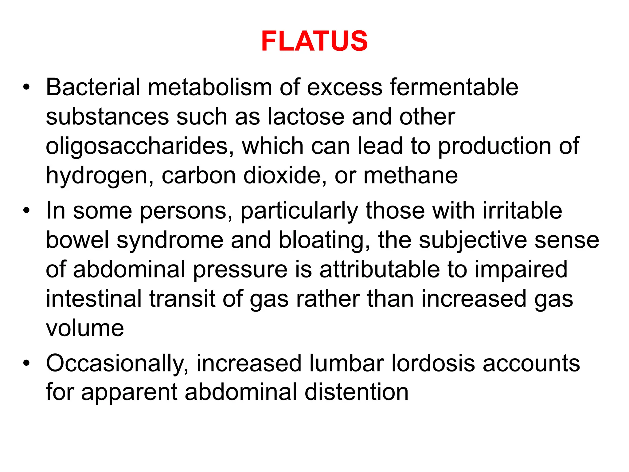
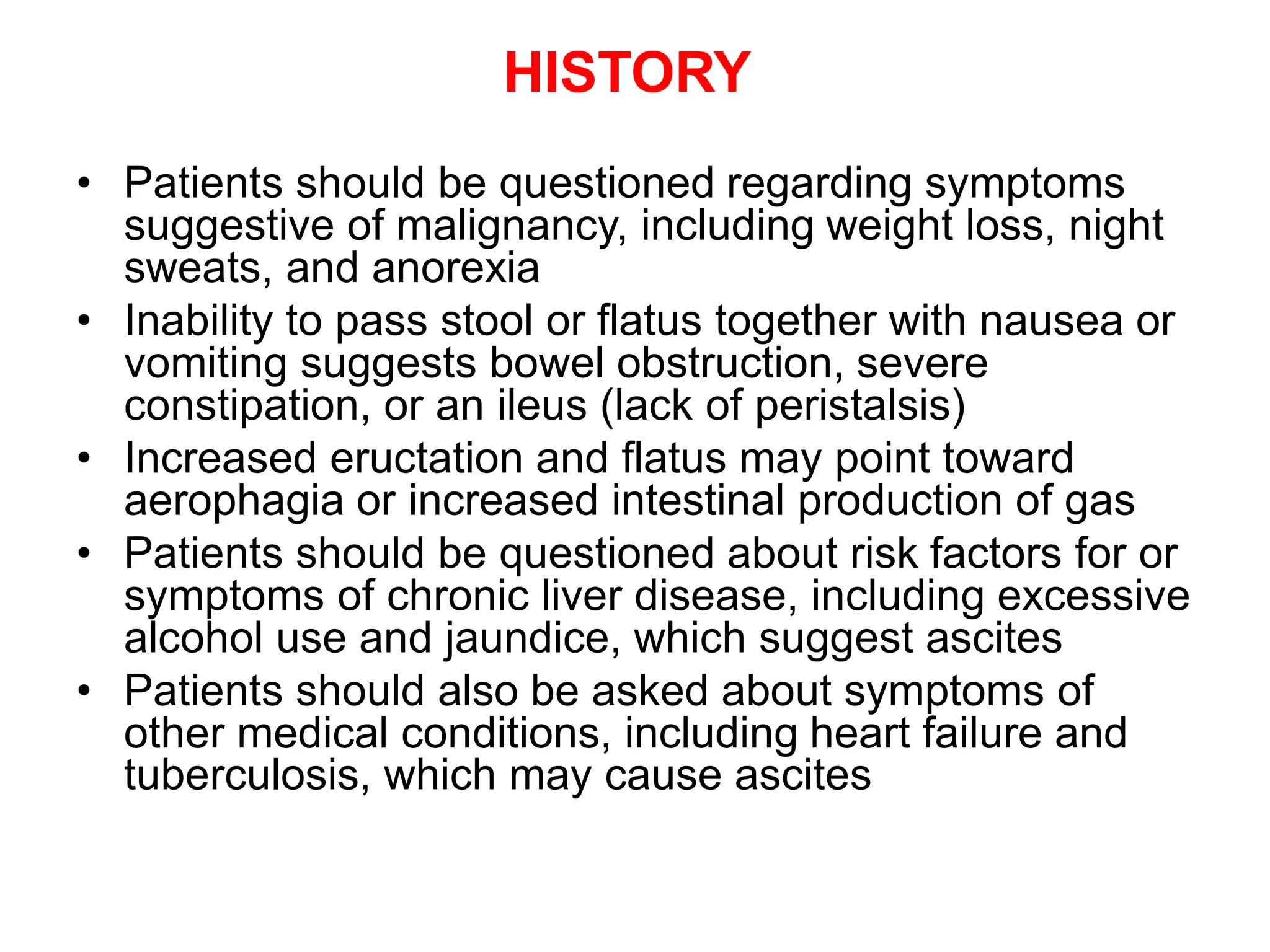
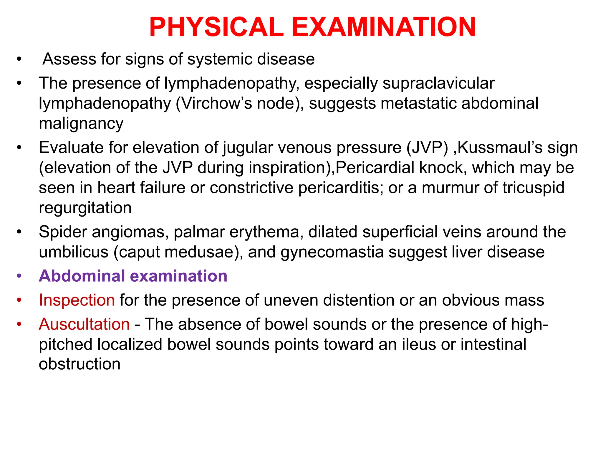
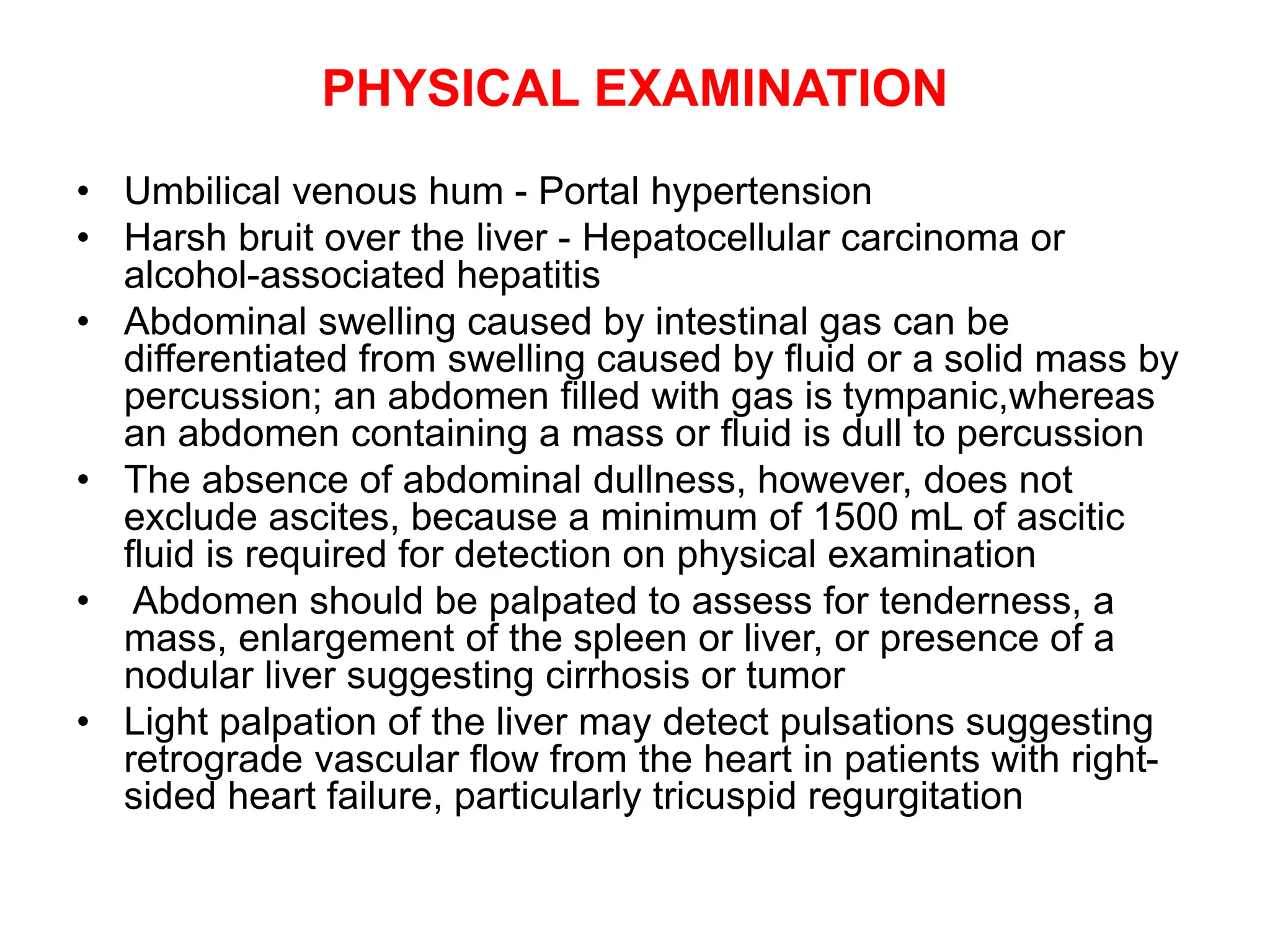
This document discusses abdominal swelling and ascites. It defines ascites as the accumulation of fluid within the abdominal cavity, which often results in abdominal distention. The document outlines various causes of abdominal swelling including fat, fluid, feces, fetus, flatus, and fatal growth. It then focuses specifically on ascites, describing the pathogenesis of ascites in the presence and absence of cirrhosis. Evaluation, treatment, and management of ascites is also discussed including paracentesis, diuretic therapy, and refractory ascites.
Any deviation in the normal nasal septum is called DNS (Deviated Nasal Septum).
Deviated Nasal Septum may be caused by mechanical trauma and may be
associated with some developmental defects.
Myringotomy (from Latin myringa "eardrum") is a surgical procedure in which a tiny incision is created in the eardrum to relieve pressure caused by excessive buildup of fluid, or to drain pus from the middle ear. A tympanostomy tube is inserted into the eardrum to keep the middle ear aerated for a prolonged time and to prevent reaccumulation of fluid. Without the insertion of a tube, the incision usually heals spontaneously in two to three weeks. Depending on the type, the tube is either naturally extruded in 6 to 12 months or removed during a minor procedure.
Strangulated intestinal obstruction is a relatively common type of acute abdomen and requires urgent surgical treatment. The causes of strangulated intestinal obstruction are many including primary volvulus, hernias, adhesions, bands, and intussusceptions.
Some slides are taken from different textbooks of medicine like Davidson, Kumar and Clark and Oxford, and some from other presentations made by respected tutors. I'm barely responsible for compilation of various resources per my interest. These resources are free for use, and I do not claim any copyright. Hoping knowledge remains free for all, forever.
CHAPTER 1 SEMESTER V PREVENTIVE-PEDIATRICS.pdfSachin Sharma
This content provides an overview of preventive pediatrics. It defines preventive pediatrics as preventing disease and promoting children's physical, mental, and social well-being to achieve positive health. It discusses antenatal, postnatal, and social preventive pediatrics. It also covers various child health programs like immunization, breastfeeding, ICDS, and the roles of organizations like WHO, UNICEF, and nurses in preventive pediatrics.
R3 Stem Cells and Kidney Repair A New Horizon in Nephrology.pptxR3 Stem Cell
R3 Stem Cells and Kidney Repair: A New Horizon in Nephrology" explores groundbreaking advancements in the use of R3 stem cells for kidney disease treatment. This insightful piece delves into the potential of these cells to regenerate damaged kidney tissue, offering new hope for patients and reshaping the future of nephrology.
Navigating Challenges: Mental Health, Legislation, and the Prison System in B...Guillermo Rivera
This conference will delve into the intricate intersections between mental health, legal frameworks, and the prison system in Bolivia. It aims to provide a comprehensive overview of the current challenges faced by mental health professionals working within the legislative and correctional landscapes. Topics of discussion will include the prevalence and impact of mental health issues among the incarcerated population, the effectiveness of existing mental health policies and legislation, and potential reforms to enhance the mental health support system within prisons.
Health Education on prevention of hypertensionRadhika kulvi
Hypertension is a chronic condition of concern due to its role in the causation of coronary heart diseases. Hypertension is a worldwide epidemic and important risk factor for coronary artery disease, stroke and renal diseases. Blood pressure is the force exerted by the blood against the walls of the blood vessels and is sufficient to maintain tissue perfusion during activity and rest. Hypertension is sustained elevation of BP. In adults, HTN exists when systolic blood pressure is equal to or greater than 140mmHg or diastolic BP is equal to or greater than 90mmHg. The
Deep Leg Vein Thrombosis (DVT): Meaning, Causes, Symptoms, Treatment, and Mor...The Lifesciences Magazine
Deep Leg Vein Thrombosis occurs when a blood clot forms in one or more of the deep veins in the legs. These clots can impede blood flow, leading to severe complications.
Antibiotic Stewardship by Anushri Srivastava.pptxAnushriSrivastav
Stewardship is the act of taking good care of something.
Antimicrobial stewardship is a coordinated program that promotes the appropriate use of antimicrobials (including antibiotics), improves patient outcomes, reduces microbial resistance, and decreases the spread of infections caused by multidrug-resistant organisms.
WHO launched the Global Antimicrobial Resistance and Use Surveillance System (GLASS) in 2015 to fill knowledge gaps and inform strategies at all levels.
ACCORDING TO apic.org,
Antimicrobial stewardship is a coordinated program that promotes the appropriate use of antimicrobials (including antibiotics), improves patient outcomes, reduces microbial resistance, and decreases the spread of infections caused by multidrug-resistant organisms.
ACCORDING TO pewtrusts.org,
Antibiotic stewardship refers to efforts in doctors’ offices, hospitals, long term care facilities, and other health care settings to ensure that antibiotics are used only when necessary and appropriate
According to WHO,
Antimicrobial stewardship is a systematic approach to educate and support health care professionals to follow evidence-based guidelines for prescribing and administering antimicrobials
In 1996, John McGowan and Dale Gerding first applied the term antimicrobial stewardship, where they suggested a causal association between antimicrobial agent use and resistance. They also focused on the urgency of large-scale controlled trials of antimicrobial-use regulation employing sophisticated epidemiologic methods, molecular typing, and precise resistance mechanism analysis.
Antimicrobial Stewardship(AMS) refers to the optimal selection, dosing, and duration of antimicrobial treatment resulting in the best clinical outcome with minimal side effects to the patients and minimal impact on subsequent resistance.
According to the 2019 report, in the US, more than 2.8 million antibiotic-resistant infections occur each year, and more than 35000 people die. In addition to this, it also mentioned that 223,900 cases of Clostridoides difficile occurred in 2017, of which 12800 people died. The report did not include viruses or parasites
VISION
Being proactive
Supporting optimal animal and human health
Exploring ways to reduce overall use of antimicrobials
Using the drugs that prevent and treat disease by killing microscopic organisms in a responsible way
GOAL
to prevent the generation and spread of antimicrobial resistance (AMR). Doing so will preserve the effectiveness of these drugs in animals and humans for years to come.
being to preserve human and animal health and the effectiveness of antimicrobial medications.
to implement a multidisciplinary approach in assembling a stewardship team to include an infectious disease physician, a clinical pharmacist with infectious diseases training, infection preventionist, and a close collaboration with the staff in the clinical microbiology laboratory
to prevent antimicrobial overuse, misuse and abuse.
to minimize the developme
CRISPR-Cas9, a revolutionary gene-editing tool, holds immense potential to reshape medicine, agriculture, and our understanding of life. But like any powerful tool, it comes with ethical considerations.
Unveiling CRISPR: This naturally occurring bacterial defense system (crRNA & Cas9 protein) fights viruses. Scientists repurposed it for precise gene editing (correction, deletion, insertion) by targeting specific DNA sequences.
The Promise: CRISPR offers exciting possibilities:
Gene Therapy: Correcting genetic diseases like cystic fibrosis.
Agriculture: Engineering crops resistant to pests and harsh environments.
Research: Studying gene function to unlock new knowledge.
The Peril: Ethical concerns demand attention:
Off-target Effects: Unintended DNA edits can have unforeseen consequences.
Eugenics: Misusing CRISPR for designer babies raises social and ethical questions.
Equity: High costs could limit access to this potentially life-saving technology.
The Path Forward: Responsible development is crucial:
International Collaboration: Clear guidelines are needed for research and human trials.
Public Education: Open discussions ensure informed decisions about CRISPR.
Prioritize Safety and Ethics: Safety and ethical principles must be paramount.
CRISPR offers a powerful tool for a better future, but responsible development and addressing ethical concerns are essential. By prioritizing safety, fostering open dialogue, and ensuring equitable access, we can harness CRISPR's power for the benefit of all. (2998 characters)
2. ABDOMINAL SWELLING
• Patients may complain of bloating or abdominal
fullness and may note increasing abdominal
girth on the basis of increased clothing or belt
size
• CAUSES
• Fat
• Fluid
• Feces
• Fetus
• Flatus
• Fatal growth
3. FLATUS
• Abdominal swelling may be the result of increased intestinal
gas
• The normal small intestine contains ~200 mL of gas made up
of nitrogen, oxygen, carbon dioxide, hydrogen, and methane
• Nitrogen and oxygen are consumed (swallowed), whereas
carbon dioxide, hydrogen, and methane are produced
intraluminally by bacterial fermentation
• CAUSES
• Aerophagia, the swallowing of air, can result in increased
amounts of oxygen and nitrogen in the small intestine and
lead to abdominal swelling
• Results from gulping food, chewing gum,smoking or as a
response to anxiety, which can lead to repetitive belching
4. FLATUS
• Bacterial metabolism of excess fermentable
substances such as lactose and other
oligosaccharides, which can lead to production of
hydrogen, carbon dioxide, or methane
• In some persons, particularly those with irritable
bowel syndrome and bloating, the subjective sense
of abdominal pressure is attributable to impaired
intestinal transit of gas rather than increased gas
volume
• Occasionally, increased lumbar lordosis accounts
for apparent abdominal distention
5. • FAT
• Weight gain with an increase in abdominal fat can result in an
increase in abdominal girth and can be perceived as
abdominal swelling
• Caused by an imbalance between caloric intake and energy
expenditure associated with a poor diet and sedentary
lifestyle
• It also can be a manifestation of certain diseases, such as
Cushing’s syndrome
• Excess abdominal fat has been associated with an increased
risk of insulin resistance and cardiovascular disease
• FLUID
• The accumulation of fluid within the abdominal cavity (ascites)
often results in abdominal distention
• Grade 1 ascites is detectable only by ultrasonography
• Grade 2 ascites is detectable by physical examination
• Grade 3 ascites results in marked abdominal distention
6. • FETUS
• Pregnancy results in increased abdominal girth
• Typically, an increase in abdominal size is first
noted at 12–14 weeks of gestation, when the
uterus moves from the pelvis into the abdomen
• FECES
• In the setting of severe constipation or
intestinal obstruction, increased stool in the
colon leads to increased abdominal girth
• These conditions are often accompanied by
abdominal discomfort or pain, nausea, and
vomiting and can be diagnosed by imaging
studies
7. • FATAL GROWTH
• An abdominal mass can result in abdominal swelling
• Neoplasms, abscesses, or cysts can grow to sizes that lead to
increased abdominal girth
• Enlargement of the intraabdominal organs, specifically the
liver (hepatomegaly) or spleen (splenomegaly), or an
abdominal aortic aneurysm can result in abdominal distention
• Bladder distention also may result in abdominal swelling
8. HISTORY
• Patients should be questioned regarding symptoms
suggestive of malignancy, including weight loss, night
sweats, and anorexia
• Inability to pass stool or flatus together with nausea or
vomiting suggests bowel obstruction, severe
constipation, or an ileus (lack of peristalsis)
• Increased eructation and flatus may point toward
aerophagia or increased intestinal production of gas
• Patients should be questioned about risk factors for or
symptoms of chronic liver disease, including excessive
alcohol use and jaundice, which suggest ascites
• Patients should also be asked about symptoms of
other medical conditions, including heart failure and
tuberculosis, which may cause ascites
9. PHYSICAL EXAMINATION
• Assess for signs of systemic disease
• The presence of lymphadenopathy, especially supraclavicular
lymphadenopathy (Virchow’s node), suggests metastatic abdominal
malignancy
• Evaluate for elevation of jugular venous pressure (JVP) ,Kussmaul’s sign
(elevation of the JVP during inspiration),Pericardial knock, which may be
seen in heart failure or constrictive pericarditis; or a murmur of tricuspid
regurgitation
• Spider angiomas, palmar erythema, dilated superficial veins around the
umbilicus (caput medusae), and gynecomastia suggest liver disease
• Abdominal examination
• Inspection for the presence of uneven distention or an obvious mass
• Auscultation - The absence of bowel sounds or the presence of high-
pitched localized bowel sounds points toward an ileus or intestinal
obstruction
10. PHYSICAL EXAMINATION
• Umbilical venous hum - Portal hypertension
• Harsh bruit over the liver - Hepatocellular carcinoma or
alcohol-associated hepatitis
• Abdominal swelling caused by intestinal gas can be
differentiated from swelling caused by fluid or a solid mass by
percussion; an abdomen filled with gas is tympanic,whereas
an abdomen containing a mass or fluid is dull to percussion
• The absence of abdominal dullness, however, does not
exclude ascites, because a minimum of 1500 mL of ascitic
fluid is required for detection on physical examination
• Abdomen should be palpated to assess for tenderness, a
mass, enlargement of the spleen or liver, or presence of a
nodular liver suggesting cirrhosis or tumor
• Light palpation of the liver may detect pulsations suggesting
retrograde vascular flow from the heart in patients with right-
sided heart failure, particularly tricuspid regurgitation
11. IMAGING AND LABORATORY EVALUATION
• Abdominal x-rays -Dilated loops of bowel in intestinal obstruction
or ileus
• Abdominal ultrasonography can detect as little as 100 mL of
ascitic fluid, hepatosplenomegaly, a nodular liver, or a mass
• If malignancy or pancreatic disease is suspected, CT can be
performed
• CT may also detect changes associated with advanced cirrhosis
and portal hypertension
• Laboratory evaluation
• Liver biochemical testing, serum albumin level measurement,
and prothrombin time determination (international normalized
ratio) to assess hepatic function as well as a complete blood
count to evaluate for the presence of cytopenias that may result
from portal hypertension or of leukocytosis, anemia, and
thrombocytosis that may result from systemic infection
• Serum amylase and lipase levels – for pancreatitis
12. • Urinary protein quantitation is indicated when nephrotic
syndrome, which may cause ascites, is suspected
• Hydrogen and methane absorbed from the intestine are
not metabolized by the host and are excreted in expired
air, and detection of increased amounts of these gases
in expired breath is the basis for tests used to diagnose
carbohydrate (e.g., lactose) malabsorption and small
intestinal bacterial overgrowth
• In selected cases, the hepatic venous pressure gradient
(pressure across the liver between the portal and hepatic
veins) can be measured via cannulation of the hepatic
vein to confirm that ascites is caused by cirrhosis
• In some cases, a liver biopsy may be necessary to
confirm cirrhosis
13. ASCITES PATHOGENESIS IN THE PRESENCE OF
CIRRHOSIS
• Ascites in patients with cirrhosis is the result of portal
hypertension and renal salt and water retention. Similar
mechanisms contribute to ascites formation in heart failure
• Portal hypertension signifies elevation of the pressure within the
portal vein
• According to Ohm’s law, pressure is the product of resistance
and flow
• Increased hepatic resistance occurs by several mechanisms
• First, the development of hepatic fibrosis, which defines
cirrhosis, disrupts the normal architecture of the hepatic
sinusoids and impedes normal blood flow through the liver
• Second, activation of hepatic stellate cells, which mediate
fibrogenesis, leads to smooth-muscle contraction and fibrosis
• Finally, cirrhosis is associated with a decrease in endothelial
nitric oxide synthetase (eNOS) production, which results in
decreased nitric oxide production and increased intrahepatic
vasoconstriction
14. • The development of cirrhosis is also associated with increased
systemic circulating levels of nitric oxide (in contrast to the decrease
seen intrahepatically), as well as increased levels of vascular
endothelial growth factor and tumor necrosis factor, that result in
splanchnic arterial vasodilation
• Vasodilation of the splanchnic circulation results in pooling of blood
and a decrease in the effective circulating volume, which is
perceived by the kidneys as hypovolemia
• Compensatory vasoconstriction via release of antidiuretic hormone
ensues; the consequences are free water retention and activation of
the sympathetic nervous system and the renin-angiotensin-
aldosterone system, which lead in turn to renal sodium and water
retention
15. PATHOGENESIS IN THE ABSENCE OF CIRRHOSIS
• Ascites in the absence of cirrhosis generally results from
peritoneal carcinomatosis, peritoneal infection, or pancreatic
disease
• Peritoneal carcinomatosis can result from primary peritoneal
malignancies such as mesothelioma or sarcoma, abdominal
malignancies such as gastric or colonic adenocarcinoma, or
metastatic disease from breast or lung carcinoma or
melanoma
• The tumor cells lining the peritoneum produce a protein-rich
fluid that contributes to the development of ascites
• Fluid from the extracellular space is drawn into the
peritoneum, further contributing to the development of ascites
• Tuberculous peritonitis causes ascites via a similar
mechanism; tubercles deposited on the peritoneum exude a
proteinaceous fluid
• Pancreatic ascites results from leakage of pancreatic
enzymes into the peritoneum
16. CAUSES
• Cirrhosis accounts for 84% of cases of ascites
• Cardiac ascites, peritoneal carcinomatosis, and
“mixed” ascites resulting from cirrhosis and a
second disease account for 10–15% of cases
• Less common causes of ascites include massive
hepatic metastasis, infection (tuberculosis,
Chlamydia infection), pancreatitis, and renal
disease (nephrotic syndrome)
• Rare causes of ascites include hypothyroidism
and familial Mediterranean fever
17. EVALUATION
• Do paracentesis, a bedside procedure in which a
needle or small catheter is passed transcutaneously
to extract ascitic fluid from the peritoneum
• Sites - Lower quadrants
• The left lower quadrant is preferred because of the
greater depth of ascites and the thinner abdominal
wall.
• Safe procedure even in patients with coagulopathy
• Complications
• Abdominal wall hematomas
• Hypotension
• Hepatorenal syndrome
• Infection
18. • Gross appearance
• Turbid fluid -presence of infection or tumor cells
• White, milky fluid - chylous ascites,triglyceride level >200
mg/dL (and often >1000 mg/dL)
• Chylous ascites results from lymphatic disruption that may
occur with trauma, cirrhosis, tumor, tuberculosis, or certain
congenital abnormalities
• Dark brown fluid – due to high bilirubin concentration,Biliary
tract perforation
• Black fluid - Pancreatic necrosis or metastatic melanoma
• Bloody – Malignant disease
• Albumin
• Total protein levels
• Cell and differential counts
• Gram’s stain and culture
• Serum albumin to calculate serum-ascites albumin gradient
19. • The SAAG is useful for distinguishing ascites caused by portal
hypertension from nonportal hypertensive ascites
• SAAG reflects the pressure within the hepatic sinusoids and
correlates with the hepatic venous pressure gradient
calculated by subtracting the ascitic albumin concentration
from the serum albumin level and does not change with
diuresis
• A SAAG ≥1.1 g/dL reflects the presence of portal hypertension
and indicates that the ascites is due to increased pressure in
the hepatic sinusoids
• According to Starling’s law, a high SAAG reflects the oncotic
pressure that counterbalances the portal pressure
• Possible causes include cirrhosis, cardiac asciteshepatic vein
thrombosis (Budd-Chiari syndrome), sinusoidal obstruction
syndrome (veno-occlusive disease), or massive liver
metastases.
• A SAAG <1.1 g/dL indicates that the ascites is not related to
portal hypertension, as in tuberculous peritonitis, peritoneal
carcinomatosis,or pancreatic ascites
20. • For high-SAAG (≥1.1) ascites, the ascitic protein level
can provide further clues to the etiology
• An ascitic protein level of ≥2.5 g/dL indicates that the
hepatic sinusoids are normal and are allowing
passage of protein into the ascites, as occurs in
cardiac ascites, early Budd-Chiari syndrome, or
sinusoidal obstruction syndrome
• An ascitic protein level <2.5 g/dL indicates that the
hepatic sinusoids have been damaged and scarred
and no longer allow passage of protein, as occurs with
cirrhosis, late Budd-Chiari syndrome, or massive liver
metastases
• Pro-brain-type natriuretic peptide (BNP) is a natriuretic
hormone released by the heart as a result of increased
volume and ventricular wall stretch
• High levels of BNP in serum occur in heart failure and
may be useful in identifying heart failure as the cause
of high-SAAG ascites
21.
22. • When secondary peritonitis resulting from a
perforated hollow viscus is suspected, ascitic
glucose and lactate dehydrogenase (LDH) levels
can be measured
• Secondary peritonitis is suggested by an ascitic
glucose level <50 mg/ dL, an ascitic LDH level
higher than the serum LDH level, and the
detection of multiple pathogens on ascitic fluid
culture
• When pancreatic ascites is suspected, the ascitic
amylase level should be measured and is typically
>1000 U/L
• Cytology can be useful in the diagnosis of
peritoneal carcinomatosis
• At least 50 mL of fluid should be obtained and sent
for immediate processing
23. • Tuberculous peritonitis is typically associated with ascitic
fluid lymphocytosis
• A smear for acid-fast bacilli has a diagnostic sensitivity of
only 0–3%; a culture increases the sensitivity to 35–50%
• In patients without cirrhosis, an elevated ascitic
adenosine deaminase level has a sensitivity of >90% for
tuberculous ascites when a cut-off value of 30–45 U/L is
used
• When the cause of ascites remains uncertain,
laparotomy or laparoscopy with peritoneal biopsies for
histology and culture remains the gold standard
24. TREATMENT
• The initial treatment for cirrhotic ascites is restriction of
sodium intake to 2 g/d
• When sodium restriction alone is inadequate to control
ascites, oral diuretics—typically the combination of
spironolactone and furosemide—are used to increase
urinary sodium excretion
• Spironolactone is an aldosterone antagonist that inhibits
sodium reabsorption in the distal convoluted tubule of
the kidney
• Use of spironolactone may be limited by
hyponatremia,hyperkalemia, and painful gynecomastia
• If the gynecomastia is distressing, amiloride (5–40
mg/d) may be substituted for spironolactone
25. • Furosemide is a loop diuretic that is generally
combined with spironolactone in a ratio of 40:100;
maximal daily doses of spironolactone and
furosemide are 400 mg and 160 mg, respectively
• Fluid intake may be restricted in patients with
hyponatremia
• Diuretics should be titrated to remove no more than
1 L of fluid daily, so body weight should not fall by
more than 1 kg daily to avoid excessive fluid
depletion
• Refractory cirrhotic ascites is defined by the
persistence of ascites despite sodium restriction and
maximal (or maximally tolerated) diuretic use
26. • Pharmacologic therapy for refractory ascites
Addition of midodrine, an α1-adrenergic agonist, or
clonidine, an α2-adrenergic agonist, to diuretic
therapy
• These agents act as vasoconstrictors,
counteracting splanchnic vasodilation
• Midodrine alone or in combination with clonidine
improves systemic hemodynamics and control of
ascites over that obtained with diuretics alone
• Use of beta blockers in patients with refractory
ascites may be associated with decreased survival
rates
27. • When medical therapy alone is insufficient, refractory
cirrhotic ascites can be managed by repeated large-
volume paracentesis (LVP) or a transjugular intrahepatic
peritoneal shunt (TIPS)—a radiologically placed
portosystemic shunt that decompresses the hepatic
sinusoids
• Intravenous (IV) infusion of albumin accompanying LVP
decreases the risk of “post paracentesis circulatory
dysfunction” and death
• Patients undergoing LVP should receive IV albumin
infusions of 6–8 g/L of ascitic fluid removed
• TIPS placement is superior to LVP in reducing the
reaccumulation of ascites but is associated with an
increased frequency of hepatic encephalopathy, with no
difference in mortality rates
29. • The peritoneo-venous shunt
• It is a long tube with a non-return valve running
subcutaneously from the peritoneum to the
internal jugular vein in the neck
• It allows ascitic fluid to pass directly into the
systemic circulation
• Complications
• Infection,
• Superior vena caval thrombosis
• pulmonary oedema
• Bleeding from oesophageal varices
• Disseminated intravascular coagulopathy
• These complications limit its use and insertion of
these shunts is now rare
31. • The Alfapump system, which consists of an automated
pump and tunneled peritoneal catheter that transports
ascites from the peritoneal cavity to the urinary bladder
• Drawbacks: A higher frequency of technical difficulties and
renal dysfunction
• Malignant ascites does not respond to sodium restriction or
diuretics
• Patients must undergo serial LVPs,transcutaneous
drainage catheter placement, or, rarely, creation of a
peritoneovenous shunt (a shunt from the abdominal cavity
to the vena cava) or placement of the Alfapump system, if
available
• Ascites caused by tuberculous peritonitis is treated with
standard antituberculosis therapy
• Noncirrhotic ascites of other causes is treated by correction
of the precipitating condition
33. COMPLICATIONS
• Spontaneous bacterial peritonitis is a common
and potentially lethal complication of cirrhotic
ascites
• Occasionally, SBP also complicates ascites
caused by nephrotic syndrome, heart failure, acute
hepatitis, and acute liver failure but is rare in
malignant ascites
• Patients with SBP generally note an increase in
abdominal girth; however, abdominal tenderness
is found in only 40% of patients, and rebound
tenderness is uncommon
• Clinical features: Fever, nausea, vomiting, or the
new onset or an exacerbation of preexisting
hepatic encephalopathy
34. • SBP is defined by a polymorphonuclear neutrophil
(PMN) count of ≥250/ μL in the ascitic fluid
• Cultures of ascitic fluid should be performed in blood
culture bottles and typically reveal one bacterial
pathogen
• The presence of multiple pathogens in the setting of an
elevated ascitic PMN count suggests secondary
peritonitis from a ruptured viscus or abscess
• The presence of multiple pathogens without an elevated
PMN count suggests bowel perforation from the
paracentesis needle
• SBP is generally the result of enteric bacteria that have
translocated across an edematous bowel wall
• The most common pathogens are gram-negative rods,
including Escherichia coli and Klebsiella, as well as
streptococci and enterococci
• Treatment of SBP
• IV cefotaxime for 5 days
35. • Nosocomial or health care–acquired SBP is
frequently caused by multidrug- resistant
bacteria, and initial antibiotic therapy should
be guided by the local bacterial epidemiology
• Cirrhotic patients with a history of SBP, an
ascitic fluid total protein concentration <1
g/dL, or active gastrointestinal bleeding
should receive prophylactic antibiotics to
prevent SBP
• Oral daily Ciprofloxacin 500mg/day or
Norfloxacin 400mg/day
• IV ceftriaxone may be used in hospitalized
patients
• Diuresis increases the activity of ascitic fluid
protein opsonins and may decrease the risk
of SBP
36. • Hepatorenal syndrome
• There are two clinical types; both are mediated by renal
vasoconstriction due to underfilling of the arterial circulation
• Type 1 hepatorenal syndrome is characterised by progressive
oliguria, a rapid rise of the serum creatinine and a very poor
prognosis (without treatment, median survival is less than 1
month)
• There is usually no proteinuria, a urine sodium excretion of less
than 10 mmol/day and a urine/ plasma osmolarity ratio of more
than 1.5
• Other non-functional causes of renal failure must be excluded
before the diagnosis is made
• Treatment consists of albumin infusions in combination with
terlipressin and is effective in about two-thirds of patients
• Haemodialysis should not be used routinely because it does not
improve the outcome
• Patients who survive should be considered for liver
transplantation.
• Type 2 hepatorenal syndrome usually occurs in patients with
refractory ascites, is characterised by a moderate and stable
increase in serum creatinine, and has a better prognosis.
37. • Renal failure
• Renal failure can occur in patients with ascites
• It can be pre-renal due to vasodilatation from sepsis
and/or diuretic therapy, or due to hepatorenal
syndrome
• Hepatic hydrothorax
• occurs when ascites, often caused by cirrhosis,
migrates via fenestrae in the diaphragm into the
pleural space
• Result in shortness of breath, hypoxia, and infection
• Treatment
• Sodium restriction, diuretics, and, if needed,
thoracentesis or TIPS placement
• Chest tube placement should be avoided