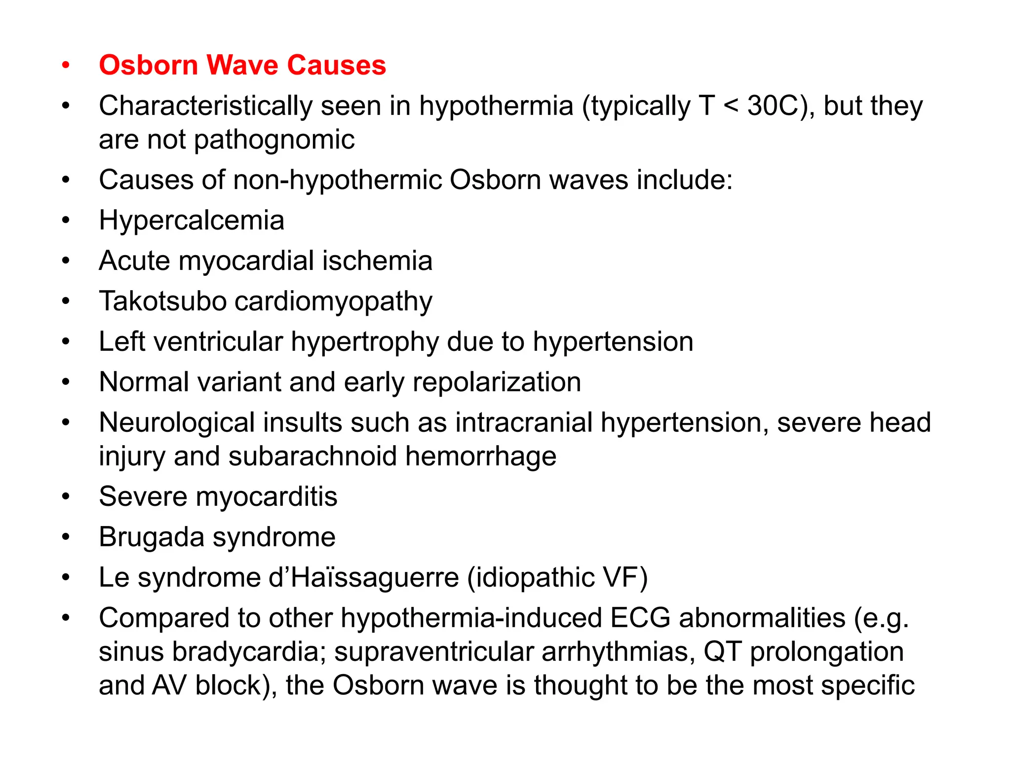
The Osborn wave, also known as a J wave, appears as a positive deflection at the J point in ECG leads. It is most commonly associated with hypothermia below 30 degrees Celsius. Other potential causes include hypercalcemia, myocardial ischemia, Takotsubo cardiomyopathy, hypertension, early repolarization, neurological insults, myocarditis, and genetic conditions. The height of the J wave tends to increase with greater severity of hypothermia. Osborn waves are a specific sign of hypothermia compared to other ECG changes like bradycardia and QT prolongation.


![Osborn Wave ECG examples
Subtle J waves in mild hypothermia [Temp: 32.5°C (90.5°F)]
The height of the J wave is roughly proportional to the degree of
hypothermia](https://image.slidesharecdn.com/osbornwavejwave-240412051427-64e5878a/75/OSBORN-WAVE-J-Wave-IN-ECG-AND-ITS-INTERPRETATION-5-2048.jpg)
![•J waves in moderate hypothermia [Temp: 30°C (86°F)]](https://image.slidesharecdn.com/osbornwavejwave-240412051427-64e5878a/75/OSBORN-WAVE-J-Wave-IN-ECG-AND-ITS-INTERPRETATION-6-2048.jpg)
![•J waves in moderate hypothermia. [Temp: 28°C (82.4°F)]](https://image.slidesharecdn.com/osbornwavejwave-240412051427-64e5878a/75/OSBORN-WAVE-J-Wave-IN-ECG-AND-ITS-INTERPRETATION-7-2048.jpg)
![•Marked J waves in severe hypothermia [Temp: 26°C (78.8°F)]
•Note “negative” J waves seen in aVR and V1](https://image.slidesharecdn.com/osbornwavejwave-240412051427-64e5878a/75/OSBORN-WAVE-J-Wave-IN-ECG-AND-ITS-INTERPRETATION-8-2048.jpg)


