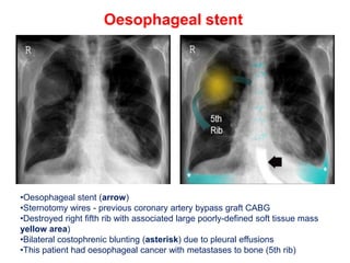
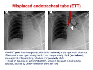
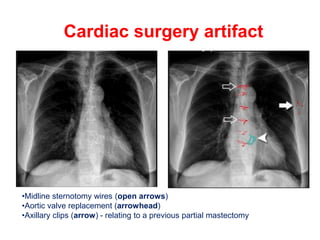
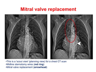
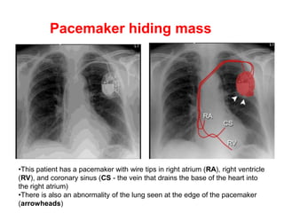
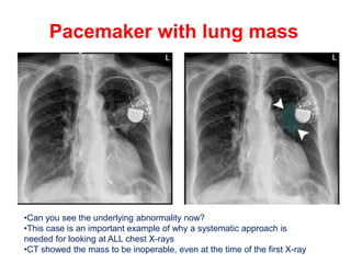
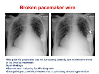
2. PCP and nipple ring
•This X-ray shows patchy shadowing throughout both lungs in a patient with
PCP - Pneumocystis pneumonia - due to underlying HIV.
•There is nipple ring made of metal and a less dense closure device
(arrowhead)
•PCP typically causes bilateral symmetrical hilar interstitial shadowing, but it can
cause any pattern of shadowing
•A normal chest X-ray does not exclude the diagnosis
3. Jugular line for chemotherapy
•A tunnelled line is inserted via the internal jugular vein
•Its tip (arrow) lies at the cavoatrial junction - the junction of the superior vena cava and
the right atrium
•The entry site (E) is where the line enters the vein,Red section is therefore intravenous
•The exit site (X) is where it exits the skin
•Between these two points the line is tunnelled through the subcutaneous tissues over
the front of the chest wall (orange section)
•The purpose of inserting a tunnelled line is so that it can stay in situ for a long period of
time. This patient with cancer was receiving long term chemotherapy, via the two access
points which lie outside the skin (green section)
4. Portacath
•This tube, known as a portacath lies entirely under the skin
•The line enters the subclavian vein just below the clavicle
• At the 'drum' end there is a membrane (asterisk) to allow delivery of drugs via a
needle
•Used for delivery of long term antibiotics, in conditions such as cystic fibrosis
•Note the underlying lungs are abnormal with a coarse, reticular (net-like) shadowing,
and parallel 'tram-lines,' indicating bronchiectasis, a typical feature of cystic fibrosis -
the diagnosis in this case
5. Tunnelled line
•This cancer patient has another type of tunnelled line which has been placed
via the left subclavian route
6. Oesophageal stent
•Oesophageal stent (arrow)
•Sternotomy wires - previous coronary artery bypass graft CABG
•Destroyed right fifth rib with associated large poorly-defined soft tissue mass
yellow area)
•Bilateral costophrenic blunting (asterisk) due to pleural effusions
•This patient had oesophageal cancer with metastases to bone (5th rib)
7. Misplaced endotracheal tube (ETT)
•The ETT (red) has been placed with its tip (asterisk) in the right main bronchus
•The arrow shows open airways which are comparatively black (arrowhead),
seen against collapsed lung, which is comparatively white
• This is an example of 'air bronchogram,' which in this case is due to lung
collapse, caused by under-ventilation of the left lung
8. Cardiac surgery artifact
•Midline sternotomy wires (open arrows)
•Aortic valve replacement (arrowhead)
•Axillary clips (arrow) - relating to a previous partial mastectomy
9. Mitral valve replacement
•This is a 'scout view' (planning view) for a chest CT scan
•Midline sternotomy wires (red ring)
•Mitral valve replacement (arrowhead)
10. Pacemaker hiding mass
•This patient has a pacemaker with wire tips in right atrium (RA), right ventricle
(RV), and coronary sinus (CS - the vein that drains the base of the heart into
the right atrium)
•There is also an abnormality of the lung seen at the edge of the pacemaker
(arrowheads)
11. Pacemaker with lung mass
•Can you see the underlying abnormality now?
•This case is an important example of why a systematic approach is
needed for looking at ALL chest X-rays
•CT showed the mass to be inoperable, even at the time of the first X-ray
12. Broken pacemaker wire
•This patient's pacemaker was not functioning correctly due to a fracture of one
of the wires (arrowhead)
Other findings
•Massive heart - allowing for AP sitting view
•Enlarged upper zone blood vessels due to pulmonary venous hypertension