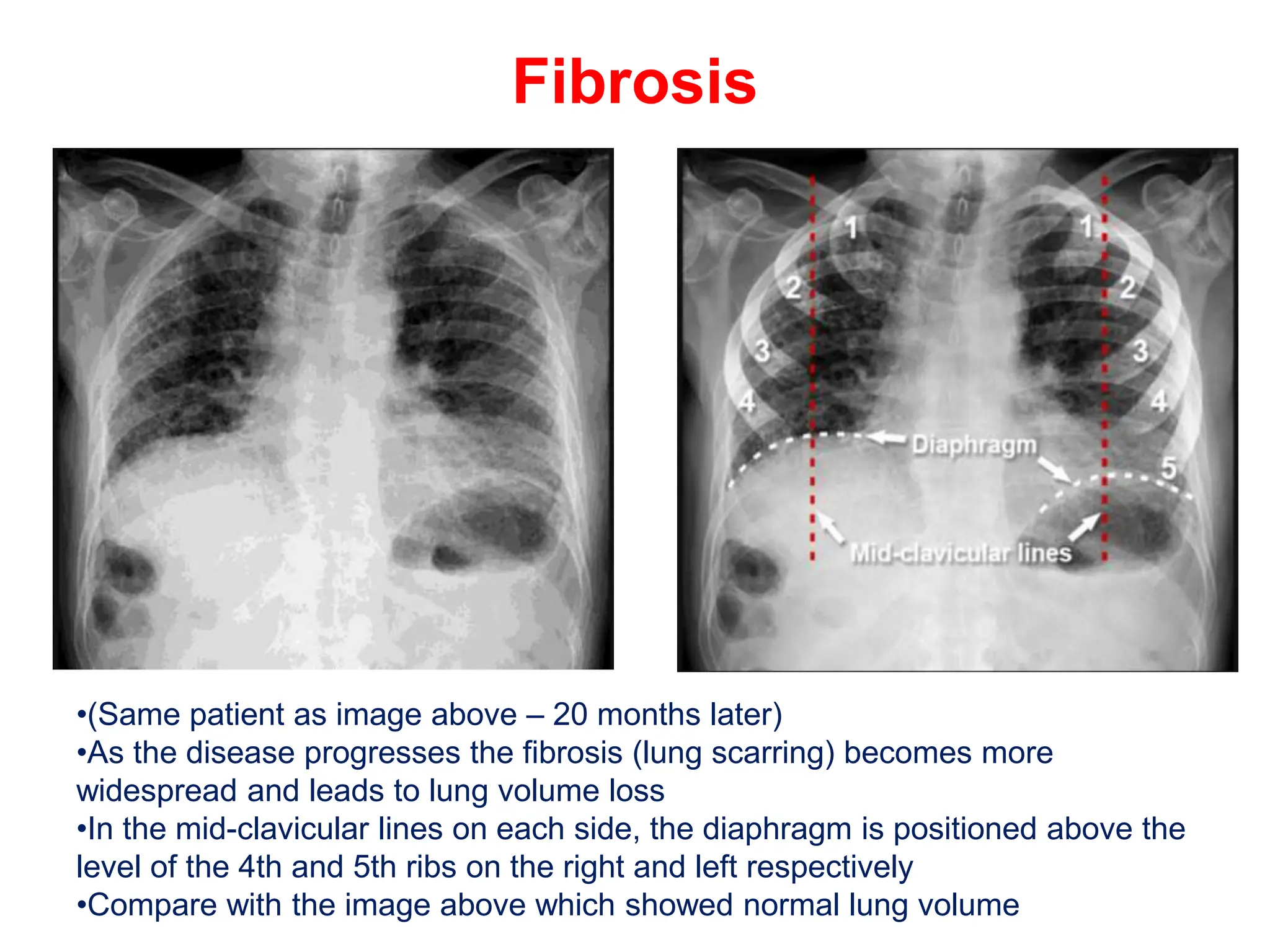
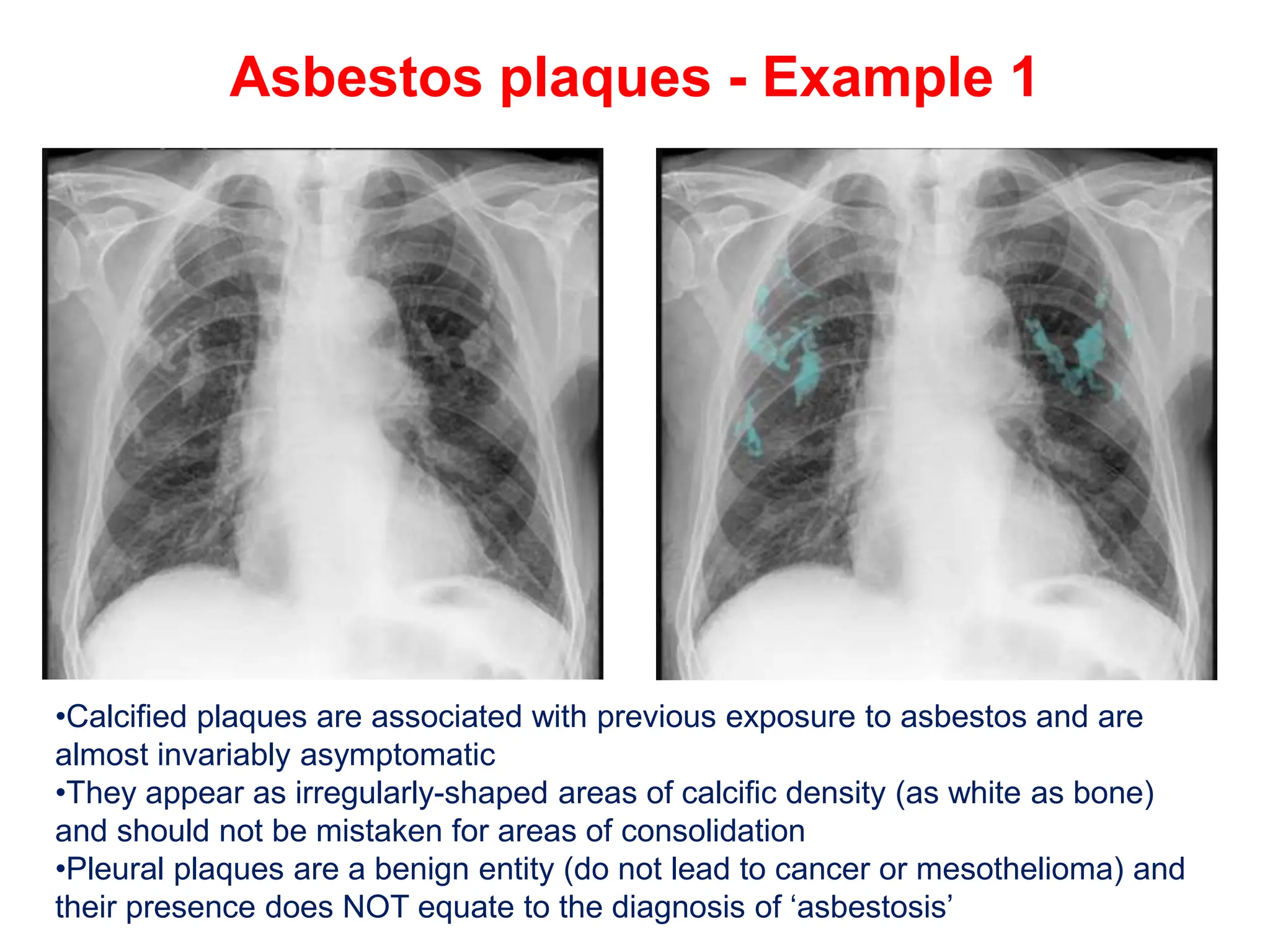
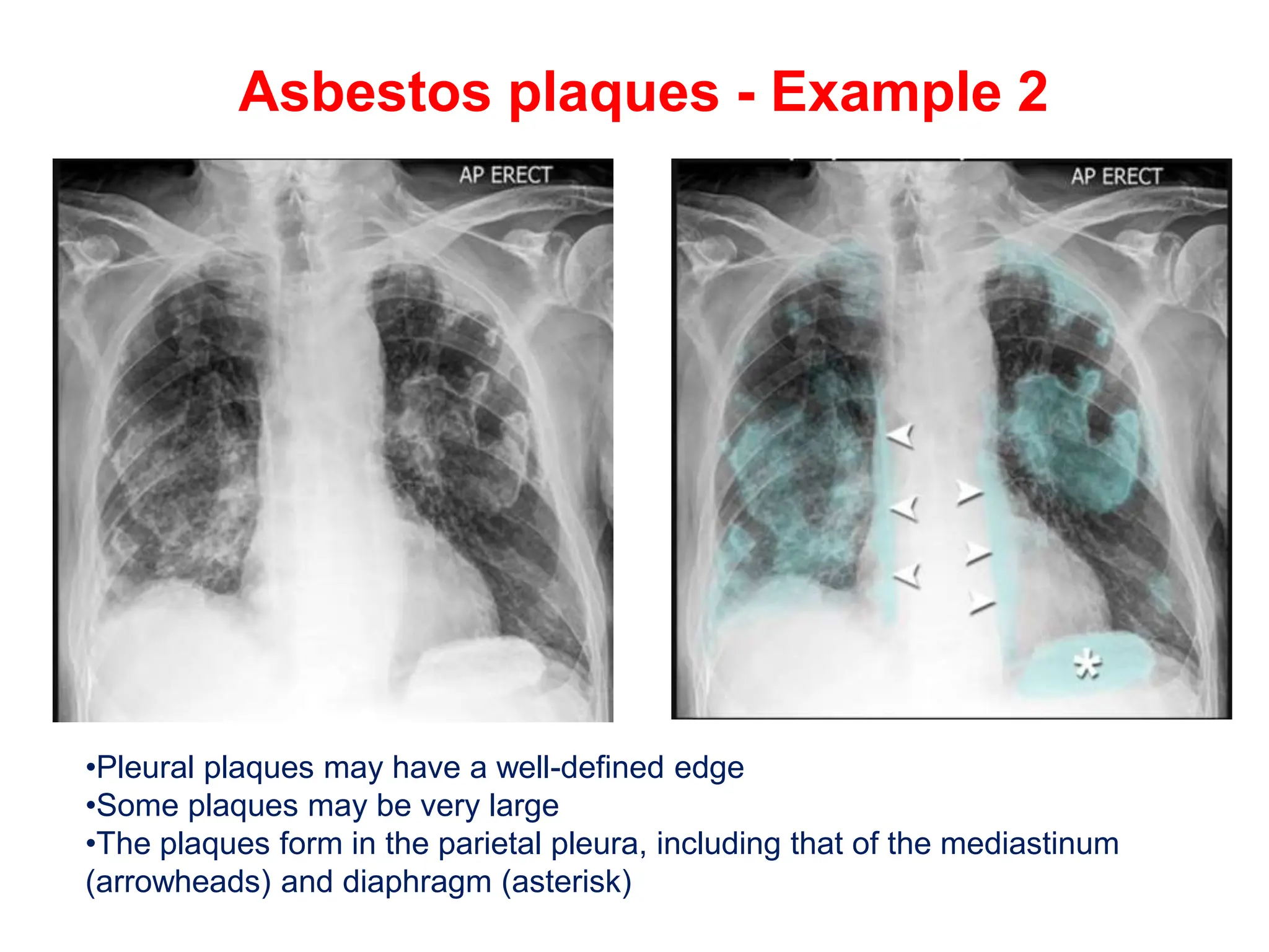
CHEST X-RAY
PULMONARY DISEASE
Basal lung consolidation
•This image shows subtle consolidation at the left lung base, partly obscured by
the heart
Air bronchogram - Example 1
•‘Air bronchogram’ is a characteristic sign of consolidation – here is an example
in a patient with pneumonia
•The black lines represent patent airways within consolidated lung (highlighted
area)
Air bronchogram - Example 2
•Another example of consolidation with air bronchogram due to pneumonia
•Remember that the term 'consolidation' does not only refer to infection
Consolidation - Right upper lobe
•Consolidation of the right upper lobe which is confined inferiorly by the horizontal
fissure
•If the consolidation is due to infection, then the term ‘lobar pneumonia’ is correctly
used
Consolidation - Right middle lobe
•The right middle lobe is located below the horizontal fissure which confines
the area of consolidation in this image
•The right middle lobe is also next to the right heart border which is obscured
in this image
Consolidation - Right lower lobe
•The right lower lobe is located adjacent to the right hemidiaphragm which is
not clearly visible in this image
•The right heart border is still visible which indicates that the consolidation is
not in the middle lobe
Consolidation - Right lower zone
•The patchy consolidation in this image is located in the right lower zone
•It is possible to determine the consolidation is not in the right upper lobe as it is
below the anatomical level of the horizontal fissure
•It is not possible to determine if the consolidation is in the right lower or middle
lobe – there is no positive silhouette sign present
Consolidation - Multiple zones
•This image shows a large area of dense consolidation located in the right middle
zone – air bronchogram is visible
•Subtle consolidation of the left lower zone with reduced definition of the left
hemidiaphragm indicates involvement of a small area of the left lower lobe
•The left heart border remains well-defined indicating the consolidation is not in
the lingula – part of the left upper lobe
RMZ pneumonia and pleural effusion
• An area of consolidation is seen in the right middle zone air bronchogram is
visible
• Below this there is an area of whiteness due to a parapneumonic effusion – no
air bronchogram is visible in this area
Lobar pneumonia - Fungal infection
•Lobar pneumonia is usually caused by typical organisms – such as Streptococcus
pneumoniae – but may also be caused by atypical organisms – as in this patient
•The consolidation obscures the left heart border indicating it is in the adjacent
lingula of the left upper lobe
•Pneumonia caused by atypical organisms is more common in individuals who are
immunocompromised
•This patient was immunocompromised due to chemotherapy treatment for
leukaemia
•Fungal elements were found following bronchoscopy and bronchial washing
Pneumocystis pneumonia
•Consolidation seen in a non-lobar distribution should raise the suspicion of
atypical organisms
•This patient with known HIV infection has subtle consolidation in the mid zones
bilaterally
•Although the consolidation appears minor, this patient was extremely unwell
with low oxygen saturation which worsened on minor effort (walking down the
ward)
•Note: Initially the chest X-ray can be entirely normal in patients with PCP
Aspergillosis
•This patient was taking immunosuppressing drugs following a renal transplant
•Dense consolidation is seen throughout both lungs due to fungal infection –
proven to be invasive aspergillosis in this case
Primary TB
•There are no radiological features which are in themselves diagnostic of primary
mycobacterium tuberculosis infection (TB) but a chest X-ray may provide some
clues to the diagnosis
•This image shows consolidation of the upper zone with ipsilateral hilar
enlargement due to lymphadenopathy
•These are typical features of primary TB
•Note: The chest X-ray may be normal in primary TB, in fact most patients
infected are never unwell enough to require a chest X-ray
Healed primary TB
•Following an immune response to primary infection, a caseating granuloma
forms which calcifies over time – this is known as a ‘Ghon focus’
•A Ghon focus is a rounded, well-defined focus of calcific density (as dense as
bone) usually located in the periphery of the lung
•This chest X-ray shows a large, rounded calcified focus near the right hilum
•The CT (not usually necessary) shows it is located in the lung peripherally
•This is a particularly large Ghon focus
Post-primary TB
•Post-primary TB (secondary TB or reactivation TB) is more common in
immunocompromised individuals – for example those with HIV/AIDS, those on
immunosuppressing drugs, or those with malnutrition or diabetes
•The upper lobes are more commonly affected
•Consolidation often extends to the hilum
•The hilar structures may be distorted due to volume loss of the upper lobe
Post-primary TB – Lung cavity
•(Same patient as image above – 4 months later)
•Cavities are a common finding in mycobacterial infection
Healed post-primary TB
•Following an immune response to post-primary infection, the affected area
often becomes scarred (fibrotic) and calcified
•The combined fibrosis and calcification can be described as ‘fibro-calcific
change’
Miliary TB
•Miliary TB is due to disseminated spread of mycobacterial infection
•It can occur either at the time of primary infection or on disease reactivation –
prognosis is poor
•Very fine nodules are typically seen scattered throughout the lungs
Lung contusion – displaced rib fractures
•Lung contusion (bleeding into the airways of the lungs) has identical
appearances to any other cause of consolidation
•It is associated with a history of direct trauma to the chest wall
•This image shows several displaced rib fractures
Lung contusion – undisplaced rib
fractures
•Rib fractures are often invisible or very difficult to see on a chest X-ray
•The consolidation seen in the right lower zone is due to lung contusion
•Note: Clinical suspicion of rib fractures is not generally considered an
indication for a chest X-ray unless there is also suspicion of complications
such as a pneumothorax
Lung cavity/abscess
•Lung cavities can be caused by disease processes other than infection
•This patient has a lung cavity which was initially thought to be a cancer but
which disappeared after a course of antibiotics
•This cavity was, therefore, correctly termed a 'lung abscess'
Lung abscess with gas/fluid level
•This patient had a history of breast cancer – (it would be unusual for a
metastatic breast lesion to cavitate)
•The cavity was an abscess which arose due to the patient being
immunocompromised by chemotherapy drugs
•A fluid level represents a collection of pus in the abscess
Bronchiectasis
•Bronchiectasis may be present even if the X-ray is normal – most patients
suspected of having bronchiectasis will need a high resolution CT to confirm
the diagnosis
•Severe bronchiectasis causes coarsening of the lung markings – very
extensive in this case
Bronchiectasis - Cystic fibrosis
•Patients with cystic fibrosis typically develop bronchiectasis
•Chest X-rays are not required to make the diagnosis, but are helpful to
monitor progression of the disease and to look for complications such as
pneumonia
•The ‘portacath’ device is used for long-term delivery of intravenous drugs
COPD – Hyperexpansion
•Flattening of the diaphragm (red lines) is often a more reliable feature of lung
hyperexpansion
•The green dotted lines indicate the predicted normal diaphragm shape and
position
COPD – Floating heart sign
•The lungs may be so hyper-expanded that the inferior border of the heart
becomes visible – the heart appears to float above the diaphragm
•Note the flattened hemidiaphragms (white dotted lines)
COPD – bullous emphysema
•Bullous emphysema manifests on a chest X-ray with areas of low density
(black) with thinning of the pulmonary vessels, predominantly affecting the
upper zones
•The lower part of the lungs may appear denser (whiter) in normal subjects
because of overlying breast tissue, but in this individual the pulmonary vessels
appear normal in this area
COPD – bullous emphysema
•Occasionally bullae are seen as discretely outlined holes in the lungs which
resemble bubbles
COPD – Large bullae
•Bullae can get very large and must not be mistaken for a pneumothorax –
inserting a chest drain can have catastrophic consequences in this setting
COPD – infective exacerbation
•The lungs are hyper-expanded – the diaphragm is flattened and too low
•There are large areas of black due to bullae
•A large area of consolidation in the left lung is due to pneumonia
Reticular shadowing - Fibrosis
•Pulmonary fibrosis causes reticular (net-like) shadowing of the lung peripheries
which is typically more prominent towards the lung bases
•It may cause the contours of the heart to be less distinct or ‘shaggy’
•Chest X-rays can be helpful in monitoring the progression of pulmonary fibrosis
Fibrosis
•(Same patient as image above – 20 months later)
•As the disease progresses the fibrosis (lung scarring) becomes more
widespread and leads to lung volume loss
•In the mid-clavicular lines on each side, the diaphragm is positioned above the
level of the 4th and 5th ribs on the right and left respectively
•Compare with the image above which showed normal lung volume
Asbestos plaques - Example 1
•Calcified plaques are associated with previous exposure to asbestos and are
almost invariably asymptomatic
•They appear as irregularly-shaped areas of calcific density (as white as bone)
and should not be mistaken for areas of consolidation
•Pleural plaques are a benign entity (do not lead to cancer or mesothelioma) and
their presence does NOT equate to the diagnosis of ‘asbestosis’
Asbestos plaques - Example 2
•Pleural plaques may have a well-defined edge
•Some plaques may be very large
•The plaques form in the parietal pleura, including that of the mediastinum
(arrowheads) and diaphragm (asterisk)
Asbestos plaques - Example 3
•When seen en face they may be difficult to see – as is the left upper zone
plaque in this image
•The diaphragm is often the best place to look for plaques where they lie in
the plane of the X-ray beam
Mesothelioma - Image 1 - Pleural effusion
•Mesothelioma frequently presents as a pleural effusion – often a lot
smaller than the effusion in this image
Mesothelioma - Image 2 - Post chest
drain
•(Same patient as image above)
•The effusion in the image above was drained
•Lobulated thickening of the pleura became visible
•The left lung is reduced in volume
•These are the typical features of mesothelioma
•Note: Pleural metastases (usually from an adenocarcinoma) may have similar
appearances to mesothelioma

CHEST X-RAY PULMONARY DISEASE pptx.pptx

  • 1.
  • 2.
    Basal lung consolidation •Thisimage shows subtle consolidation at the left lung base, partly obscured by the heart
  • 3.
    Air bronchogram -Example 1 •‘Air bronchogram’ is a characteristic sign of consolidation – here is an example in a patient with pneumonia •The black lines represent patent airways within consolidated lung (highlighted area)
  • 4.
    Air bronchogram -Example 2 •Another example of consolidation with air bronchogram due to pneumonia •Remember that the term 'consolidation' does not only refer to infection
  • 5.
    Consolidation - Rightupper lobe •Consolidation of the right upper lobe which is confined inferiorly by the horizontal fissure •If the consolidation is due to infection, then the term ‘lobar pneumonia’ is correctly used
  • 6.
    Consolidation - Rightmiddle lobe •The right middle lobe is located below the horizontal fissure which confines the area of consolidation in this image •The right middle lobe is also next to the right heart border which is obscured in this image
  • 7.
    Consolidation - Rightlower lobe •The right lower lobe is located adjacent to the right hemidiaphragm which is not clearly visible in this image •The right heart border is still visible which indicates that the consolidation is not in the middle lobe
  • 8.
    Consolidation - Rightlower zone •The patchy consolidation in this image is located in the right lower zone •It is possible to determine the consolidation is not in the right upper lobe as it is below the anatomical level of the horizontal fissure •It is not possible to determine if the consolidation is in the right lower or middle lobe – there is no positive silhouette sign present
  • 9.
    Consolidation - Multiplezones •This image shows a large area of dense consolidation located in the right middle zone – air bronchogram is visible •Subtle consolidation of the left lower zone with reduced definition of the left hemidiaphragm indicates involvement of a small area of the left lower lobe •The left heart border remains well-defined indicating the consolidation is not in the lingula – part of the left upper lobe
  • 10.
    RMZ pneumonia andpleural effusion • An area of consolidation is seen in the right middle zone air bronchogram is visible • Below this there is an area of whiteness due to a parapneumonic effusion – no air bronchogram is visible in this area
  • 11.
    Lobar pneumonia -Fungal infection •Lobar pneumonia is usually caused by typical organisms – such as Streptococcus pneumoniae – but may also be caused by atypical organisms – as in this patient •The consolidation obscures the left heart border indicating it is in the adjacent lingula of the left upper lobe •Pneumonia caused by atypical organisms is more common in individuals who are immunocompromised •This patient was immunocompromised due to chemotherapy treatment for leukaemia •Fungal elements were found following bronchoscopy and bronchial washing
  • 12.
    Pneumocystis pneumonia •Consolidation seenin a non-lobar distribution should raise the suspicion of atypical organisms •This patient with known HIV infection has subtle consolidation in the mid zones bilaterally •Although the consolidation appears minor, this patient was extremely unwell with low oxygen saturation which worsened on minor effort (walking down the ward) •Note: Initially the chest X-ray can be entirely normal in patients with PCP
  • 13.
    Aspergillosis •This patient wastaking immunosuppressing drugs following a renal transplant •Dense consolidation is seen throughout both lungs due to fungal infection – proven to be invasive aspergillosis in this case
  • 14.
    Primary TB •There areno radiological features which are in themselves diagnostic of primary mycobacterium tuberculosis infection (TB) but a chest X-ray may provide some clues to the diagnosis •This image shows consolidation of the upper zone with ipsilateral hilar enlargement due to lymphadenopathy •These are typical features of primary TB •Note: The chest X-ray may be normal in primary TB, in fact most patients infected are never unwell enough to require a chest X-ray
  • 15.
    Healed primary TB •Followingan immune response to primary infection, a caseating granuloma forms which calcifies over time – this is known as a ‘Ghon focus’ •A Ghon focus is a rounded, well-defined focus of calcific density (as dense as bone) usually located in the periphery of the lung •This chest X-ray shows a large, rounded calcified focus near the right hilum •The CT (not usually necessary) shows it is located in the lung peripherally •This is a particularly large Ghon focus
  • 16.
    Post-primary TB •Post-primary TB(secondary TB or reactivation TB) is more common in immunocompromised individuals – for example those with HIV/AIDS, those on immunosuppressing drugs, or those with malnutrition or diabetes •The upper lobes are more commonly affected •Consolidation often extends to the hilum •The hilar structures may be distorted due to volume loss of the upper lobe
  • 17.
    Post-primary TB –Lung cavity •(Same patient as image above – 4 months later) •Cavities are a common finding in mycobacterial infection
  • 18.
    Healed post-primary TB •Followingan immune response to post-primary infection, the affected area often becomes scarred (fibrotic) and calcified •The combined fibrosis and calcification can be described as ‘fibro-calcific change’
  • 19.
    Miliary TB •Miliary TBis due to disseminated spread of mycobacterial infection •It can occur either at the time of primary infection or on disease reactivation – prognosis is poor •Very fine nodules are typically seen scattered throughout the lungs
  • 20.
    Lung contusion –displaced rib fractures •Lung contusion (bleeding into the airways of the lungs) has identical appearances to any other cause of consolidation •It is associated with a history of direct trauma to the chest wall •This image shows several displaced rib fractures
  • 21.
    Lung contusion –undisplaced rib fractures •Rib fractures are often invisible or very difficult to see on a chest X-ray •The consolidation seen in the right lower zone is due to lung contusion •Note: Clinical suspicion of rib fractures is not generally considered an indication for a chest X-ray unless there is also suspicion of complications such as a pneumothorax
  • 22.
    Lung cavity/abscess •Lung cavitiescan be caused by disease processes other than infection •This patient has a lung cavity which was initially thought to be a cancer but which disappeared after a course of antibiotics •This cavity was, therefore, correctly termed a 'lung abscess'
  • 23.
    Lung abscess withgas/fluid level •This patient had a history of breast cancer – (it would be unusual for a metastatic breast lesion to cavitate) •The cavity was an abscess which arose due to the patient being immunocompromised by chemotherapy drugs •A fluid level represents a collection of pus in the abscess
  • 24.
    Bronchiectasis •Bronchiectasis may bepresent even if the X-ray is normal – most patients suspected of having bronchiectasis will need a high resolution CT to confirm the diagnosis •Severe bronchiectasis causes coarsening of the lung markings – very extensive in this case
  • 25.
    Bronchiectasis - Cysticfibrosis •Patients with cystic fibrosis typically develop bronchiectasis •Chest X-rays are not required to make the diagnosis, but are helpful to monitor progression of the disease and to look for complications such as pneumonia •The ‘portacath’ device is used for long-term delivery of intravenous drugs
  • 26.
    COPD – Hyperexpansion •Flatteningof the diaphragm (red lines) is often a more reliable feature of lung hyperexpansion •The green dotted lines indicate the predicted normal diaphragm shape and position
  • 27.
    COPD – Floatingheart sign •The lungs may be so hyper-expanded that the inferior border of the heart becomes visible – the heart appears to float above the diaphragm •Note the flattened hemidiaphragms (white dotted lines)
  • 28.
    COPD – bullousemphysema •Bullous emphysema manifests on a chest X-ray with areas of low density (black) with thinning of the pulmonary vessels, predominantly affecting the upper zones •The lower part of the lungs may appear denser (whiter) in normal subjects because of overlying breast tissue, but in this individual the pulmonary vessels appear normal in this area
  • 29.
    COPD – bullousemphysema •Occasionally bullae are seen as discretely outlined holes in the lungs which resemble bubbles
  • 30.
    COPD – Largebullae •Bullae can get very large and must not be mistaken for a pneumothorax – inserting a chest drain can have catastrophic consequences in this setting
  • 31.
    COPD – infectiveexacerbation •The lungs are hyper-expanded – the diaphragm is flattened and too low •There are large areas of black due to bullae •A large area of consolidation in the left lung is due to pneumonia
  • 32.
    Reticular shadowing -Fibrosis •Pulmonary fibrosis causes reticular (net-like) shadowing of the lung peripheries which is typically more prominent towards the lung bases •It may cause the contours of the heart to be less distinct or ‘shaggy’ •Chest X-rays can be helpful in monitoring the progression of pulmonary fibrosis
  • 33.
    Fibrosis •(Same patient asimage above – 20 months later) •As the disease progresses the fibrosis (lung scarring) becomes more widespread and leads to lung volume loss •In the mid-clavicular lines on each side, the diaphragm is positioned above the level of the 4th and 5th ribs on the right and left respectively •Compare with the image above which showed normal lung volume
  • 34.
    Asbestos plaques -Example 1 •Calcified plaques are associated with previous exposure to asbestos and are almost invariably asymptomatic •They appear as irregularly-shaped areas of calcific density (as white as bone) and should not be mistaken for areas of consolidation •Pleural plaques are a benign entity (do not lead to cancer or mesothelioma) and their presence does NOT equate to the diagnosis of ‘asbestosis’
  • 35.
    Asbestos plaques -Example 2 •Pleural plaques may have a well-defined edge •Some plaques may be very large •The plaques form in the parietal pleura, including that of the mediastinum (arrowheads) and diaphragm (asterisk)
  • 36.
    Asbestos plaques -Example 3 •When seen en face they may be difficult to see – as is the left upper zone plaque in this image •The diaphragm is often the best place to look for plaques where they lie in the plane of the X-ray beam
  • 37.
    Mesothelioma - Image1 - Pleural effusion •Mesothelioma frequently presents as a pleural effusion – often a lot smaller than the effusion in this image
  • 38.
    Mesothelioma - Image2 - Post chest drain •(Same patient as image above) •The effusion in the image above was drained •Lobulated thickening of the pleura became visible •The left lung is reduced in volume •These are the typical features of mesothelioma •Note: Pleural metastases (usually from an adenocarcinoma) may have similar appearances to mesothelioma