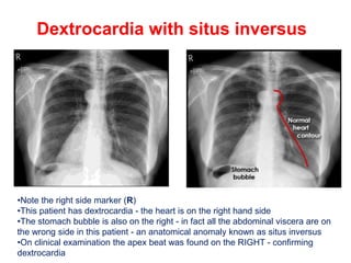
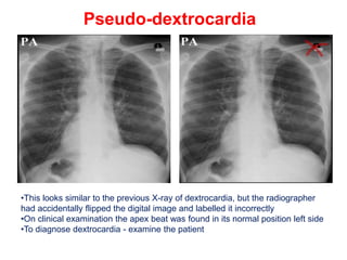
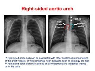
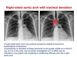
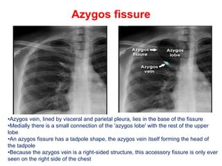
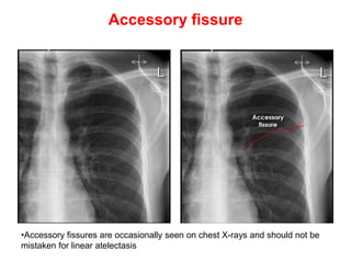
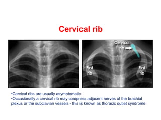
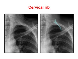
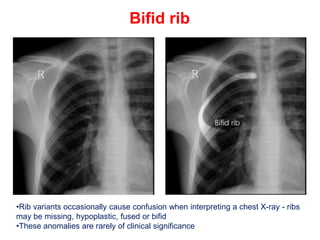
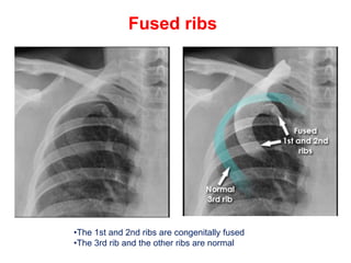
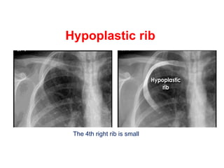
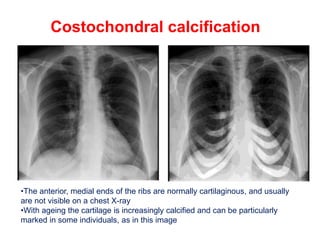
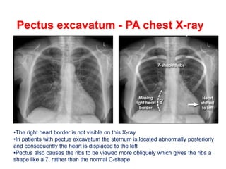
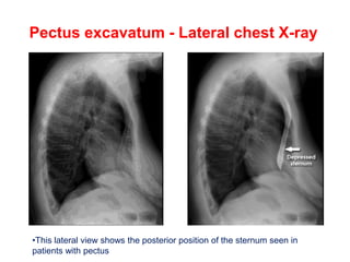
CHEST X-RAY
ANATOMICAL VARIANTS
Dextrocardia with situs inversus
•Note the right side marker (R)
•This patient has dextrocardia - the heart is on the right hand side
•The stomach bubble is also on the right - in fact all the abdominal viscera are on
the wrong side in this patient - an anatomical anomaly known as situs inversus
•On clinical examination the apex beat was found on the RIGHT - confirming
dextrocardia
Pseudo-dextrocardia
•This looks similar to the previous X-ray of dextrocardia, but the radiographer
had accidentally flipped the digital image and labelled it incorrectly
•On clinical examination the apex beat was found in its normal position left side
•To diagnose dextrocardia - examine the patient
Right-sided aortic arch
•A right-sided aortic arch can be associated with other anatomical abnormalities
of the great vessels, or with congenital heart diseases such as tetralogy of Fallot
•A right-sided aortic arch may also be an asymptomatic and incidental finding,
as in this case
Right-sided aortic arch with tracheal deviation
•A right-sided aortic arch may produce symptoms related to tracheal or
oesophageal compression
•Compression or deviation of these structures is not usually visible on a chest X-
ray, as it is in this case, and so further investigation (CT or MRI) may be
indicated in any patient with breathing or swallowing difficulty who has a right
aortic arch
Azygos fissure
•Azygos vein, lined by visceral and parietal pleura, lies in the base of the fissure
•Medially there is a small connection of the 'azygos lobe' with the rest of the upper
lobe
•An azygos fissure has a tadpole shape, the azygos vein itself forming the head of
the tadpole
•Because the azygos vein is a right-sided structure, this accessory fissure is only ever
seen on the right side of the chest
Accessory fissure
•Accessory fissures are occasionally seen on chest X-rays and should not be
mistaken for linear atelectasis
Cervical rib
•Cervical ribs are usually asymptomatic
•Occasionally a cervical rib may compress adjacent nerves of the brachial
plexus or the subclavian vessels - this is known as thoracic outlet syndrome
Cervical rib
Bifid rib
•Rib variants occasionally cause confusion when interpreting a chest X-ray - ribs
may be missing, hypoplastic, fused or bifid
•These anomalies are rarely of clinical significance
Fused ribs
•The 1st and 2nd ribs are congenitally fused
•The 3rd rib and the other ribs are normal
Hypoplastic rib
The 4th right rib is small
Costochondral calcification
•The anterior, medial ends of the ribs are normally cartilaginous, and usually
are not visible on a chest X-ray
•With ageing the cartilage is increasingly calcified and can be particularly
marked in some individuals, as in this image
Pectus excavatum - PA chest X-ray
•The right heart border is not visible on this X-ray
•In patients with pectus excavatum the sternum is located abnormally posteriorly
and consequently the heart is displaced to the left
•Pectus also causes the ribs to be viewed more obliquely which gives the ribs a
shape like a 7, rather than the normal C-shape
Pectus excavatum - Lateral chest X-ray
•This lateral view shows the posterior position of the sternum seen in
patients with pectus
Scoliosis
•Scoliosis may make the interpretation of a chest X-ray more difficult
•Thoracic scoliosis usually has a rotational element and so assessment of
heart size may not be accurate
•Note: A chest X-ray is not usually used to assess a scoliosis. If treatment is
being considered then specific scoliosis views are required

CHESTX-RAY ANATOMICAL VARIANTS......pptx

  • 1.
  • 2.
    Dextrocardia with situsinversus •Note the right side marker (R) •This patient has dextrocardia - the heart is on the right hand side •The stomach bubble is also on the right - in fact all the abdominal viscera are on the wrong side in this patient - an anatomical anomaly known as situs inversus •On clinical examination the apex beat was found on the RIGHT - confirming dextrocardia
  • 3.
    Pseudo-dextrocardia •This looks similarto the previous X-ray of dextrocardia, but the radiographer had accidentally flipped the digital image and labelled it incorrectly •On clinical examination the apex beat was found in its normal position left side •To diagnose dextrocardia - examine the patient
  • 4.
    Right-sided aortic arch •Aright-sided aortic arch can be associated with other anatomical abnormalities of the great vessels, or with congenital heart diseases such as tetralogy of Fallot •A right-sided aortic arch may also be an asymptomatic and incidental finding, as in this case
  • 5.
    Right-sided aortic archwith tracheal deviation •A right-sided aortic arch may produce symptoms related to tracheal or oesophageal compression •Compression or deviation of these structures is not usually visible on a chest X- ray, as it is in this case, and so further investigation (CT or MRI) may be indicated in any patient with breathing or swallowing difficulty who has a right aortic arch
  • 6.
    Azygos fissure •Azygos vein,lined by visceral and parietal pleura, lies in the base of the fissure •Medially there is a small connection of the 'azygos lobe' with the rest of the upper lobe •An azygos fissure has a tadpole shape, the azygos vein itself forming the head of the tadpole •Because the azygos vein is a right-sided structure, this accessory fissure is only ever seen on the right side of the chest
  • 7.
    Accessory fissure •Accessory fissuresare occasionally seen on chest X-rays and should not be mistaken for linear atelectasis
  • 8.
    Cervical rib •Cervical ribsare usually asymptomatic •Occasionally a cervical rib may compress adjacent nerves of the brachial plexus or the subclavian vessels - this is known as thoracic outlet syndrome
  • 9.
  • 10.
    Bifid rib •Rib variantsoccasionally cause confusion when interpreting a chest X-ray - ribs may be missing, hypoplastic, fused or bifid •These anomalies are rarely of clinical significance
  • 11.
    Fused ribs •The 1stand 2nd ribs are congenitally fused •The 3rd rib and the other ribs are normal
  • 12.
    Hypoplastic rib The 4thright rib is small
  • 13.
    Costochondral calcification •The anterior,medial ends of the ribs are normally cartilaginous, and usually are not visible on a chest X-ray •With ageing the cartilage is increasingly calcified and can be particularly marked in some individuals, as in this image
  • 14.
    Pectus excavatum -PA chest X-ray •The right heart border is not visible on this X-ray •In patients with pectus excavatum the sternum is located abnormally posteriorly and consequently the heart is displaced to the left •Pectus also causes the ribs to be viewed more obliquely which gives the ribs a shape like a 7, rather than the normal C-shape
  • 15.
    Pectus excavatum -Lateral chest X-ray •This lateral view shows the posterior position of the sternum seen in patients with pectus
  • 16.
    Scoliosis •Scoliosis may makethe interpretation of a chest X-ray more difficult •Thoracic scoliosis usually has a rotational element and so assessment of heart size may not be accurate •Note: A chest X-ray is not usually used to assess a scoliosis. If treatment is being considered then specific scoliosis views are required