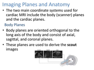
HEART EMBRYOLOGY AND
RADIOLOGICAL ANATOMY
• Mesenchymal cells in the splanchnic mesoderm
proliferate and form isolated cell clusters known
as angiogenic clusters.
• The angiogenic clusters acquire lumen.
• The intraembryonic coleomic cavity located over
the plexus later form pericardial cavity
• The embryo folds in cephalocaudal and
transversely bringing the two heart tubes closer.
• The two endocardial heart tube fuse in cephalo-
caudal direction.
• The tube is attached to the dorsal side of the
pericardial cavity by dorsal mesocardium.
• The primitive heart tube elongates and develops
alternate dilatations and constrictions.
1-truncus arteriosus.
2-bulbus cordis.
3-primitive ventricle.
4-primitive atrium.
5-sinus venosus.
• Bulbus cordis & ventricle grow faster than other
regions, so the heart bends upon itself,forming
U-shaped bulboventricular loop.
• The atrium & sinus venosus also come to lie
dorsal to truncus arteriosus, bulbus cordis &
ventricle .(S-shaped heart tube)
Formation of cardiac loop
• Heart tube elongates and bends.
• The cehpalic portion: bends in ventral and caudal
direction to the right.
• The caudal portion: shifts in a dorsocranial
direction and to the left.The bendings creates a
cardiac loop.
• Truncus arteriosus is continous cranially with
aortic sac ,from which aortic arches develop.
• Sinus venosus has right & left horns.
• Each horn receives umbilical, vitelline ,&
common cardinal veins.
• The conus cordis forms the outflow tract of
both ventricles.
• The proximal portion of the bulbus form the
primitive right ventricle.
• The primitive ventricle becomes trabeculated
and form the primitive left ventricle.
Partitioning
• Includes dividing of A-V canal , primitive atrium
& primitive ventricle.
• Endocardial cushions: these are masses of cells
and extracellular matrices develop in the
atrioventricular and conotruncal regions .
• In the atrioventricular region they are : – Dorsal
& ventral swellings
– Fuse, dividing the single AV canal into paired
canals.
Persistant AV canal
• The persistent atrioventricular canal results
from the failure of the superior and inferior
cushions to fuse.
• There is a single atrioventricular canal in
which all four chambers communicate.
• The atrial and ventricular septa cannot fully
form as they rely on the cushions to form the
membranous portions of these septa.
Septum formation in common atrium
• A sickle- shaped crest grows from the roof of the
common atrium into the lumen.
• This crest is the first portion of the septum
primum.
• The opening between the lower rim of the
septum primum and the endocardial cushions is
the ostium primum.
• Endocardial cushions grow along the edge of
the septum primum, closing the ostium primum.
• Before closure is complete, perforations develop
in the upper portion of the septum primum
forming ostium secundum.
• A new crescent-shaped fold appears, the septum
secundum,never forms a complete partition in
the atrial cavity .
• The free concave edge of the septum secundum
begins to overlap the ostium secundum.The
opening left by the septum secundum is called
the oval foramen (foramen ovale)
Atrial Septal Defect
• Primum type ASD
Ostium primum is patent because septum primum
doesn’t fuse with endocardial cushions
Secundum ASD
• It results from either excessive resorption of
septum primum or short septum secundum
leaving large patent foramen ovale.
Septum formation in ventricles
• The medial walls of the expanding ventricles
become apposed and gradually merge, forming
the muscular interventricular septum.
• The interventricular foramen, above the muscular
portion of the interventricular septum, is closed
by endocardial cushion thus forming
membranous part of septum.
Partioning of Truncus Arteriosus
• Continuous set of ridges develop in bulbus
cordis(bulbar ridges) and truncus arteriosus
(truncal ridges).
• Grow toward each other, spiraling 180º
• Fuse to form spiraling aorticopulmonaryseptum,
dividing aorta & pulmonary trunk
Heart on X-ray
• On Xray heart size, shape and contours of heart
are seen.
Position of valves on frontal and lateral xray
Bright Blood Imaging
• Bright blood imaging describes the high signal
intensity of fast-flowing blood and is typically
used to evaluate cardiac function.
• The main pulse sequences used for bright blood
imaging include GRE or SSFP.
Imaging Planes and Anatomy
• The two main coordinate systems used for
cardiac MRI include the body (scanner) planes
and the cardiac planes.
Body Planes
• Body planes are oriented orthogonal to the
long axis of the body and consist of axial,
sagittal, and coronal planes.
• These planes are used to derive the scout
images
• These are useful for the evaluation of thoracic
vascular structures cardiac morphology, the
pericardium and para cardiac masses,
• The obliquity (≈ 45°) of these planes to the walls of
the heart precludes accurate anatomic and
functional characterization.
• Such information should be obtained from the
specialized cardiac planes.
• Standard CMR planes of the heart are comparable to
the standard cardiac views, well-known and
established in other non-invasive imaging techniques
such as echocardiography, cardiac CT, x-ray LV
angiography and nuclear medicine
Cardiac Planes
• The standard cardiac planes are established
using the scout images and include short axis,
horizontal long axis (four-chamber view), and
vertical long axis (two-chamber view).
Vertical long
axis plane
Horizontal long axis
Or 4 chamber plane
3 chamber plane
Chamber wise anatomy on CT and MR
Right atrium
• Develops from
Sinus venosus – forms smooth part, receives
blood from SVC,IVC, coronary sinus
Primitive heart- forms trabeculated atrial
appendage which is broad based and triangular in
shape,
• Rough trabeculations are due to pectinate
muscles.
Crista terminalis
• It is a vertically orientated smooth muscle ridge
extending from SVC to IVC.
• It represents the fusion line between the primitive
RA and the sinus venosus portion of the right
atrium.
Eustachian valve
Thebesian valve
Left atrium
• It is the most posterosuperior chamber.
• Develops from
Absorption of pulmonary veins- forms posterior
smooth portion.
Primitive heart- forms trabeculated atrial
appendage which has
narrow attachment and
tubular in shape.
Sluggish flow in LAA
simulates thrombus
filled on the delayed image
Lipomatous hypertrophy of the
interatrial septum
Dumbbell-shaped fatty mass and
sparing of the fossa ovalis
The interatrial septum is thin containing fat ms 0.9-9.9mm
The interatrial septum separates the right and left atria and
features the fossa ovalis, which appears as an
area of focal thinning.
Right ventricle
• The right ventricle is located in the most anterior
portion of the heart behind sternum, triangular
in shape.
• It is more trabeculated and thinner than the left
ventricle.
• Unique features of the right ventricle include
-heavy trabeculation
- moderator band
• The right ventricle contains three small papillary
muscles:anterior, posterior, and medial papillary
muscles.
• If prominent, this structure can be mistaken for an
intracardiac mass or thrombus.
Moderator band
• It is a tissue ridge that extends across the right
ventricular apex from the anterior papillary
muscle to the interventricular septum.
• It contains the right bundle branch.
Left ventricle
• The left ventricle contains
two papillary muscles, the
anterior and posterior
papillary muscles.
• These are larger than the
papillary muscles of the
right ventricle.
• The posterior papillary
muscle can have a
globular shape and can be
misinterpreted as a mass
or thrombus
• Two unique features of the left ventricle are
-smooth wall
-shared annulus between mitral and aortic valves.
Physiologic thinning of the LV apex
Normal wall thickness of the
left ventricular myocardium is
6–11 mm.
Normal thinning of the interventricular septum of the
membranous portion (arrow) which can be mistaken as
a ventricular septal defect.
Coronary Arteries
Left Coronary Artery
• The left coronary artery (LCA) arises from the left
sinus of Valsalva.
RCA
LAD
LCX
LAD artery
(arrows) coursing
in the epicardial
fat of the
interventricular
groove toward
the LV apex.
LAD gives septal branches medially
and diagonal branches laterally
Septal branches supply IVS
Diagonal branches supply LV wall.
septal
diagonal
LCx artery courses in left AV groove
and gives obtuse marginal branches
Trifurcationof LCA
Right coronary artery
• The RCA arises from the right coronary sinus
and courses in the right AV groove towards the
crux of the heart.
• The first branch of the RCA is
conus artery supplies RVOT.
• In 58% of patients, the
sinoatrial nodal artery arises
from RCA; in the remaining
it arises from the LCx artery.
RCA
Acute
marginal
branch
• Multiple ventricular branches arise from RCA, the
largest is called the acute marginal branch.
Dominance
• The coronary artery that gives rise to the PDA
and posterolateral branch is referred to as the
“dominant” artery.
• RCA is dominant in 70% of cases.
• LCA is dominant in 10% of cases.
• In the remaining cases, the RCA and LCA are
codominant.
Echocardiography
• Trans-thoracic echocardiography
Standard views:
Parasternal view
Apical view
Sub xiphoid view
Suprasternal view
• Trans oesophageal echocardiography
Parasternal long axis view:
• This view is used to assess end-diastolic and
end-systolic LV dimensions.
• Parasternal short axis view
heart ppt.pptx
heart ppt.pptx
heart ppt.pptx
heart ppt.pptx

heart ppt.pptx

  • 1.
  • 3.
    • Mesenchymal cellsin the splanchnic mesoderm proliferate and form isolated cell clusters known as angiogenic clusters. • The angiogenic clusters acquire lumen. • The intraembryonic coleomic cavity located over the plexus later form pericardial cavity
  • 4.
    • The embryofolds in cephalocaudal and transversely bringing the two heart tubes closer. • The two endocardial heart tube fuse in cephalo- caudal direction. • The tube is attached to the dorsal side of the pericardial cavity by dorsal mesocardium.
  • 5.
    • The primitiveheart tube elongates and develops alternate dilatations and constrictions. 1-truncus arteriosus. 2-bulbus cordis. 3-primitive ventricle. 4-primitive atrium. 5-sinus venosus.
  • 6.
    • Bulbus cordis& ventricle grow faster than other regions, so the heart bends upon itself,forming U-shaped bulboventricular loop. • The atrium & sinus venosus also come to lie dorsal to truncus arteriosus, bulbus cordis & ventricle .(S-shaped heart tube)
  • 7.
    Formation of cardiacloop • Heart tube elongates and bends. • The cehpalic portion: bends in ventral and caudal direction to the right. • The caudal portion: shifts in a dorsocranial direction and to the left.The bendings creates a cardiac loop.
  • 9.
    • Truncus arteriosusis continous cranially with aortic sac ,from which aortic arches develop. • Sinus venosus has right & left horns. • Each horn receives umbilical, vitelline ,& common cardinal veins.
  • 10.
    • The conuscordis forms the outflow tract of both ventricles. • The proximal portion of the bulbus form the primitive right ventricle. • The primitive ventricle becomes trabeculated and form the primitive left ventricle.
  • 11.
    Partitioning • Includes dividingof A-V canal , primitive atrium & primitive ventricle. • Endocardial cushions: these are masses of cells and extracellular matrices develop in the atrioventricular and conotruncal regions .
  • 12.
    • In theatrioventricular region they are : – Dorsal & ventral swellings – Fuse, dividing the single AV canal into paired canals.
  • 13.
    Persistant AV canal •The persistent atrioventricular canal results from the failure of the superior and inferior cushions to fuse. • There is a single atrioventricular canal in which all four chambers communicate. • The atrial and ventricular septa cannot fully form as they rely on the cushions to form the membranous portions of these septa.
  • 14.
    Septum formation incommon atrium • A sickle- shaped crest grows from the roof of the common atrium into the lumen. • This crest is the first portion of the septum primum. • The opening between the lower rim of the septum primum and the endocardial cushions is the ostium primum.
  • 15.
    • Endocardial cushionsgrow along the edge of the septum primum, closing the ostium primum. • Before closure is complete, perforations develop in the upper portion of the septum primum forming ostium secundum.
  • 16.
    • A newcrescent-shaped fold appears, the septum secundum,never forms a complete partition in the atrial cavity . • The free concave edge of the septum secundum begins to overlap the ostium secundum.The opening left by the septum secundum is called the oval foramen (foramen ovale)
  • 18.
    Atrial Septal Defect •Primum type ASD Ostium primum is patent because septum primum doesn’t fuse with endocardial cushions
  • 19.
    Secundum ASD • Itresults from either excessive resorption of septum primum or short septum secundum leaving large patent foramen ovale.
  • 20.
    Septum formation inventricles • The medial walls of the expanding ventricles become apposed and gradually merge, forming the muscular interventricular septum.
  • 21.
    • The interventricularforamen, above the muscular portion of the interventricular septum, is closed by endocardial cushion thus forming membranous part of septum.
  • 23.
    Partioning of TruncusArteriosus • Continuous set of ridges develop in bulbus cordis(bulbar ridges) and truncus arteriosus (truncal ridges). • Grow toward each other, spiraling 180º
  • 24.
    • Fuse toform spiraling aorticopulmonaryseptum, dividing aorta & pulmonary trunk
  • 25.
    Heart on X-ray •On Xray heart size, shape and contours of heart are seen.
  • 30.
    Position of valveson frontal and lateral xray
  • 33.
    Bright Blood Imaging •Bright blood imaging describes the high signal intensity of fast-flowing blood and is typically used to evaluate cardiac function. • The main pulse sequences used for bright blood imaging include GRE or SSFP.
  • 34.
    Imaging Planes andAnatomy • The two main coordinate systems used for cardiac MRI include the body (scanner) planes and the cardiac planes. Body Planes • Body planes are oriented orthogonal to the long axis of the body and consist of axial, sagittal, and coronal planes. • These planes are used to derive the scout images
  • 35.
    • These areuseful for the evaluation of thoracic vascular structures cardiac morphology, the pericardium and para cardiac masses,
  • 37.
    • The obliquity(≈ 45°) of these planes to the walls of the heart precludes accurate anatomic and functional characterization. • Such information should be obtained from the specialized cardiac planes. • Standard CMR planes of the heart are comparable to the standard cardiac views, well-known and established in other non-invasive imaging techniques such as echocardiography, cardiac CT, x-ray LV angiography and nuclear medicine
  • 38.
    Cardiac Planes • Thestandard cardiac planes are established using the scout images and include short axis, horizontal long axis (four-chamber view), and vertical long axis (two-chamber view).
  • 40.
    Vertical long axis plane Horizontallong axis Or 4 chamber plane 3 chamber plane
  • 41.
  • 42.
    Right atrium • Developsfrom Sinus venosus – forms smooth part, receives blood from SVC,IVC, coronary sinus Primitive heart- forms trabeculated atrial appendage which is broad based and triangular in shape, • Rough trabeculations are due to pectinate muscles.
  • 44.
    Crista terminalis • Itis a vertically orientated smooth muscle ridge extending from SVC to IVC. • It represents the fusion line between the primitive RA and the sinus venosus portion of the right atrium.
  • 46.
  • 47.
    Left atrium • Itis the most posterosuperior chamber. • Develops from Absorption of pulmonary veins- forms posterior smooth portion. Primitive heart- forms trabeculated atrial appendage which has narrow attachment and tubular in shape.
  • 49.
    Sluggish flow inLAA simulates thrombus filled on the delayed image
  • 50.
    Lipomatous hypertrophy ofthe interatrial septum Dumbbell-shaped fatty mass and sparing of the fossa ovalis The interatrial septum is thin containing fat ms 0.9-9.9mm The interatrial septum separates the right and left atria and features the fossa ovalis, which appears as an area of focal thinning.
  • 51.
    Right ventricle • Theright ventricle is located in the most anterior portion of the heart behind sternum, triangular in shape. • It is more trabeculated and thinner than the left ventricle. • Unique features of the right ventricle include -heavy trabeculation - moderator band
  • 52.
    • The rightventricle contains three small papillary muscles:anterior, posterior, and medial papillary muscles. • If prominent, this structure can be mistaken for an intracardiac mass or thrombus.
  • 53.
    Moderator band • Itis a tissue ridge that extends across the right ventricular apex from the anterior papillary muscle to the interventricular septum. • It contains the right bundle branch.
  • 54.
    Left ventricle • Theleft ventricle contains two papillary muscles, the anterior and posterior papillary muscles. • These are larger than the papillary muscles of the right ventricle. • The posterior papillary muscle can have a globular shape and can be misinterpreted as a mass or thrombus
  • 56.
    • Two uniquefeatures of the left ventricle are -smooth wall -shared annulus between mitral and aortic valves. Physiologic thinning of the LV apex Normal wall thickness of the left ventricular myocardium is 6–11 mm.
  • 57.
    Normal thinning ofthe interventricular septum of the membranous portion (arrow) which can be mistaken as a ventricular septal defect.
  • 58.
    Coronary Arteries Left CoronaryArtery • The left coronary artery (LCA) arises from the left sinus of Valsalva. RCA LAD LCX
  • 59.
    LAD artery (arrows) coursing inthe epicardial fat of the interventricular groove toward the LV apex. LAD gives septal branches medially and diagonal branches laterally Septal branches supply IVS Diagonal branches supply LV wall. septal diagonal
  • 60.
    LCx artery coursesin left AV groove and gives obtuse marginal branches Trifurcationof LCA
  • 61.
    Right coronary artery •The RCA arises from the right coronary sinus and courses in the right AV groove towards the crux of the heart. • The first branch of the RCA is conus artery supplies RVOT. • In 58% of patients, the sinoatrial nodal artery arises from RCA; in the remaining it arises from the LCx artery. RCA Acute marginal branch
  • 62.
    • Multiple ventricularbranches arise from RCA, the largest is called the acute marginal branch. Dominance • The coronary artery that gives rise to the PDA and posterolateral branch is referred to as the “dominant” artery. • RCA is dominant in 70% of cases. • LCA is dominant in 10% of cases. • In the remaining cases, the RCA and LCA are codominant.
  • 63.
    Echocardiography • Trans-thoracic echocardiography Standardviews: Parasternal view Apical view Sub xiphoid view Suprasternal view • Trans oesophageal echocardiography
  • 64.
    Parasternal long axisview: • This view is used to assess end-diastolic and end-systolic LV dimensions.
  • 65.