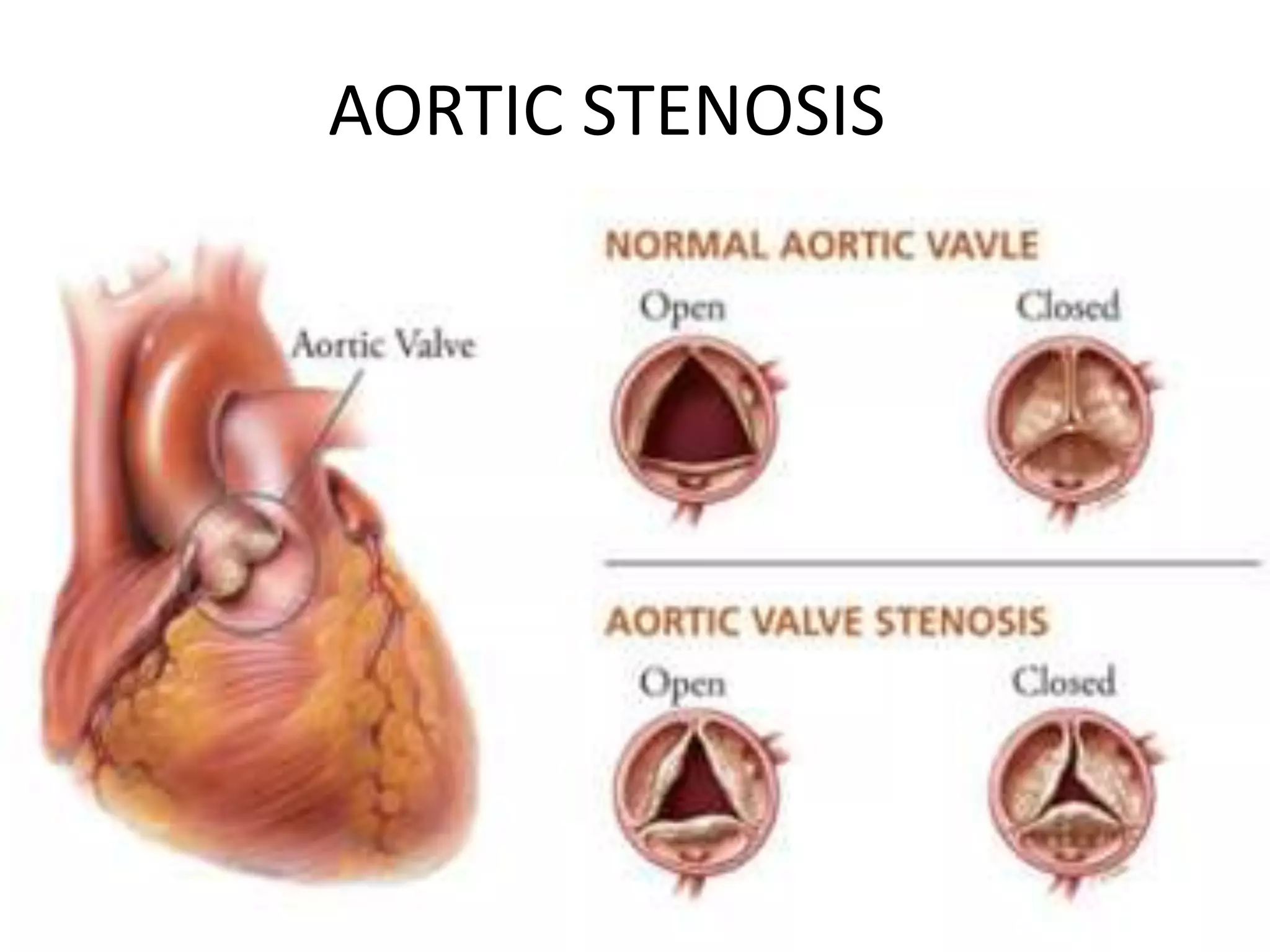
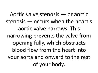
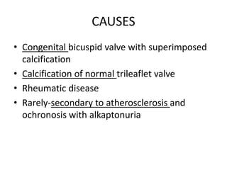
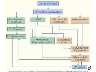
AORTIC STENOSIS
Aortic valve stenosis — or aortic
stenosis — occurs when the heart's
aortic valve narrows. This
narrowing prevents the valve from
opening fully, which obstructs
blood flow from the heart into
your aorta and onward to the rest
of your body.
TYPES
CAUSES
• Congenital bicuspid valve with superimposed
calcification
• Calcification of normal trileaflet valve
• Rheumatic disease
• Rarely-secondary to atherosclerosis and
ochronosis with alkaptonuria
1)CONGENITAL AORTIC VALVE DISEASE
• CHILDREN < 1 YEAR  MOSTLY UNICUSPID
VALVULAR STENOSIS SEEN  PRODUCE
SEVERE OBSTRUCTION OFTEN FATAL
2)CALCIFIC AORTIC VALVE DISEASE
• MCC of AS in adults
• New concept says that disease process
represents proliferative and inflammatory
changes + lipid accumulation , upregulation of
ACE activity , increased oxidative stress and
inflitration of T lymphocytes and macrophages
ultimately leading to bone formation .
• When it progresses  calcification of base of the
cusps  immobilizationof cusps
• So seen in MC in pagets disease of bone and
ESRD.
• ? Possible genetic predesposition to valve
calcification .
• Risk factors = same as that of vascular
atherosclerosis
• Statins  reduce the progression but not in
advanced disease.
3)RHEUMATIC AORTIC VALVE DISEASE
• d/t adhesions & fusion of commisures & cusps
+ vascularization of of valve leaflets 
stiffening of cusp borders  calcific nodules
develop on both sides  orifice narrows  so
often it is stenotic as well as regurgitant.
• 1.5 to 2.0 cm  mild stenosis
1.0 to 1.5 cm  severe stenosis .
CLINICAL PRESENTATION
• Exertional dyspnoea , angina , syncope ,
ultimately heart failure.
• Presentation typically @ 50 – 70 years .
• Late findings  AF, Pulm HTN , SYST Venous
HTN .
• TIA –d/t microthrombi on cusps
• Microangiopathic hemolytic anemia 
schistocytes  hemoglobinuria
PHYSICAL EXAMINATION
• Carotid upstroke
• Slow rising , late peaking ,low amplitude carotid
pulse –parvus and tardus  severe AS
• BP reduced in severe AS
• Radiation to murmur to carotid  results in
palpable thrill or carotid shudder .
• Cardiac impulse is shifted to down and left .
• Systolic thrill when patient leans forward during
full expiration  mc in R 2nd IC space radiating to
carotids.
AUSCULTATION
• ESM (crescendo decrescendo) at base of heart
radiating to carotids
• Gallavardin phenomenon  in calcific AS , murmur is
heard at base of heart but high frequency components
may radiate to apex .
• Louder and late peaking murmur indicates more severe
stenosis .
• S1 is normal .
• S2 may be single in severe AS  calcification and
immobility of aortic valve makes it inaudible.
• S4 may be heard  d/t vigorous atrial contraction.
• Murmur accentuated by squatting .
• Ejection click may be heard
ECHOCARDIOGRAPHY
• Cause and severity can be found out .
• Doppler echo measures transaortic jet
velocity  most useful in assessing severity
and clinical outcome.
CHEST CT
• Evidence of aortic root disease by measuring
the dimensions
CLINICAL OUTCOME
• 1) SYMPTOMATIC PATIENTS
• 2)ASYMPTOMATIC PATIENTS
ASYMPTOMATIC PATIENTS
• Severity increases gradually over 10 -15 years
 long latent period.
• Prognosis remains excellent in patient is
asymptomatic
• Strongest predictor of progression to
symptoms is doppler aortic jet velocity .
• Sudden death in asymptomatic patients is
unlikely .
SYMPTOMATIC PATIENTS
• Even if mild symptoms are present , survival is
poor unless outflow obstruction is relieved.
• Interval from onset of symptoms to time of death
is
2yrs  in pts with heart failure
3yrs  in pts with syncope
5yrs  in pts with angina
Recent studies shows that avg survival is 1 to 3 yrs
after symptom onset .
HEMODYNAMIC PROGRESSION
• Factors associated with hemodynamic
progression include older age , more severe
leaflet calcification ,renal insufficiency , HTN ,
smoking , and hyperlipidemia
• BNP and exercise testing  used to evaluate
disease progression .
TREATMENT
• -MEDICAL
• -SURGICAL
MEDICAL
• Diuretics
• Avoid beta blockers and nitrates
• Manage concurrent cardiac conditions
• Cautious use of ACE inhibitors
• If HF occurs  digoxin and diuretics
SURGICAL
• Balloon aortic valvotomy
• AVR – if symptomatic.
• TAVI – new technology

Aortic stenosis- Dr Shaz Pamangadan

  • 1.
  • 2.
    Aortic valve stenosis— or aortic stenosis — occurs when the heart's aortic valve narrows. This narrowing prevents the valve from opening fully, which obstructs blood flow from the heart into your aorta and onward to the rest of your body.
  • 3.
  • 4.
    CAUSES • Congenital bicuspidvalve with superimposed calcification • Calcification of normal trileaflet valve • Rheumatic disease • Rarely-secondary to atherosclerosis and ochronosis with alkaptonuria
  • 5.
  • 6.
    • CHILDREN <1 YEAR  MOSTLY UNICUSPID VALVULAR STENOSIS SEEN  PRODUCE SEVERE OBSTRUCTION OFTEN FATAL
  • 7.
    2)CALCIFIC AORTIC VALVEDISEASE • MCC of AS in adults • New concept says that disease process represents proliferative and inflammatory changes + lipid accumulation , upregulation of ACE activity , increased oxidative stress and inflitration of T lymphocytes and macrophages ultimately leading to bone formation . • When it progresses  calcification of base of the cusps  immobilizationof cusps • So seen in MC in pagets disease of bone and ESRD.
  • 8.
    • ? Possiblegenetic predesposition to valve calcification . • Risk factors = same as that of vascular atherosclerosis • Statins  reduce the progression but not in advanced disease.
  • 9.
    3)RHEUMATIC AORTIC VALVEDISEASE • d/t adhesions & fusion of commisures & cusps + vascularization of of valve leaflets  stiffening of cusp borders  calcific nodules develop on both sides  orifice narrows  so often it is stenotic as well as regurgitant. • 1.5 to 2.0 cm  mild stenosis 1.0 to 1.5 cm  severe stenosis .
  • 11.
    CLINICAL PRESENTATION • Exertionaldyspnoea , angina , syncope , ultimately heart failure. • Presentation typically @ 50 – 70 years . • Late findings  AF, Pulm HTN , SYST Venous HTN . • TIA –d/t microthrombi on cusps • Microangiopathic hemolytic anemia  schistocytes  hemoglobinuria
  • 12.
    PHYSICAL EXAMINATION • Carotidupstroke • Slow rising , late peaking ,low amplitude carotid pulse –parvus and tardus  severe AS • BP reduced in severe AS • Radiation to murmur to carotid  results in palpable thrill or carotid shudder . • Cardiac impulse is shifted to down and left . • Systolic thrill when patient leans forward during full expiration  mc in R 2nd IC space radiating to carotids.
  • 13.
    AUSCULTATION • ESM (crescendodecrescendo) at base of heart radiating to carotids • Gallavardin phenomenon  in calcific AS , murmur is heard at base of heart but high frequency components may radiate to apex . • Louder and late peaking murmur indicates more severe stenosis . • S1 is normal . • S2 may be single in severe AS  calcification and immobility of aortic valve makes it inaudible. • S4 may be heard  d/t vigorous atrial contraction. • Murmur accentuated by squatting . • Ejection click may be heard
  • 14.
    ECHOCARDIOGRAPHY • Cause andseverity can be found out . • Doppler echo measures transaortic jet velocity  most useful in assessing severity and clinical outcome.
  • 15.
    CHEST CT • Evidenceof aortic root disease by measuring the dimensions
  • 16.
    CLINICAL OUTCOME • 1)SYMPTOMATIC PATIENTS • 2)ASYMPTOMATIC PATIENTS
  • 17.
    ASYMPTOMATIC PATIENTS • Severityincreases gradually over 10 -15 years  long latent period. • Prognosis remains excellent in patient is asymptomatic • Strongest predictor of progression to symptoms is doppler aortic jet velocity . • Sudden death in asymptomatic patients is unlikely .
  • 18.
    SYMPTOMATIC PATIENTS • Evenif mild symptoms are present , survival is poor unless outflow obstruction is relieved. • Interval from onset of symptoms to time of death is 2yrs  in pts with heart failure 3yrs  in pts with syncope 5yrs  in pts with angina Recent studies shows that avg survival is 1 to 3 yrs after symptom onset .
  • 19.
    HEMODYNAMIC PROGRESSION • Factorsassociated with hemodynamic progression include older age , more severe leaflet calcification ,renal insufficiency , HTN , smoking , and hyperlipidemia • BNP and exercise testing  used to evaluate disease progression .
  • 20.
  • 21.
    MEDICAL • Diuretics • Avoidbeta blockers and nitrates • Manage concurrent cardiac conditions • Cautious use of ACE inhibitors • If HF occurs  digoxin and diuretics
  • 22.
    SURGICAL • Balloon aorticvalvotomy • AVR – if symptomatic. • TAVI – new technology