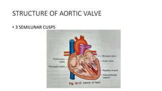
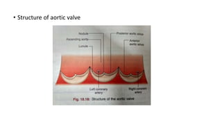
AORTIC VALVE – ANATOMY
&
AORTIC STENOSIS
STRUCTURE OF AORTIC VALVE
• 3 SEMILUNAR CUSPS
• Cusps are attached directly to the vessel wall
• Nodule
• Lunule
• Aortic sinuses - coronary arteries arise from anterior and left
posterior aortic sinuses
• Structure of aortic valve
• Narrowing of the valve orifice due to fusion of cusps is known as –
stenosis
• Dilatation of the valve orifice or stiffening of the cusps causes
imperfect closure of valves leading to back flow of blood –
incompetence or regurgitation
AORTIC STENOSIS
• Occurs in one fourth of all patients with chronic valvular heart disease
• 80% of adult patients with symptomatic AS are male
CAUSES
Infants, children, adoloscents
• Congenital aortic stenosis
• Congenital subvalvular aortic stenosis
• Congenital supravalvular aortic stenosis
Young adults to middle aged
• Calcification and fibrosis of congenitally bicuspid aortic valve
• Rheumatic aortic stenosis
Middle aged to elderly
• Senile degenerative aortic stenosis
• Calcification of bicuspid valve
• Rheumatic aortic stenosis
• Congenital (bicuspid, unicuspid)
• Degenerative calcific
• Rheumatic fever
• Radiation
Pathogeensis
• Initially cardiac output is maintained by steady increase in pressure
gradient across aortic valve
• LV hypertrophy and coronary blood flow inadequate to supply the
myocardium- angina
• LV failure and pulmonary edema
Symptoms
• Cardinal symptoms are angina,breathlessness and syncope
• Mild or moderate stenosis: usually asymptomatic
• Exertional dyspnoea
• Angina
• Exertional syncope
• Sudden death
• Episodes of acute pulmonary edema
Signs
• Ejection systolic murmur
• Pulsus parvus et tardus
• Slow rising carotid pulse
• A thrill or anacrotic shudder over carotid arteries
• a wave in the jvp is accentuated
• Thrusting apex beat
• Narrow pulse pressure
• Signs of pulmonary venous congestion
• LV impulse is usually displaced laterally
• Double apical impulse (with a palpable S4)
• Auscultation
oEjection systolic murmur
oParadoxical splitting of S2
oS4 is audible at the apex
Investigations
ECG
Left ventricular hypertrophy – ST segment depression and T wave inversion
(LV strain) in lead I and aVL
Left bundle branch block
Chest X ray
May be normal; sometimes enlarged left ventricle and dilated ascending
aorta on postero-anterior view, calcified valve on lateral view
Echocardiogram
Calcified valve with restricted opening, hypertrophied left ventricle
Doppler
Measurement of severity of stenosis(systolic gradient across the aortic valve)
Detection of associated aortic regurgitation
Cardiac catheterisation
Mainly to identify associated coronary artery disease
May be used to measure gradient between left ventricle and aorta
Natural history
• Average time to death after the onset of various symptoms
• Angina – 3 years
• Syncope – 3 years
• Dyspnoea – 2 years
• Congestive heart failure – 1.5 to 2 years
Medical treatment
• Nitroglycerin
• Beta blockers , ACE inhibitors
• Statins
• Surgical – aortic valve replacement
• Percutaneous balloon aortic valvuloplasty
• Children and young adults
• Bridge to operation
• Percutaneous aortic valve replacement
Aortic stenosis in oldage
• Most common form of valve disease affecting very old
• Syncope, angina, heart failure
• Because of increasing stiffening in the central arteries, low pulse
pressure and a slow rising pulse may not be present
• Trans catheter aortic valve implantation (TAVI)
• Biological valve is preferable
THANK YOU

Aortic stenosis

  • 1.
    AORTIC VALVE –ANATOMY & AORTIC STENOSIS
  • 2.
    STRUCTURE OF AORTICVALVE • 3 SEMILUNAR CUSPS
  • 3.
    • Cusps areattached directly to the vessel wall • Nodule • Lunule • Aortic sinuses - coronary arteries arise from anterior and left posterior aortic sinuses
  • 4.
    • Structure ofaortic valve
  • 5.
    • Narrowing ofthe valve orifice due to fusion of cusps is known as – stenosis • Dilatation of the valve orifice or stiffening of the cusps causes imperfect closure of valves leading to back flow of blood – incompetence or regurgitation
  • 6.
    AORTIC STENOSIS • Occursin one fourth of all patients with chronic valvular heart disease • 80% of adult patients with symptomatic AS are male
  • 7.
    CAUSES Infants, children, adoloscents •Congenital aortic stenosis • Congenital subvalvular aortic stenosis • Congenital supravalvular aortic stenosis
  • 8.
    Young adults tomiddle aged • Calcification and fibrosis of congenitally bicuspid aortic valve • Rheumatic aortic stenosis Middle aged to elderly • Senile degenerative aortic stenosis • Calcification of bicuspid valve • Rheumatic aortic stenosis
  • 9.
    • Congenital (bicuspid,unicuspid) • Degenerative calcific • Rheumatic fever • Radiation
  • 10.
    Pathogeensis • Initially cardiacoutput is maintained by steady increase in pressure gradient across aortic valve • LV hypertrophy and coronary blood flow inadequate to supply the myocardium- angina • LV failure and pulmonary edema
  • 11.
    Symptoms • Cardinal symptomsare angina,breathlessness and syncope • Mild or moderate stenosis: usually asymptomatic • Exertional dyspnoea • Angina • Exertional syncope • Sudden death • Episodes of acute pulmonary edema
  • 12.
    Signs • Ejection systolicmurmur • Pulsus parvus et tardus • Slow rising carotid pulse • A thrill or anacrotic shudder over carotid arteries • a wave in the jvp is accentuated • Thrusting apex beat • Narrow pulse pressure • Signs of pulmonary venous congestion
  • 13.
    • LV impulseis usually displaced laterally • Double apical impulse (with a palpable S4)
  • 14.
    • Auscultation oEjection systolicmurmur oParadoxical splitting of S2 oS4 is audible at the apex
  • 16.
    Investigations ECG Left ventricular hypertrophy– ST segment depression and T wave inversion (LV strain) in lead I and aVL Left bundle branch block Chest X ray May be normal; sometimes enlarged left ventricle and dilated ascending aorta on postero-anterior view, calcified valve on lateral view
  • 17.
    Echocardiogram Calcified valve withrestricted opening, hypertrophied left ventricle Doppler Measurement of severity of stenosis(systolic gradient across the aortic valve) Detection of associated aortic regurgitation Cardiac catheterisation Mainly to identify associated coronary artery disease May be used to measure gradient between left ventricle and aorta
  • 18.
    Natural history • Averagetime to death after the onset of various symptoms • Angina – 3 years • Syncope – 3 years • Dyspnoea – 2 years • Congestive heart failure – 1.5 to 2 years
  • 19.
    Medical treatment • Nitroglycerin •Beta blockers , ACE inhibitors • Statins
  • 20.
    • Surgical –aortic valve replacement • Percutaneous balloon aortic valvuloplasty • Children and young adults • Bridge to operation • Percutaneous aortic valve replacement
  • 21.
    Aortic stenosis inoldage • Most common form of valve disease affecting very old • Syncope, angina, heart failure • Because of increasing stiffening in the central arteries, low pulse pressure and a slow rising pulse may not be present • Trans catheter aortic valve implantation (TAVI) • Biological valve is preferable
  • 22.